NOTE: Transgenic lines used in this study include macrophage-specific lineage mpeg1:mCherry (constructed in-house), neutrophil-specific mpo: GFP ,erythroid-specific gata1:dsRed and ubiq:secAnnexinV-mVenus, a reporter for cell death (re-derived in house)on wild-type, nacre (mitfaw2/w2) and mutant (bbhm292) backgrounds.
1. Day 0: Egg production and collection
- Collect fertilized embryos from natural spawning in breeding boxes produced from 1 male and 1-2 female adult zebrafish.
NOTE: For atorvastatin protocol, any wildtype/transgenic animals can be used; however, hemorrhage rates differ slightly between strains.
- Incubate 100 embryos at 28 °C in standard E3 embryo medium per Petri dish and stage according to standard guidelines.
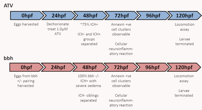
- At ~6 hours post fertilization (hpf), remove dead and unfertilized embryos from the dish using a Pasteur pipette.Figure 1 shows the experimental timeline.
2. Day 1: Atorvastatin treatment at 24 hpf
- Dechorionate embryos for atorvastatin treatment using sharp, ultra-thin dissection forceps. Numbers required for experimentation can be adjusted accordingly.
- Add 30 mL of E3 embryo medium to two clean Petri dishes. Use one dish for 100 embryos.
NOTE: If plates are designed for cell culture, dechorionated zebrafish at this early stage often stick to the bottom. To avoid this, rinse the plates thoroughly in clean water before use.
- Remove 60 µL of embryo water from the treatment plate and add 60 µL of 0.5 mM atorvastatin (ATV). At a 0.5 mM stock concentration, the above dilution will result in a final concentration of 1 µM, resulting in ~20% of larvae non-hemorrhaged (ICH-) and ~80% of larvae hemorrhaged (ICH+). Use the other plate for untreated controls.
NOTE: Atorvastatin is solubilized in distilled water (3 mg into 10 mL) to make a 0.5 mM stock solution. Incubate overnight at room temperature in the dark with agitation as solubilization takes some time. Complete solubilization can take up to 1 week. Do not use DMSO(Dimethyl sulfoxide). Solution is aliquoted and stored at -20°C. Do not freeze-thaw.
- Using a Pasteur pipette, transfer 100 embryos in as little water as possible to the treatment plates.
- Incubate the plates at 28 °C.
NOTE: ICH(intracerebral hemorrhage) will occur between 33 and 48 hpf. Atorvastatin does not need to be removed as incubation longer than 24 h does not cause any further developmental issues.
3. Day 2: Separating ICH- and ICH+ populations at 50 hpf
- Separate ICH+ fish from ICH- populations and transfer to new dishes for ease.
- If using the ATV model at a concentration of 1 µM, 75-100% of larvae will exhibit hemorrhage (ICH+) at this time point.
NOTE: The response of the larvae differs between strains, if larvae are not hemorrhaging at the desired frequencies, use a fresh batch of atorvastatin or a higher concentration. If larvae have not exhibited hemorrhage by 48 hpf then consider them ICH-.
- If using the bbh model, all homozygous mutants will exhibit hemorrhage by 48 hpf. If using a heterozygous incross, the ICH- heterozygous and wild-type siblings can be used as control animals for experiments.
- If necessary, anesthetize the larvae by adding 0.02% MS222 to the E3 media. Using a Pasteur pipette, sort the larvae for the presence of blood in the head into fresh E3 media.See Figure 2.
NOTE: Blood in the head may appear in the fore, mid, or hindbrain or in combination, and bleed volume can vary between animals. In the bbh mutants, ICH is often associated with severe edema, which is recognizable by larger heads, wider space between the eyes, and a more diffuse bleed. However, not all ICH+ bbh larvae exhibit edema.