All procedures involving animal models have been reviewed by the local institutional animal care committee and the JoVE veterinary review board.
1. Recombinant Adeno-Associated Virus (rAAV) Production and Purification
Note: rAAV vector production and purification were performed by the Leuven Viral Vector Core (LVVC).
- Briefly, transfect subconfluent low (<50) passage adherent HEK 293T cells using a 25kD linear polyethylenimine 150 nM NaCl transfection solution and three different plasmids in a ratio of 1:1:1 in DMEM medium 2% fetal bovine serum. After 24 hr of incubation at 37 °C in a 5% CO2, replace the medium with fresh DMEM medium 2% fetal bovine serum.
Note: The plasmids include the constructs for the AAV7 serotype, the AAV transfer plasmid encoding the human A53T mutant alpha-synuclein (α-SYN) under the control of the CMVie enhanced synapsin1 promoter, and the pAdvDeltaF6 adenoviral helper plasmid.
- Harvest the medium 5 days after transient transfection and concentrate using tangential flow filtration.
- Purify the rAAV vector particles from the concentrated medium using an iodixanol step gradient.
- Use standard techniques of real-time PCR for genomic copy (GC) determination. In this protocol, a vector titer of 3.0 E11 GC/ml was used to develop an α-SYN based rat model for Parkinson's disease (PD).
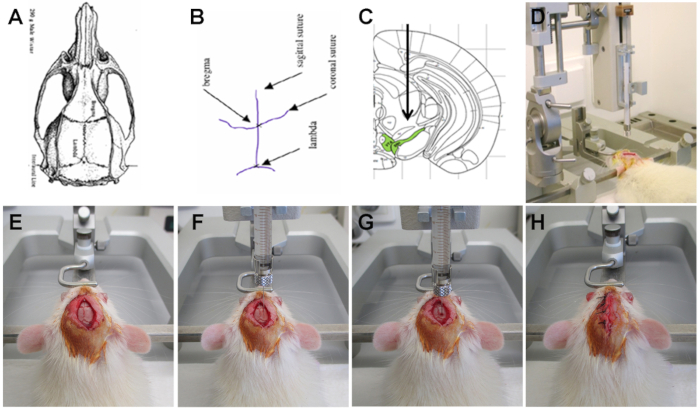
2. Stereotactic injection of rAAV α-SYN Vector in the substantia nigra (SN) of the Rat (Figure 1)
- House eight-week-old female Wistar rats weighing about 200-250 g under a normal 12 hr light/dark cycle with free access to pelleted food and tap water.
- Submit the rat to intraperitoneal (i.p.) anesthesia containing a mixture of ketamine (60 mg/kg) and medetomidine (0.4 mg/kg). Once the rat is anesthetized and doesn't react when squeezing the different paws, administer a micro-transponder subcutaneously on the back of the rat for further recognition using a micro-transponder implanter. Check if the micro-transponder is positioned correctly and can be read out by the reading device.
- Cut the hair on top of the scalp. Apply a local anesthetic on both the scalp and the ears. Perform the rest of the surgical procedure under a laminar flow using aseptic techniques.
- Place the rats in a stereotactic head frame using two ear bars, a mouth, and a nose bar. Cover the body of the rat with a paper blanket to avoid a drop in body temperature. Apply an ocular lubricant to prevent the eyes from drying.
- Disinfect the scalp with jodium 1% in isopropanol 70% and make a small incision in the midline of the scalp. Gently scrape away the membranes on the skull and rinse with saline. Let the skull dry for several minutes. Observe the cranial sutures and the two reference points: Bregma and Lambda.
- To inject the rAAV vector into the SN, define the coordinates towards Bregma (anteroposterior: 5.3 mm; mediolateral: 2.0 mm; and dorsoventral: 7.2 mm calculated from the dura).
Note: The three-dimensional coordinates for each region of interest can be calculated using a stereotaxic atlas of the rat brain, applying Bregma as anatomical reference point.
- Fill a 10 µl microinjection syringe (30-gauge 20 mm) with the rAAV vector and place it in the stereotaxic instrument connected to a motorized microinjection pump. Control the volume by releasing a drop of vector and eliminate in a polyvalent cleaning detergent pH 9 (e.g. RBS).
- Visually check if the head is fixed straight in the head frame and evaluate the left-right axis. Carefully visually define the anteroposterior and mediolateral coordinates for Bregma and Lambda and measure their height using a 30-gauge 20 mm needle in the dorsoventral arm of the stereotactic frame.
- Allow a maximum of 0.3 mm difference in height between Bregma and Lambda. Place the needle back on Bregma and apply the anteroposterior and mediolateral coordinates by moving the anteroposterior and the mediolateral arm of the stereotactic frame.
- At the place of injection, measure the height of the skull and ensure that it does not differ more than 0.3 mm from the height of Bregma. Drill a hole in the skull with a diameter of approximately 2 mm. Measure the height of the dura, which will serve as a reference for applying the dorsoventral coordinate. Alternatively, subtract a fixed thickness for the skull (0.9 mm).
- Penetrate the dura using a 26-gauge needle and absorb the blood with sterile tissue. Wait until all bleeding has stopped before proceeding.
- Slowly insert the 10 µl microinjection syringe pre-loaded with vector solution into the brain to the pre-determined depth (dorsoventral coordinate). Wait 1 min with the needle in place. Inject 3 µl of vector solution (3.0 E11 genome copies/ml (medium vector dose) or 1.0 E12 GC/ml (high vector dose) of rAAV2/7 α-SYN or eGFP control vector) using the motorized microinjection pump with a throughput of 0.25 µl/min.
- After injection, keep the needle in place for another 5 min before slowly removing it. Stitch the scalp using coated braided polyester 3.0, disinfect with 1% jodium in 70% isopropanol and gently remove the animal from the stereotactic instrument. First loosen the nose and mouth bar, then the two ear bars.
- To reverse the anesthesia, inject the rat intraperitoneally with 0.5 mg/kg atipamezole and place the rat in a clean cage on a heating plate of 38 °C until it wakes up. Cover the rat with a paper blanket to prevent a drop in body temperature.
- Provide easy access to food and water for the first hours. Monitor the rat for the first few days. If necessary, apply analgesia.
Note: There is no need to remove the stitches from the skull. After 1-2 weeks the skull is completely repaired, and the stiches come loose.