Method Article

Induction of a Subarachnoid Hemorrhage in a Mouse Model through Endovascular Perforation

July 8th, 2025

In This Article

Abstract

Loading...
$$\rightleftharpoonup{xx}$$ $$\longleftharp{xx}$$, $$\longrightharp{xx}$$,

Source: Liu, S., et al. Endovascular Perforation Model for Subarachnoid Hemorrhage Combined with Magnetic Resonance Imaging (MRI). J. Vis. Exp. (2021).

The video demonstrates the induction of subarachnoid hemorrhage via endovascular perforation in a mouse model. An incision is made in the neck region of the anesthetized mouse to access the common carotid artery and its bifurcation. A filament is inserted through the arteries to perforate an intracranial artery branch, inducing subarachnoid hemorrhage.
Protocol

Loading...
$$\rightleftharpoonup{xx}$$ $$\longleftharp{xx}$$, $$\longrightharp{xx}$$,

All procedures involving animal models have been reviewed by the local institutional animal care committee and the JoVE veterinary review board.

1. Animal preparation

  1. Induce anesthesia by injecting ketamine (70 mg/kg) and xylazine (16 mg/kg) intraperitoneally. Maintain normal body temperature, contributing to quick induction of deep anesthesia. Test for adequate sedation with a pain stimulus, such as a toe pinch, and verify the absence of a reaction.
  2. Carefully shave the neck hair of the mouse with a razor, clean it with 70% ethanol followed by betadine/chlorhexidine, and apply 1% lidocaine on the skin surface for local pain control.
  3. Place the mouse in a supine position. Use tape to fix the limbs and tail, gently stretching the skin of the neck to the opposite side of the surgery. Simultaneously, elevate the neck slightly.
  4. Use ophthalmic ointment (e.g., 5% dexpanthenol) to prevent dehydration of the eyes during the operation.

2. SAH induction

  1. Open the neck skin with a sterile scalpel, from the chin to the upper edge of the breastbone (1.5 cm), and bluntly separate salivary glands from their surrounding connective tissue.
  2. Separate the muscle group along one side [in this case, the right side] of the trachea, exposing the common carotid artery (CCA) sheath covered with nourishing blood vessels and venules. The CCA and the nerves are located in close proximity to each other.
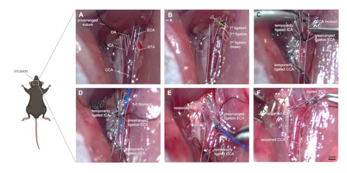
  3. Dissociate the CCA and leave a free 8-0 silk suture around the CCA without ligating it in advance. Pay attention to the protection of the vagal nerve, as it is easily damaged (Figure 1A).
  4. A triple bifurcation of the CCA, the internal carotid artery (ICA), and the external carotid artery (ECA) is visible along the lower posterior third of the diastasis. Dissect the distal end of the ECA and ligate the vessel twice as far distaally as possible.
  5. Disconnect the ECA at the midpoint of the twice ligated segment, creating a vessel stump.
  6. Prearrange one ligation for the filament around the ECA stump, do not close it until successful filament insertion.
  7. Use a suture or micro clip to occlude the ICA and CCA temporarily (Figure 1B).
  8. Make a small incision (approximately half of the ECA diameter) in the ECA using microvascular scissors. Insert a 5-0 (alternatively 4-0) prolene filament into the ECA and advance it into the CCA.
  9. Close the ligature on the ECA slightly while loosening the micro clip on the ICA and CCA (Figure 1C).
  10. Gently pull back on the filament and adjust the ECA stump in the cranial direction, invaginating the filament through the bifurcation into the ICA (Figure 1D).
  11. Point the filament tip medially at an angle of ~30° to the tracheal midline and ~30° to the horizontal plane. Push the filament forward inside the ICA. After reaching the anterior cerebral artery (ACA)- middle cerebral artery (MCA) bifurcation, resistance is encountered (~9 mm).
  12. Advance the filament 3 mm further, perforating the right ACA. Promptly withdraw the filament to the ECA stump, allowing blood flow into the subarachnoid space.
  13. Keep the filament in this position for about 10 s (Figure 1E). The presence of muscle tremors, ipsilateral miosis, gasping for breath, altered heart rhythm, and urinary incontinence can be supporting evidence of successful surgery.
  14. Temporarily close the CCA to avoid excess blood loss. Pull out the filament instantly and ligate the ECA with the prearranged suture. Reopen the CCA and allow reperfusion and further effusion of blood into the subarachnoid space (Figure 1F).
  15. After checking for bleeding leakage, disinfect the skin surrounding the wound to prevent postoperative skin infections, and suture the wound with a non-absorbable 4-0 polyester fiber suture.
  16. Place the mouse in a thermal box until consciousness is regained. Wait until the animal is fully awake and ensure it has regained sufficient consciousness to maintain sternal recumbency. Do not return animals to the company of other mice until fully recovered.
  17. Administer 200-300 mg/kg body weight paracetamol for postoperative pain relief.
  18. Check on the mice daily after surgery.

Access restricted. Please log in or start a trial to view this content.

Results

Loading...
$$\rightleftharpoonup{xx}$$ $$\longleftharp{xx}$$, $$\longrightharp{xx}$$,

Surgical procedure on rat for ischemic stroke; prearranged sutures, ligation steps, incision sites.
Figure 1: Step-by-step images of surgical technique. (A) Depiction of the exposed right carotid artery anatomy: the CCA and its bifurcation into ICA and ECA are identified, as well as the small branches of the ECA (OA and STA). (...

Access restricted. Please log in or start a trial to view this content.

Disclosures

Loading...
$$\rightleftharpoonup{xx}$$ $$\longleftharp{xx}$$, $$\longrightharp{xx}$$,
No conflicts of interest declared.

Materials

List of materials used in this article
NameCompanyCatalog NumberComments
Eye creamBayer815529836Bepanthen
Ligation suture (5-0)SMISilk black USP
Light source for microscopeZeissCL 6000 LED
KetamineCP-pharma797-037100 mg/mL
Paracetamol (40 mg/mL)bene Arzneimittel4993736
Prolene filament (5-0)ErhiconEH7255
RazorWellaHS61
Surgical instrument (Fine Scissors)FST14060-09
Surgical instrument (forceps#1)AESCULAPFM001R
Surgical instrument (forceps#2)AESCULAPFD2855R
Surgical instrument (forceps#3)HammacherHCS 082-12
Surgical instrument (Needle holder)FST91201-13
Surgical instrument (Vannas Spring Scissors)FST15000-08
Surgical microscopeZeissStemi 2000 C
Wounding suture(4-0)ErhiconCB84D
XylavetCP-pharma797-06220 mg/mL

Reprints and Permissions

Request permission to reuse the text or figures of this JoVE article

Request Permission

Tags

Common Carotary ArteryInternal Carotid ArteryExternal Carotid ArteryAnterior Cerebral ArteryFilament InsertionVascular OcclusionHemorrhage Induction

Related Articles