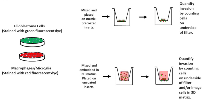
1. Fluorescent Labeling of Cells
NOTE: Label glioblastoma cell lines and microglia with fluorescent dyes 9. Alternatively, generate cell lines that constitutively express fluorescent proteins such as GFP/RFP.
- Plate cells on a 6 well plate such that they will be 70 - 80% confluent on the day of staining. For the murine glioblastoma cell line GL261 and human glioblastoma cell line U87, plate 1 x 10⁶ and 1.5 x 10⁶ cells, respectively, on a 6 cm dish 24 hr before staining.
- Prepare fluorescent cell stain dye solution in DMSO (Dimethyl sulfoxide) and add 5 µM dye to media, either Roswell Park Memorial Institute medium (RPMI) or Macrophage Serum Free Medium (MSFM), vortex well.
- Incubate cells with dye for 30 min at 37 °C and 5% CO2.
- Remove dye containing media and add fresh media (RPMI or MSFM) to the cells.
- Incubate cells for 30 min. Note: Cells are now stained and ready to be used in the experiment.
2. Invasion Assay "3D"-embedding Glioma and Macrophages/Microglia in Matrix
- Thaw matrix mix at 4 °C overnight. Perform all further manipulations using a matrix on ice in the hood.
- Prepare 10 mg/ml concentration of matrix in media (MSFM or RPMI) with 0.3% BSA on ice.
NOTE: The stock concentration of the matrix is typically around 15 mg/ml.
- To prepare cells (labeled in step 1) for suspension in the matrix, remove media from cells by aspiration. Wash cells on a dish with 2 mM EDTA (Ethylenediamine tetraacetic acid)/PBS (Phosphate-buffered saline). Add 500 µl of 2 mM EDTA/PBS to cells and incubate for 5 - 10 min at 37 °C and 5% CO2.
- Resuspend cells in 5 ml of media containing 0.3% BSA.
- Centrifuge for 5 min at 120 x g.
- Aspirate the supernatant from the pellet. Resuspend pellet in media/0.3% BSA such that the concentration of cells is equal to 1 x 10⁶ cells/ml. Use a hemocytometer to count the cells.
- Add 1.5 x 10⁵ GL261 cells (in 150 µl) + 5 x 10⁴ microglia cells (in 50 µl) into a 1.5 ml microfuge tube. Add 2 x 10⁵ GL261 cells (200 µl) into a separate 1.5 ml microfuge tube.
NOTE: GL261 cells alone without microglia serve as a control.
- Spin cells in a microfuge for 5 min at 120 x g.
- Resuspend cells in 200 µl cold matrix on ice.
- Plate 50 µl (50,000 cells) onto the center of the top compartment of the 8 µM pore size chamber insert.
- Incubate at 37 °C for 30 min for polymerization to occur.
- Add 200 µl serum-free medium to the upper chamber and 700 µl serum-containing cell growth medium to the lower well.
- Incubate at 37 °C, 5% CO2 for 48 hr.
- After incubation, remove cells from the top of the chamber by gentle aspiration and fix chambers by placing them in 3.7% formaldehyde in PBS. Allow chambers to remain in fixative for 15 min at room temperature (or overnight at 4 °C). Remove fixative by gentle aspiration and replace with 500 µl 1x PBS. Proceed to section 4 for image analysis.