A subscription to JoVE is required to view this content. Sign in or start your free trial.

Method Article

Assessing Transendothelial Electrical Resistance in an In Vitro Blood-Brain Barrier Model

1K views

July 8th, 2025

In This Article

Abstract

Source: Kuzmanov, I., et al. An In Vitro Model of the Blood-brain Barrier Using Impedance Spectroscopy: A Focus on T Cell-endothelial Cell Interaction. J. Vis. Exp. (2016)

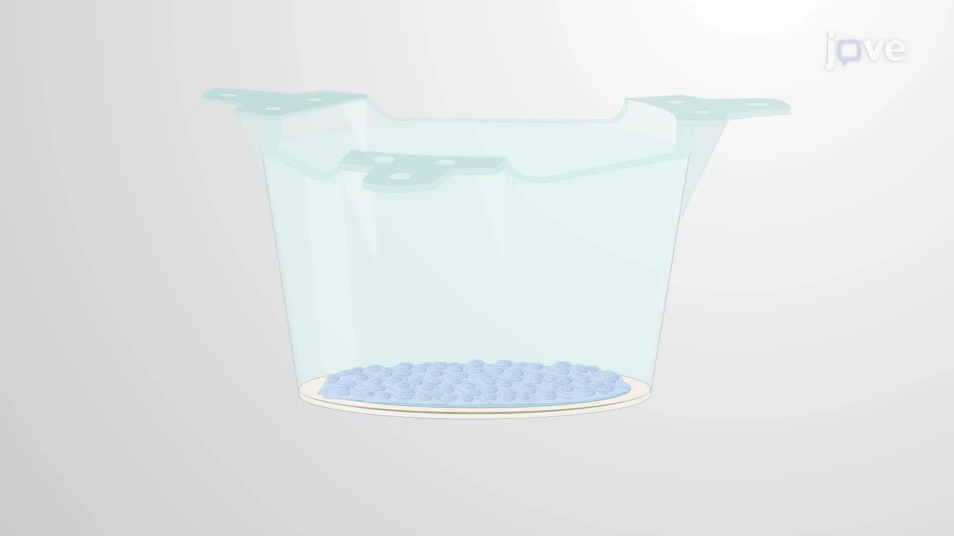
This video demonstrates a method for assessing transendothelial electrical resistance (TEER) in mouse brain microvascular endothelial cells in vitro. A high TEER value indicates intact endothelial cell junctions, while the addition of activated T cells lowers the TEER value, reflecting disruption of the junctional barrier.
Protocol

1. Setting Up and Performing Transendothelial Electrical Resistance (TEER) Measurement

  1. Place the 24-well module of the transendothelial electrical resistance (TEER) instrument under the laminar flow hood. Remove the lids and place inserts with primary mouse brain microvascular endothelial cells (MBMECs) in the instrument using forceps.
  2. Pipette 810 µl of fresh medium to the lower compartment of the module wells: add it carefully between the insert and the wall of the module well.
  3. Close the lids and place the instrument in the incubator. Connect the instrument to its computer; turn on the instrument controller and op....

Access restricted. Please log in or start a trial to view this content.

Disclosures

No conflicts of interest declared.

Materials

List of materials used in this article
NameCompanyCatalog NumberComments
cellZscopenanoAnalytics GmbHwww.nanoanalytics.comincluding: 24-well Cell Module, Controller, PC with cellZscope software v2.2.2
Transwell membrane inserts - pore size 0.4 µmCorning3470for TEER measurement as the main readout
Transwell membrane inserts - pore size 3 µmCorning3472for TEER measurement as the quality control prior to T-cell transmigration assay
24-well cell culture plateGreiner650 180flat-bottom; for MBMEC culture
96-well cell culture plateCostar3526round-bottom; for immune cell culture
Neubauer counting chamberMarienfeldMF-0640010for cell counting
Collagen type IV from human placentaSigmaC5533for MBMEC coating solution
Fibronectin from bovine plasmaSigmaF1141-5MGfor MBMEC coating solution
DMEM (+ GlutaMAX)Gibco31966-021for MBMEC isolation and MBMEC culture medium
Penicillin/StreptomycinSigmaP4333for MBMEC isolation and MBMEC culture medium
HeparinSigmaH3393for MBMEC culture medium
Human Basic Fibroblast Growth Factor (bFGF)PeproTech100-18Bfor MBMEC culture medium
IMDM + 1% L-GlutaminGibco21980-032for T cell culture medium
Recombinant Murine IFN-γPeproTech315-05for T-cell transmigration assays
Recombinant Murine TNF-αPeproTech315-01Afor T-cell transmigration assays
NA/LE purified anti-mouse IFN-γ antibodyBD Biosciences554408clone XMG1.2; recommended final concentration: 20 µg/ml
Granzyme B Inhibitor IICalbiochem368055recommended final concentration: 10 µM
PE anti-mouse CD4 antibodyBiolegend116005clone RM4-4; for analysis of T cell transmigration
Trypan Blue solution, 0.4%Thermo Fisher Scientific15250061for cell counting

Tags

Mouse Brain Microvascular Endothelial CellsT Cell Endothelial Cell InteractionImpedance SpectroscopyEndothelial Cell JunctionsJunctional Barrier DisruptionTEER Measurement ProtocolIn Vitro BBB ModelActivated T Cells