This protocol describes the controlled infusion of autologous blood into the pontine region of a rat's brain to model massive pontine hemorrhage. The purpose is to simulate a localized brain bleed by increasing localized pressure.
A subscription to JoVE is required to view this content. Sign in or start your free trial.
Method Article
Inducing Massive Pontine Hemorrhage in a Rat Model by Autologous Blood Injection
1.1K views
⸱
April 28th, 2025
In This Article
Abstract
Protocol
All procedures involving animal models have been reviewed by the local institutional animal care committee and the JoVE veterinary review board.
- Animal and instruments
- Use 8-week-old male Sprague-Dawley rats weighing 250 ± 10 g.
- House the rats for at least 7 days before surgery under controlled environmental conditions with an ambient temperature of 25 °C, relative humidity of 65%, and a 12/12-h light-dark cycle.
- Provide food and water with no limit.
- Prepare the instruments (Figure 1A-E).
2. Inject the blood in the pons
- Weigh the rats again 3 days before surgery to select rats with suitable body weight for the experiments.
- During the 3 days before modeling, train the rats to walk on balance beam 3 times per day to ensure normal rats could pass the balance beam without pause.
- Preheat the heating pad to 37 °C before anesthesia.
- Attach a microdrill to the holder on the stereotaxic frame.
- Inject the rats with 50 mg/kg ketamine and 5 mg/kg xylazine intraperitoneally. Wait until there is no toe-pinch response.
- Transport the rat into the stereotaxic frame in a prone position. Put the ear bars above the ear canal to secure the head. Fix the skull in a horizontal position to avoid skewing of the injection (Figure 1F).
- Maintain anesthesia by isoflurane (97.5% oxygen and 2.5% isoflurane) through a stereotaxic nose cone with an inlet and outlet port.
- Use eye ointment to keep the cornea moist.
- Shave the hair above the skull with a micro shaver.
- Draw a 3 cm mid-line with a marker pen in the skull from the line of the bilateral lateral canthus to 0.5 cm behind the posterior fontanelle, as shown in Figure 1F.
- Apply Entoiodine surgical scrub in a circular fashion, starting at the middle of the marked mid-line and rotating outward.
- Place the surgical drape.
- Make an incision with a scalpel along the marked mid-line.
- Use a cotton swab to remove any potential blood.
- Place a piece of forceps on each side of the scalp flap to expose the skull (Figure 1G).
- Dip a cotton swab in 0.9% saline and gently remove the connective tissues from the skull bone to avoid the connective tissues getting caught in the microdrill.
- Mark the central point of the bregma as the origin point via a marker pen.
- Perform a craniotomy (1 mm in diameter) using a microdrill at AP-9.0 mm, Lat 0 mm (Figure 2B and Table 1). Proceed carefully because this point is very close to the venous sinus (Figure 1G).
- Remove the microdrill from the stereotaxic frame.
- Put a 100 µL Hamilton syringe into the stereotaxic holder (Figure 1K). Turn on the injection pump switch, click the Rapid Inhalation button, aspirate heparin solution (12500 U diluted in 100 mL of saline) to 100 µL and then drain it completely to prevent blood clotting too fast.
- Apply chlorhexidine and 75% alcohol surgical scrub to the whole tail from root to tip at least 3 times to disinfect the skin, soften the horniness, and dilate the tail vein to increase the success rate of injection.
- Attach a scalp acupuncture to a 1 mL syringe.
- Insert the scalp acupuncture into a lateral tail vein 3 cm from the tail tip and take 150 µL of blood (Figure 1H).
- Remove the scalp acupuncture from the 1 mL syringe.
- Transfer the blood into a tube (Figure 1I).
NOTE: Transfer quickly to prevent blood clotting. - Change the surgical gloves.
- Choose Withdraw mode, set the volume to 100 µL, set the speed at 200 µL/min, click the RUN button, and aspirate 100 µL of blood into the Hamilton syringe (Figure 1J).
- Choose Infuse mode and set the speed at 1 µL/min.
- Advance the syringe until the tip reaches 9.2 mm below the surface of the brain (Figure 2C and Figure 1L).
- Click the Run button to inject the first 10 µL of blood at a speed of 1 µL/min (Table 1).
- Stop the injection and leave the syringe in position for 20 min to prevent blood from flowing into the subarachnoid space.
- Retract the syringe until the tip arrives at 9.0 mm below the surface of the brain.
- Restart the injection at the same speed of 1 µL/min until the residual blood has been injected completely.
- Leave the syringe in position for 10 min to avoid blood backflow.
- Remove the syringe from the brain slowly.
- Use bone cement to cover the craniotomy hole.
- When the cement dries, suture the wound with a 4-0 polyamide suture filament. After sewing 3 or 4 stitches, tie 2-1-1 standard surgical knots.
- To prevent infection, inject the rat with penicillin (0.25 mL, 80 IU diluted in 4 mL saline) intraperitoneally.
- Inject the rats subcutaneously with Butorphanol tartrate (2.5 mg/kg) and repeat the injection every 2 hours to relieve postoperative pain until 24 hours after surgery.
- Observe the rat every 15 min until it fully recovers from anesthesia. Return it to the original cage, with a heating pad underneath the cage. Provide the rat free access to food and water until sacrifice.
Table 1: Injection of autologous blood.
Mild | Moderate | Massive | |
Total volume | 30 μL | 60 μL | 100 μL |
First injection | |||
Stereotactic coordinates | AP -9.0 mm; Lat 0; Vert -9.2 mm | ||
Volume | 10 μL | 10 μL | 10 μL |
Speed | 1 μL/min | 1 μL/min | 1 μL/min |
Interval time | 20 min | 20 min | 20 min |
Second injection | |||
Stereotactic coordinates | AP -9.0 mm; Lat 0; Vert -9.2 mm | AP -9.0 mm; Lat 0; Vert -9.0 mm | |
Volume | 20 μL | 50 μL | 90 μL |
Speed | 1 μL/min | 1 μL/min | 1 μL/min |
Before withdrawal of the needle | 10 min | 10 min | 10 min |
AP: anteroposterior position | |||
Lat: lateral | |||
Vert: vertical | |||
Access restricted. Please log in or start a trial to view this content.
Results
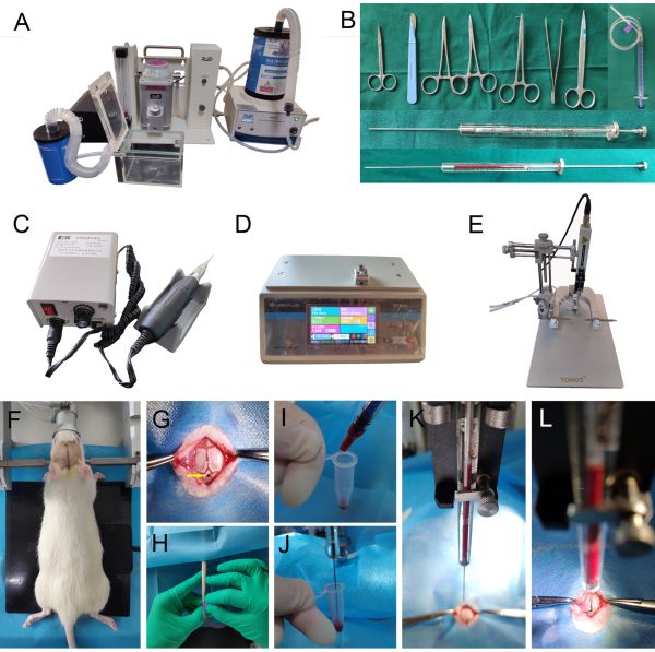
Figure 1: Instruments and procedure of surgery. (A) Anesthesia machine. (B) Surgical instruments. (C) Microdrill. (D) Micro-injection pump. (E) Stereotaxic apparatus. (F) A line is marked i...
Access restricted. Please log in or start a trial to view this content.
Materials
| Name | Company | Catalog Number | Comments |
|---|---|---|---|
| 100ml Saline solution | Guangdong yixiang | 191222201 C1 | Preparing heparin diluent |
| 100μl Microinjector | Shanghai Gaoge | Injection of autologous blood | |
| 1ml Syringe | Jiangsu Zhiyu | 20191014 | Withdraw autologous blood from the tail vein |
| 75% Alcohol | Shandong Lierkang | Disinfection of rat tail | |
| Adhesive tape | Shanghai Jinzhong | Surgicl instruments | |
| Animal anesthesia system | RWD | R510-31S-6 | Inducing and maintaining anesthesia |
| Balance beam | Jiangsu Saiangsi | For neurological deficit scores | |
| Bone cement | Shanghai Xinshiji | 20180306 | Surgicl instruments |
| Butorphanol tartrate | Jiangsu Hengrui | For pain management | |
| Electric cranial drill | Nanjing Darwin biotechnology | 20180090018 | Making a burr hole on the skull |
| EP tube | Nantong Surui | Transfer autologous blood | |
| Erythromycin eye cream | Yunnan pharmacy | Eyes protection | |
| Heating pad | Dangerous Jungle | JR01 | Keeping warm |
| Heparin sodium injection | Chengdu Haitong Pharmacal Company | 190701 | Preparing heparin diluent |
| IndoPhors | Guoyao of China | Sterilization | |
| Isoflurane | RWD | 20080701 | Inducing and maintaining anesthesia |
| Micro-injection pump | Baoding Leifu | TFD03-01-C | Injection of autologous blood |
| Needle holder | Shanghai Jinzhong | J32020 | Surgicl instruments |
| Penicilin | Guoyao of China | Infection Prevention | |
| Q-tips | Jiangxi Songhe | Surgicl instruments | |
| Scalp needle | Jiangxi Hongda | 20200313 | Withdraw autologous blood from the tail vein |
| Scalpel | Shanghai Kaiyuan | 170902 | Surgicl instruments |
| Shearing scissors | Shanghai Jinzhong | Y00040 | Surgicl instruments |
| Stereotaxic apparatus | RWD | 900-00001-00 | For surgical positioning |
| Surgical towel | Xinxiang Huakangweicai | 20070601 | Surgicl instruments |
| Suture needle | Shanghai Jinzhong | Surgicl instruments | |
| Suture scissors | Shanghai Jinzhong | J25041 | Surgicl instruments |
| Tissue holding forceps | Shanghai Jinzhong | J31080 | Surgicl instruments |
