A subscription to JoVE is required to view this content. Sign in or start your free trial.

Method Article

Generating a Middle Cerebral Artery Occlusion in a Rat Pup to Induce Local Brain Ischemia

776 views

April 28th, 2025

In This Article

Abstract

Source: Larpthaveesarp, A., et. al. Transient Middle Cerebral Artery Occlusion Model of Neonatal Stroke in P10 Rats. J. Vis. Exp. (2017)

This video demonstrates the establishment of the Middle Cerebral Artery Occlusion model in a rat pup. The internal carotid artery (ICA) is exposed, and a ligature is secured to restrict blood flood. An occlude is inserted through the ICA to reach the Middle Cerebral Artery. The pup is then allowed to recover before further analysis.

Protocol

All procedures involving animal models have been reviewed by the local institutional animal care committee and the JoVE veterinary review board.

1. Middle Cerebral Artery Occlusion

  1. Weigh the pup and ensure that it is the proper weight (19-21 g). Anesthetize the pup in 3% isoflurane in 100% oxygen (O2) and ensure that there is no response to a foot pinch. Maintain the body surface temperature between 35.5 °C and 37 °C with the use of a heating pad under the surgical stage.
  2. Secure the animal in a supine position with tape across the shoulder regions. Using sterile cotton swabs, swab the anterior cervical area with a povidone-iodine solution followed by a swab of 70% ethanol in double-distilled water, alternating between each solution for four swabs total.
  3. Locally infiltrate 0.25% bupivacaine into the planned incision site. Using a stereoscope, make a midline 5 to 7 mm anterior cervical incision to expose the common carotid artery (CCA). Place 2-4 retractors to keep the cavity open and the artery exposed.
  4. Locate the internal carotid artery (ICA), occipital artery (OA), and external carotid artery (ECA). Groom the arteries to get a clear view. Be careful not to disturb the vagus nerve.
    NOTE: For sham-operated pups, the incision is left open, and the arteries are exposed, after which the incision is sutured closed. The total anesthesia time is equivalent to that of the occlusion surgery.
  5. Cut 1.5 cm of 6-0 silk braided suture thread. Unbraid the suture, pulling out single strands. Make sure that the single strands are neat and not frayed.
    NOTE: If necessary, smooth the frayed ends by dipping the single strand in sterile water and grooming the strand with forceps tips.
  6. While holding the suture strand with 45-degree forceps, move the forceps in a sweeping arc motion to go under the ICA so that the forceps tips emerge between the ICA and OA.
    NOTE: If the dissection is done well, this step will be relatively easy. If the ICA and OA are touching, be careful not to rupture the arteries when using the forceps to isolate the ICA. If bleeding occurs, apply pressure to the artery with the forceps until the bleeding stops. Absorb the blood with a sterile swab.
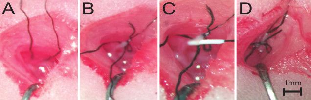
  7. Grab the end of the suture strand that is held by the forceps and pull it so that the end is easy to access. Release the strand from the forceps and back the forceps out from under the ICA, reversing the motion in step 1.5 (Figure 1A).
  8. Tie a temporary ligature around the ICA at the base, closest to where it separates from the CCA.
    NOTE: It is important to tie the knot so that the end of the strand that will be pulled to remove the knot is long enough (greater than 1 mm, less than 3 mm) to easily grasp with forceps while ensuring that there is an appropriate amount of suture strand on the other side of the knot for retraction.
  9. Carefully retract the ICA laterally and use a clip to secure the strand to excess skin near the axilla region on the side opposite the incision. Ensure that this retracting strand is taut enough to stop blood flow before proceeding to the next step to minimize the risk of uncontrolled bleeding. Observe that the artery is flat and pale.
    NOTE: Do not over-retract, as it can cause a partial or complete tear of the ICA. The retraction can be adjusted just before making the arteriotomy by pulling on the strand on the side opposite to the clip.
  10. Use 45° forceps to grasp another unbraided suture strand and loop it under and around the ICA, as in step 1.5. Position this strand lateral to the retraction strand (Figure 1B).
    NOTE: This step can also be done prior to retraction. If unsure about the quality of retraction, this knot can be very loosely tied before the next step.
  11. Cut a 0.2 mm arteriotomy midway between the tied and untied ligatures, erring closer to the tied ligature.
    NOTE: Blood remaining in the tied-off artery may empty through the arteriotomy but should not exceed 5 µL. If bleeding persists, carefully pull the retraction strand to increase retraction, with care to avoid damaging the artery from excessive tension.
  12. Using a metric ruler, measure the occluding suture and cut the suture with an extra allowance of 2-3 mm to remove the occluder during reperfusion. Hold the occluder with 45-degree forceps and use straight forceps to create a bend at the appropriate length to reach the MCA, marking a stopping point for advancement.
    NOTE: A 10 mm occlusion length from the silicone tip end to the bend is used for postnatal or P-10 Sprague Dawley or Long Evans rat pups in this weight range.
  13. Using 45° forceps, feed the silicone-coated nylon occlusion suture into the arteriotomy and advance the suture to the bend that marks the pre-determined distance to the MCA (Figure 1C). Ensure that advancement feels smooth; immediately stop advancement if resistance is felt. During advancement, aim the suture in a direction that is parallel to the CC/ECA, towards the head.
    NOTE: If the suture is advanced dorsally toward the animal's spine, it may run into the pterygopalatine artery (PTA). If resistance is felt after 3-5 mm of advancement, the suture has hit the PTA junction. Back the occluder out of the artery until the silicone head is near the arteriotomy before adjusting the advancement direction. It is not necessary to completely remove the occluder from the arteriotomy.
  14. Secure the occluder by tying a temporary ligature using the strand from step 1.10 (Figure 1D).
  15. Remove the retractor clip. Trim the strands of both of the temporary ligatures so that the strand of the knot that is pulled to remove the knot is easy to grasp with straight forceps and is longer than the strand that is pulled to tighten the knot.
    NOTE: The strands must be short enough so that they do not tangle in the cavity after closure.
  16. Remove the retractors and close the cavity using 6-0 braided silk to create three to four interrupted sutures.
  17. Remove the pup from anesthesia and place it on a heating pad in room air. Monitor the pup until it has regained sufficient consciousness to maintain sternal recumbency and ensure that it has fully recovered before returning it to the dam. Ensure that the pup maintains a body surface temperature between 35.5 °C and 37 °C.

Access restricted. Please log in or start a trial to view this content.

Results
Surgical suturing process in tissue, step-by-step diagram; close-up of suturing technique, 1mm scale.

Figure 1: Live Surgical Images of the tMCAO Procedure. (A) The first suture strand is looped around the ICA, as detailed in step 1.6. (B) The first temporary ligature is tied, and the ICA is retracted. The second suture s...

Access restricted. Please log in or start a trial to view this content.

Materials

List of materials used in this article
NameCompanyCatalog NumberComments
IsoflouraneHenry Schein50033Anesthetic, at 3%
Trinocular SurgioscopeWorld Precision InstrumentsPSMT5N
Heating padSunbeam000731-500-000Low to medium setting
IR ThermometerExtech Instruments72-5270
Retraction kit for small animalsFine Science Tools18200-20
CermaCut ScissorsFine Science Tools14958-09
Dumont #5SF ForcepsFine Science Tools112522-002x
Dumont #5/45 ForcepsFine Science Tools11251-352x
B-2 Micro ClampFine Science Tools00398-02
Forcepts for Clamp ApplicationFine Science Tools00072-14
Micro Vannas ScissorsFine Science Tools15000-032mm cutting edge
Occlusion SuturesDoccol602123PK10701712PK5Re
RulerFine Science Tools
6-0 Perma-Hand Silk Reverse CuttingSutureEthicon769G
EuthasolVirbac7101010.22 ml/kg
Cotton Tipped ApplicatorsHenry Schein100-9249
Laboratory TapeVWR89097-990

Tags

Rat Pup ModelInternal Carotid ArterySurgical ProcedureBrain Ischemia InductionOcclusion Suture InsertionLigature PlacementAnesthesia ProtocolCervical IncisionPostoperative Recovery