This video demonstrates a method to trace the origins of sensory neurons in the dura mater using fluorescence microscopy. A retrograde tracer is introduced into the exposed dura mater of an anesthetized rat. The tracer is taken up by sensory neuronal axons and travels retrogradely to their cell bodies within the trigeminal ganglion and cervical dorsal root ganglia. The ganglia are then sectioned, stained with antibodies that label the tracer and neuropeptides in the cell bodies, and analyzed using fluorescence microscopy to confirm the ganglionic origins of the sensory neurons.
A subscription to JoVE is required to view this content. Sign in or start your free trial.
Method Article
Investigating the Origins of Sensory Neurons in the Dura Mater Using Fluorescence Microscopy
1K views
⸱
May 29th, 2025
In This Article
Abstract
Protocol
All procedures involving animal models have been reviewed by the local institutional animal care committee and the JoVE veterinary review board.
1. Retrograde tracing study with fluorogold (FG)
- Surgical procedures
- Determine the coordinate area of interest in rat cranial dura mater.
- Prepare a 10 µL micro-syringe and test it with liquid paraffin.
- Anesthetize the rats with tribromoethanol solution (150 mg/kg) via intraperitoneal injection. Check for the depth in anesthesia by the lack of response to toe pinch.
- Shave the rat's head with an electric razor.
- Put blunt ear bars on the rat and place it on the stereotaxic device. Then put the mouth holder and apply ophthalmic ointment on the eyes.
- Clean the surgical site of the head skin using 10% povidone iodine followed by 75% ethanol.
- Make an incision along the midline of the scalp.
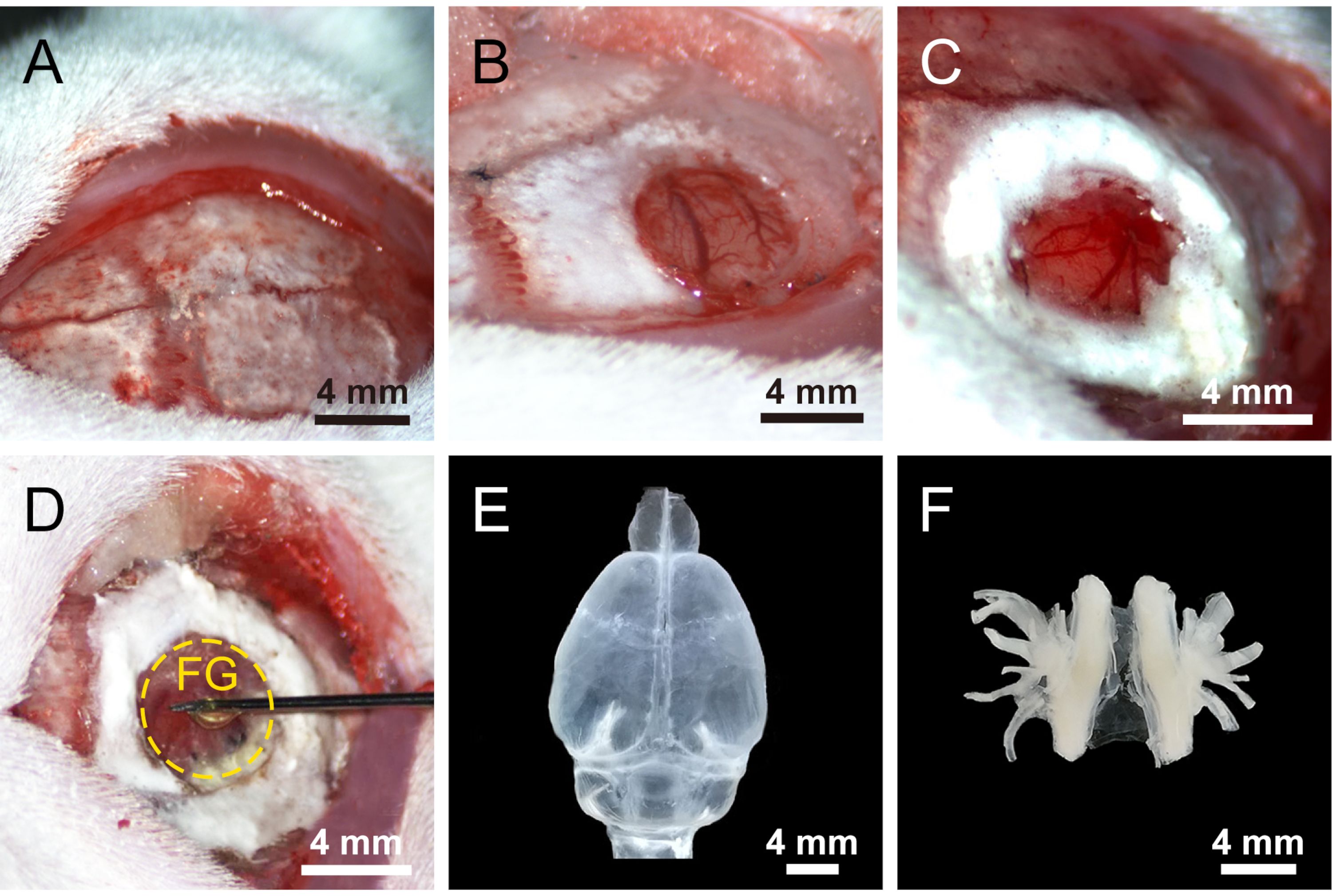
- Bluntly remove the periosteum and muscle tissues away from the skull using sterile cotton-tipped applicators (Figure 1A).
- Drill a small hole (~5-7 mm) using a burr drill with a round-tip bit (#106) on the left parietal and temporal bones above the middle meningeal artery (MMA), and make sure that the cranial dura mater was kept intact (Figure 1B).
- Build a bank around the hole with dental silicate cement to limit the spread of the tracer (Figure 1C).
- Add 2 µL of 2% FG into the hole around MMA with a 10 µL micro-syringe (Figure 1D).
- Cover the hole with a small piece of hemostatic sponge.
- Put a piece of paraffin film on the hole and seal the edges with bone wax to prevent the leakage of tracer and to avoid contaminating the surrounding tissues.
- Suture the wound with sterile thread.
- Keep the rats in a warm area until they have fully recovered.
- Return the rats back to their cages and add an antibiotic and analgesic into the drinking water.
- Perfusions and sections
- After 7 survival days, perfuse these rats.
- Dissect out the trigeminal ganglion (TG) and C1-4 dorsal root ganglia (DRGs), then post-fix and cryoprotect them (Figure 1F).
- Cut the TG and DRGs at the thickness of 30 µm on a cryostat microtome system in the sagittal direction and mounted on silane-coated glass slides.
- Double immunofluorescences for FG- and calcitonin gene-related peptide (CGRP)-labeling in the TG and DRGs
NOTE: Although the FG-labeling can be directly observed with ultraviolet (UV) illumination under mercury lamp without additional staining, the labeled neurons with FG were further examined in TG and cervical DRGs using double immunofluorescences with FG and CGRP for revealing the origins of dural CGRP-immunoreactive nerve fibers in the TG and DRGs.- Circle the sections with the histochemical pen.
- Incubate the sections for 30 min in a blocking solution containing 3% normal donkey serum and 0.5% Triton X-100 in 0.1 M phosphate buffer (PB).
- Transfer the samples into the solution of rabbit anti-fluorogold (1:1000) and mouse anti-CGRP antibody (1:1000) in 0.1 M PB containing 1% normal donkey serum and 0.5% Triton X-100 overnight at 4 °C.
- Wash the sections three times in 0.1 M PB the following day.
- Incubate in a mixed solution of donkey anti-rabbit Alexa Fluor 594 (1:500) and donkey anti-mouse Alexa Fluor 488 (1:500) secondary antibody in 0.1 M PB containing 1% normal donkey serum and 0.5% Triton X-100 for 1.5 h at room temperature.
- Wash and apply coverslips to the sections.
- Observation and recording
- Take images of the FG-labeled neurons in TG and DRGs under UV illumination by fluorescent microscope equipped with a digital camera.
- Capture images of the FG- and CGRP-labeled neurons in TG and DRGs under a fluorescent microscope equipped with a digital camera.
- Use the editing software to adjust the brightness and contrast of images and to add labels in the pictures.
Access restricted. Please log in or start a trial to view this content.
Results
Access restricted. Please log in or start a trial to view this content.
Materials
| Name | Company | Catalog Number | Comments |
|---|---|---|---|
| Alexa Fluor 488 donkey anti-mouse IgG (H+L) | |||
| Brain stereotaxis instrument | |||
| CellSens Dimension | |||
| Fluorogold (FG) | |||
| Fluorescent imaging system | |||
| Freezing microtome | |||
| Micro Drill | |||
| Mouse anti-CGRP | |||
| Normal donkey serum | |||
| Phalloidin 568 | |||
| Photoshop and Illustration | |||
| Rabbit anti- Fluorogold | |||
| Sprague Dawley | |||
| Superfrost plus microscope slides |
