This video demonstrates the use of fluorescence imaging to measure cytosolic calcium levels in short-term and long-term rat hippocampal cultures. By comparing young and aged neurons, it highlights how increased L-type voltage-gated calcium channel density and prolonged activation in aged neurons result in calcium overloading, a hallmark of neuronal aging. Fluorescent calcium indicators reveal these differences through changes in fluorescence intensity upon stimulation.
A subscription to JoVE is required to view this content. Sign in or start your free trial.
Method Article
In Vitro Calcium Imaging in Short-Term and Long-Term Rat Hippocampal Cultures
903 views
⸱
May 29th, 2025
In This Article
Abstract
Protocol
1. Short and Long-term Culture of Rat Hippocampal Neurons
- Preparation of poly-D-lysine coated, 12 mm glass coverslips.
- Sterilize 12 mm diameter glass coverslips in ethanol for at least 24 hr. Allow them to dry under sterile conditions.
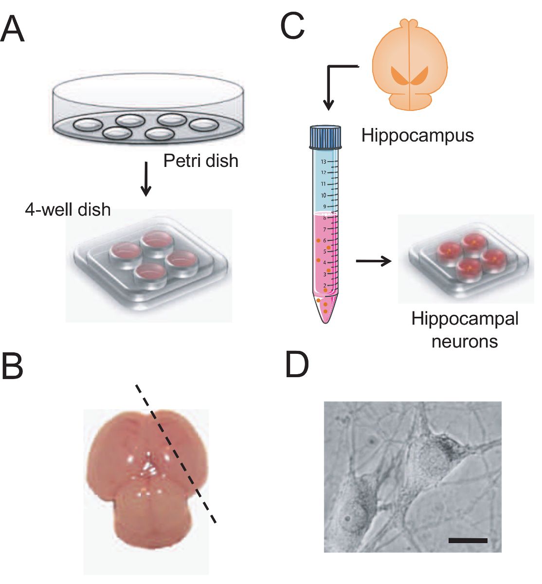
- Distribute the coverslips on a strip of parafilm in a Petri dish. Cover the surface of each coverslip with 200 µl of 1 mg/ml poly-D-lysine. Allow treating overnight (O/N).
- The next day wash the coverslips with double distilled sterile water every 15 min for 90 min under sterile conditions.
- Fill a four-well multidish plate with 500 µl of Neurobasal Culture Medium per well. Neurobasal Culture Medium should be supplemented with 10% fetal bovine serum, 2 % B27, 1 µg/ml gentamicin and 2 mM L-glutamine.
- Place the coverslips on the multidish plate. Maintain them in a humidified 37 ºC and 5% CO2 incubator until use.
- Isolation of hippocampal neurons from neonatal rats.
- Prepare 1.8 ml papain solution (directly purchased) in a vial (20 µl/ml) under sterile conditions: To obtain a final concentration of 20 µl/ml add around 40 µl papain in 1.8 ml Hank's plus 0.6% bovine serum albumin (BSA, µl should be calculated depending on the supplier conditions). Hank's 0.6% BSA is prepared by mixing 85 ml of Hanks' Balanced Salt Solution (HBSS) medium with 15 ml of 4% bovine serum albumin (BSA, prepared solving 4 g of BSA in 100 ml HBSS). Prepare it in a sterile hood.
- Incubate the papain in Hank's plus 0.6% BSA for 30 min at 37 ºC before use. Filter the solution using a 0.22 µm filter prior to use.
- Prepare Ham's F-12 medium by adding Dulbecco's Modified Eagle's Medium powder (13.5 g per L) in 900 ml of double distilled sterile water. Then add 6 g 4-(2-hydroxyethyl)-1-piperazineethanesulfonic acid (HEPES) and 336 mg NaHCO3 and adjust the pH to 7.42 using NaOH 4 N. Add sterile water to obtain a 1 L solution and filter it using a 0.20 µm polyethersulfone (PES) bottle top filter. Carry this process in a sterile hood. Finally saturate the solution with CO2 under sterile conditions before use. Store solutions at 4 ºC and use them chilled.
- Euthanize newborn (P0) rat pups by decapitation and wash the head in sterile HBS medium. Open the skull with the help of sterile scissors. Extract the brain with a spatula and wash it quickly in Ham's F-12 medium.
- Make a diagonal cut with the aid of a scalpel in each hemisphere (Figure 1B) and carry it to a Petri dish containing Ham's F-12 medium.
- Discard meninges carefully with the help of dissection forceps and under a magnifying glass.
- Identify the hippocampus in a characteristic concave location over the cortex using magnifying glass. Then, separate the hippocampus from the cortex by pulling carefully from one border and removing it from its position using eye scissors.
- Transfer the hippocampal tissue to a 4-well plate filled with sterile Hank's medium lacking Ca2+ and Mg2+ plus 0.6% BSA and wash hippocampal tissue. Without removing the media cut the tissue in small pieces (around 2 x 2 mm) using the same eye scissors.
- Transfer hippocampal pieces to a vial containing 1.8 ml pre-filtered papain solution. Then incubate them at 37 ºC with occasional, gentle shaking. 15 min later, add 90 µl of DNase I solution (50 µg/ml final) and incubate for 15 additional min.
- Transfer the tissue to a 10 ml centrifuge tube and wash the fragments with fresh Neurobasal Culture Medium. Obtain cell suspension by passing tissue fragments through a 5 ml plastic pipette.
- Centrifuge cell suspension at 160 x g for 5 min. Remove the supernatant using a plastic sterile Pasteur pipette and suspend pellet carefully with a 1 ml automated pipette in 1 ml of Neurobasal Culture Medium.
- Measure cell density using a Neubauer counting chamber. Put the glass coverslip over the Neubauer chamber. Add 10 µl of cell suspension. Make sure the cell suspension enters the chamber uniformly and makes no bubbles. Count the number of cells under the microscope and perform corresponding calculations to obtain a suitable drop of 40-80 µl containing 30 x 103 cells. The total number of cells will depend on the number of animals used.
- Short-term and long-term culture of rat hippocampal neurons.
- Over the four-well multidish plate containing 500 µl of Neurobasal Culture Medium prepared before and kept in the incubator, plate around 30 x 103 cells in a drop of about 50 µl to each well of the multidish plate containing one poly-D-lysine-coated, 12 mm diameter glass coverslip.
- Maintain primary hippocampal cells in a humidified 37 ºC and 5% CO2 incubator for 2-5 days in vitro (DIV, short-term, young cultures) or >15 DIV (Long-term, aged cultures) before experiments without changing the culture media.
2. Fluorescence Imaging of Cytosolic Ca2+ Concentration
- Preparation of test solutions for fluorescence imaging.
- Prepare an external HEPES-buffered saline (HBS) solution containing, in mM: NaCl 145, KCl 5, MgCl2 1, CaCl2 1, glucose 10, sodium-HEPES 10 (pH 7.42).
- Prepare an external, Mg2+-free, HEPES-buffered saline (HBS) solution containing, in mM: NaCl 146, KCl 5, CaCl2 1, glucose 10 and HEPES 10 (pH 7.42).
- Solve N-Methyl-D-aspartate (NMDA) in Mg2+-free, HEPES-buffered saline (HBS) at a final concentration of 100 µM and supplement it with 10 µM glycine.
- Prepare an HBS solution containing KCl instead of NaCl by solving (in mM): KCl 145, MgCl2 1, CaCl2 1, glucose 10 and HEPES 10 (pH 7.42).
- Loading of hippocampal cells with fluorescent calcium probe fura2/AM.
- Take the cell culture containing coverslips off the incubator and wash them with HBS medium at room temperature (RT) by transferring them to a new four-well multidish plate containing 500 µl of HBS per well.
- Incubate cells with fura2/AM 4 µM (prepared in the same HBS medium) for 60 min at RT (25 ºC) in a dark place.
- After 60 min, wash coverslips with fresh HBS medium.
- Recording fluorescence images of cytosolic [Ca2+] in cultured cells.
- Turn on the lamp, microscope, perfusion system, fluorescence camera and computer.
- Place the coverslips in a thermostated platform for open 12 mm glass coverslips on the stage of the inverted microscope and select a microscopic field using a 40X objective (oil, numerical aperture [NA]:1.3). Perfuse cells continuously with pre-warmed (37 ºC) HBS medium in absence or presence of test substances. Maintain the flow at a rate of about 5-10 ml/min.
Note: Cell perfusion system for living cells is mounted in a thermostated platform for open 12 mm glass coverslips. 8-lines gravity-driven perfusion system equipped with a valve controller is used to perfuse the solutions. A vacuum pump is responsible for removing any excess medium. Solutions are heated using an in-tube heating system. - Capture a background image with the shutter closed at both excitation wavelengths.
- Epi-illuminate cells alternately at 340 and 380 nm. Record light emitted at 520 nm every 5-10 sec with a fluorescence camera, which is filtered by a fura-2 dichroic mirror.
- When the recording period is finished, store the complete sequence of images emitted at 520 nm in the computer for further analysis.
- Analysis of recorded fluorescent images.
- Open the experiment file. Using the aquacosmos software, click on 'Ratio' and select the desired ratio range. Calculate the pixel-by-pixel ratio in the resulting images in order to obtain a sequence of ratio images.
- Subtract background by adjusting the 'background elimination'. Press 'start calculation'.
- Press 'all times sequence' button and erase the ancient regions of interest (ROIs). For quantitative analysis of individual cells, establish new regions of interest or ROIs corresponding to individual neurons. Average all ratio values in each pixel corresponding to each ROI and each image to obtain a recording of ratio fluorescence values for individual ROIs (cells).
- To graph the individual recordings, export the ratio fluorescence values corresponding to each ROI to a graphing program.
- Make the corresponding calculations for estimating the size of the rises in ratio fluorescence in response to each stimulation using a suitable data analysis and graphing software.
Access restricted. Please log in or start a trial to view this content.
Results
Access restricted. Please log in or start a trial to view this content.
Materials
| Name | Company | Catalog Number | Comments |
|---|---|---|---|
| Neurobasal Culture Medium | |||
| HBSS medium | |||
| Ham's F-12 medium | |||
| DNase I (from bovine pancreas) | |||
| Fetal Bobine Serum | |||
| B27 | |||
| Gentamicin | |||
| L-glutamine | |||
| 12 mm glass coverslips | |||
| Papain | |||
| 4-well multidish plaques | |||
| Petri dishes | |||
| Sterile pipettes | |||
| Fura2-AM | |||
| Lipofectamine2000 | |||
| NMDA | |||
| Glycine | |||
| Zeiss Axiovert S100 TV microscope | |||
| Xcite ilumination system | |||
| ORCA ER fluorescence camera | |||
| VC-8 valve controller | |||
| SH-27B heating system | |||
| Aquacosmos Software |
