This video demonstrates the method of magnetic resonance elastography (MRE) for assessing tissue viscoelastic properties by analyzing shear wave propagation through a gelatin phantom, mimicking brain tissue. The process involves generating and capturing shear waves with magnetic resonance imaging or MRI and analyzing wave patterns to differentiate tissue stiffness.
A subscription to JoVE is required to view this content. Sign in or start your free trial.
Method Article
Magnetic Resonance Elastography for Assessing Viscoelastic Properties
1K views
⸱
May 29th, 2025
In This Article
Abstract
Protocol
1. Gelatin phantom preparation
- Weigh gelatin, glycerol, and water according to Table 1. Mix the gelatin powder with water to obtain the gelatin solution.
NOTE: Table 1 shows the concentrations of the individual components used to prepare the two phantoms. The higher the concentration of gelatin, the stiffer the phantom. - Heat the gelatin solution to 60 °C in a water bath. Add glycerol to the gelatin solution while maintaining the temperature.
NOTE: Glycerol stabilizes gelatin mixtures by increasing their melting temperature and shear modulus17. - Stir the solution and heat it to 60 °C again. Pour the mixed solution into a container that will be used for Magnetic Resonance Elastography (MRE) and indentation tests. Cool the solution to room temperature and wait till the solution is solidified.
2. Magnetic resonance elastography or MRE test
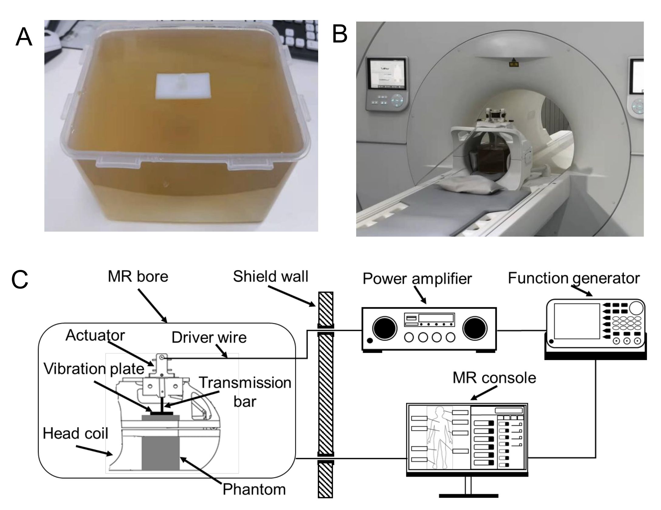
- Put the vibration plate on top of the gelatin phantom. Ensure that the contact between the phantom and the vibration plate is firm (Figure 1A).
NOTE: The vibration plate is made of Polyamide with a dimension of 50 x 50 x 5 mm3. - Place the gelatin phantom into the head coil. Put sponges and sandbags around the gelatin phantom to make sure the phantom is firmly placed. Use a custom-built electromagnetic actuator with a transmission bar. Mount an electromagnetic actuator on the head coil. Connect the transmission bar to the vibration plate (Figure 1B).
- Connect the power lines of the actuator with the amplifier. Connect the control lines with the controller (Figure 1C).
- Actuator and MRI scan parameter settings
- Set the waveform, vibration frequency, and amplitude in the function generator. Set the desired vibration amplitude by adjusting the power amplifier.
NOTE: Here, the waveform is set to sinusoidal in the function generator; the vibration frequency is set to 40 Hz or 50 Hz, and the amplitude is set to 1.5 Vpp. In the power amplifier, the amplification ratio is set to 40%. - Set the function generator to work in the trigger mode. Connect the trigger line to the external trigger port of the MRI (Magnetic Resonance Imaging) machine.
- Set the MRE scanning (actuator) frequency the same as that from the function generator, so that the motion encoding gradient is synchronized with the motion of the vibration plate.
- Set the waveform, vibration frequency, and amplitude in the function generator. Set the desired vibration amplitude by adjusting the power amplifier.
- Data measurement and analysis
- Follow the routine imaging positioning procedures. Use a 2D gradient-echo (GRE) based MRE sequence for imaging of the gelatin phantom20. Set the GRE-MRE imaging parameters as follows: Flip-angle = 30°; TR/TE = 50/31 ms; Field-of-view = 300 mm; Slice thickness = 5 mm; Voxel size = 2.34 x 2.34 mm2.
- Measure the phase images at four temporal points in one sinusoidal cycle. Apply both positive and negative motion encoding gradients at each time point.
- Based on the phase image acquired, remove the background phase by subtracting the positive and negative encoded phase images. Unwrap the phase with a reliability sorting-based algorithm.
- Extract the principal component of the motion by applying fast Fourier Transform to the unwrapped phase images. Filter the phase image with a digital bandpass filter. Estimate the shear modulus with a 2D direct inversion (DI) algorithm to obtain storage modulus G' and loss modulus G''.
NOTE: The cut-off frequency of the bandpass filter is [0.04 0.08]. The size of the fitting window of the DI algorithm is 11 x 11.
Table 1: The mass and mass concentration of the gelatin, glycerol, and water used for preparing the two gelatin phantoms. The mass unit is grams.
Access restricted. Please log in or start a trial to view this content.
Results
Access restricted. Please log in or start a trial to view this content.
Materials
| Name | Company | Catalog Number | Comments |
|---|---|---|---|
| 24-channel head & Neck coil | |||
| 3T MR Scanner | |||
| Acquisition board | |||
| Computer-Windows | |||
| Electromagnetic actuator | |||
| Function generator | |||
| Gelatin | |||
| Glycerol | |||
| Indenter control program | |||
| Laser sensor | |||
| Load cell | |||
| MATLAB | |||
| Power amplifier | |||
| Voice coil electric motor |
