This video demonstrates a fluorescent tracing technique to identify retinal ganglion cell (RGC) subtypes in a transgenic mouse retina. The process involves securing the isolated retina in a recording chamber, maintaining cell viability with oxygenated perfusion, and targeting fluorescently labeled RGCs using a pipette with a tracer. By analyzing dendritic morphology and stratification, the method enables classification of distinct RGC subtypes.
Method Article
Identification of Mouse Retinal Ganglion Cell Subtypes Through Fluorescent Tracing
762 views
⸱
May 29th, 2025
In This Article
Abstract
Protocol
All procedures involving animal samples have been reviewed and approved by the appropriate animal ethical review.
1. Preparation of Retinal Tissue (2 h)
NOTE: All procedures in this section should be performed under dim red illumination
- Dark-adapt animals for at least 1 h prior to dissection. Perform all procedures under dim red illumination.
- Euthanize the animals by CO2 asphyxiation and enucleate the eyeballs into a Petri dish with previously oxygenated extracellular solution.
- Poke the cornea with a needle and cut it away by cutting it with ophthalmic scissors at the border of the cornea and sclera.
- Remove the lens using #5 forceps. Gently make a tear in the sclera with the forceps and sever the optic nerve where the retina and sclera meet. Carefully finish removing the sclera from the retina.
- Remove the transparent vitreous using #5 forceps; once removed, the vitreous appears as a gelatinous substance stuck to the forceps. Slice the retinas in half (so that there are 4 pieces/animal) and store them in oxygenated extracellular solution at RT until use.
- When ready to mount the tissue in the recording chamber, place a piece of retina to incubate in enzyme solution diluted in 500 µL of oxygenated extracellular solution. Incubate in a Petri dish for 2 min at room temperature (RT) on a shaker.
- Wash the piece of retina in oxygenated extracellular solution and place the tissue in a glass-bottom recording chamber; use a plastic transfer pipette with the tip cut off to allow for the retina to be transferred without causing damage to the tissue.
- Use forceps to carefully flatten the tissue with the photoreceptor layer facing down. Remove excess fluid using a pipette. Anchor the tissue using a platinum ring with nylon mesh.
NOTE: This method could also be used to prepare the tissue for the isolation of RNA from labeled amacrine and bipolar cells. - Fill the chamber with oxygenated extracellular solution and mount it onto a microscope stage. Perfuse tissue with oxygenated extracellular solution at 2-4 mL/min.
3. Visualization and Targeting of GFP+ Retinal Ganglion Cells (10 min)
NOTE: All procedures in this section should be performed under dim red illumination
- Before beginning, pull glass micropipettes (outer diameter [OD]: 1.2 mm, inner diameter [ID]: 0.69 mm) for electrophysiological recordings using a micropipette puller. Use the following protocol for the electrodes (please note that the parameters should be adjusted accordingly to achieve the desired resistance and will vary across pullers and with different glass): Heat: Ramp +10; Pull: 0; Vel: 23; Delay: 1; Pressure: 500; Program loop: 5 times. Ensure that the tips are ~1 µm in diameter, with resistances of 2-4 MΩ for targeting large cells and of 5-7 MΩ for targeting smaller cells.
- Observe the ganglion cell layer using Infrared Differential Interference Contrast (IR-DIC) optics (Figure 1A). Identify Green Fluorescent Protein (GFP)+ Retinal Ganglion Cells (RGCs) using epifluorescence (~480 nm) (Figure 1B).
- Locate the pipette filled with intracellular solution in DIC. Apply a slight positive pressure and zero voltage offsets on the amplifier.
- Press the glass micropipette against a GFP+ cell and apply negative pressure to form a GΩ seal between the pipette and the cell membrane. Apply test voltage command steps (e.g., 5 mV) to monitor the seal resistance. After forming a stable seal, rupture the membrane by applying brief pulses of negative pressure to gain whole-cell access.
- Wait 1-2 min for the dendrites of the cell to fill with fluorescent tracer.
NOTE: The cell can be morphologically typed by examining the morphology in epifluorescence (Figure 1C). In the case of melanopsin-expressing RGCs, dendritic stratification in the inner plexiform layer is visualized by examining the dendrites filled with fluorescent tracer under epifluorescent illumination and determining whether they stratify far from the soma in the OFF sublamina (M1 ipRGCs), near the ganglion cell layer in the ON sublamina (M2 & M4 ipRGCs), or both (M3 ipRGCs). This observation, combined with soma size (M4s have distinctly large somas compared to all other ipRGC subtypes), allows for the identification of cell type. Thus, this technique allows for the identification of cell type in vitro prior to RNA isolation. This method could be modified for other cell type identification protocols involving either dendritic morphology or cellular physiology.
Results
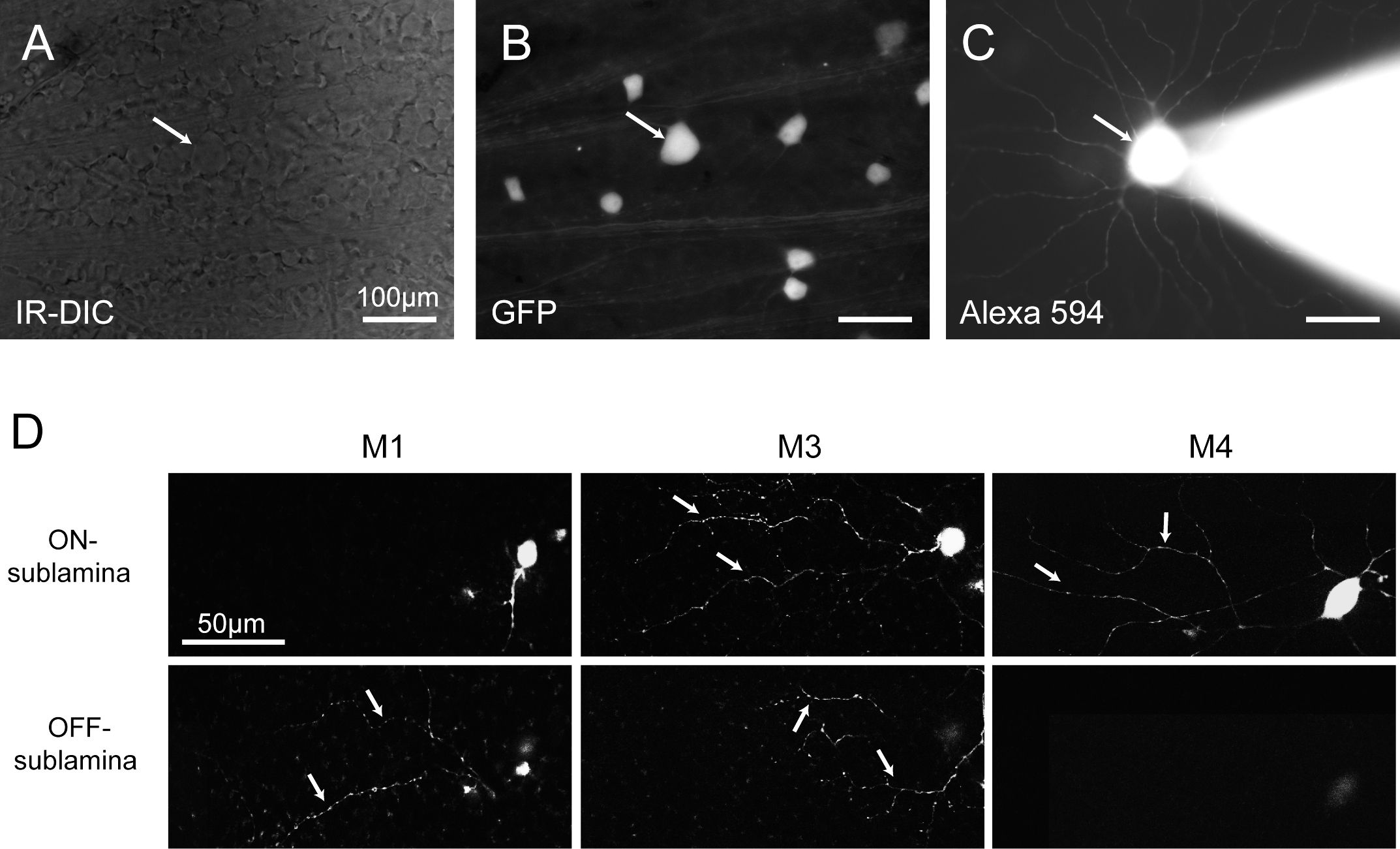
Figure 1: Identification of RGCs Types based on GFP Expression in Melanopsin-expressing ipRGCs and on Morphological Properties. (A) The ganglion-cell layer of the retina visualized using IR-DIC in the whole-mount retina preparation. (B) The same preparation visualized in epifluorescence (~480 nm) to identify GFP-expressing ganglion cells. (C) A GFP-expressing cell targeted for patch-clamp recording and filled with a fluorescent tracer. The cell can be identified as an M4 ipRGC based on its very large soma size and ON stratifying dendrites. (D) Confocal image of ipRGC dendrites imaged in the ON and/or OFF sublamina of the IPL (M1), in the ON sublamina of the IPL (M4), and in both the ON and OFF sublaminae of the IPL (M3). IR-DIC: infrared differential interference contrast.
Materials
| Name | Company | Catalog Number | Comments |
|---|---|---|---|
| Diethyl pyrocarbonate (DEPC) | |||
| Alexa Fluor 594 Hydrazide | |||
| Petri dish (35mm diameter) | |||
| Ophthalmologic scissors | |||
| #5 Forceps | |||
| Glass Micropipette | |||
| Micropipette Puller | |||
| 1mL syringe |
