This video demonstrates the procedure for imaging microtubule abnormalities in a rat brain tissue slice using a two-photon excitation microscope. It employs second harmonic generation (SHG) signals to detect tubulin defects and reduced myelin production.
A subscription to JoVE is required to view this content. Sign in or start your free trial.
Method Article
Second Harmonic Generation Imaging in a Rat Model to Study Tubulin Defects
647 views
⸱
June 17th, 2025
In This Article
Abstract
Protocol
All procedures involving animal samples have been reviewed and approved by the appropriate animal ethical review committee.
1. Transfer to the microscope
- If the microscope is in a different room, prepare an insulated box with warmed gel packs to transfer the tissue sections while maintaining the temperature. The box will also keep the sections warm until they are imaged.
- Put the sample under the microscope, and make sure it is positioned properly under the objective by direct observation through the oculars with transmitted light.
NOTE: The goal is to align the predicted emitting structures with the laser's oscillation direction to maximize the emitted (and, therefore, detected) second harmonic generation (SHG) signal. - Remove the excess HBSS so that a thin liquid film covers the entire sample. Visually check the liquid film every few minutes to avoid excessive evaporation and drying of the sample.
NOTE: Under the conditions described, a section is used for no more than 20 min. Drying the sample causes drastic artifactual effects (Figure 1A). If longer imaging times are needed, periodic soaking of the section in a warm, fresh medium is recommended rather than adding more HBSS. The use of perfusion chambers and glue or low-melting agarose to immobilize the tissue is also recommended when longer experiments are to be done. - Prepare the microscope stage for non-descanned imaging, which might include closing all the doors of the dark incubation chamber or covering the incubation chamber with a black nylon polyurethane-coated fabric.
2. Imaging
- Select the "non-descanned" imaging mode along the transmission path. This way, the capture of the weak SH signal of tubulin will be optimized.
- Select the LCI Plan-Neofluar 25x/0.8 NA objective.
- Set a laser power between 13 mW and 26 mW, with a pixel dwell time of 12.6 µs. Take images no bigger than 512 pixels x 512 pixels, with a speed of 5 and averaging 2, for an average acquisition time of about 15 s.
NOTE: Using higher laser powers and/or longer dwell times may damage the sample (Figure 1B). - Take images first using a 485 nm short-pass filter (SP485), and, in a second step, add a sharp 405 nm bandpass filter (BP405; Figure 2).
NOTE: Pseudo-brightfield images can be taken through the same detector using the residual laser light of a visible line (405 nm or 488 nm lasers work best).
Access restricted. Please log in or start a trial to view this content.
Results
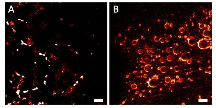
Figure 1: Examples of artifacts. (A) Artifact due to drying of the sample. (B
Access restricted. Please log in or start a trial to view this content.
Materials
| Name | Company | Catalog Number | Comments |
|---|---|---|---|
| 405/10 nm BrightLine(R) single-band bandpass filter | Semrock | FF01-405/10-25 | 32 mm diameter, with housing ring |
| Black Nylon, Polyurethane-Coated Fabric | Thorlabs | BK5 | 5' x 9' (1.5 m x 2.7 m) x 0.005" (0.12 mm) Thick |
| Confocal microscope | Zeiss | LSM710NLO AxioObserver Z1 | Inverted microscope, objective used is LCI Plan-Neofluar 25x/0.8 NA |
| Hanks′ Balanced Salt solution (HBSS) | Gibco | 14025-076 | Could be prepared from powders |
| NDD module, type C | Zeiss | 000000-1410-101 | To detect the signal, reducing light loss. Housing the 000000-1935-163 filter set with the SP485 |
| Phosphate buffered saline (PBS) | Gibco | 10010-031 | Could be prepared from powders or tabs |
| Pulsed laser | Coherent | Chameleon Vision II | 680–1080 nm tunable laser |
