This video demonstrates the intravenous delivery of an arginine-vasopressin (AVP) receptor antagonist in a mouse model. Transient middle cerebral artery occlusion induces brain edema. An AVP receptor antagonist is administered through a catheter in the left jugular vein, alleviating edema.
A subscription to JoVE is required to view this content. Sign in or start your free trial.
Method Article
Intravenous Delivery of Receptor Blockers to Reduce Brain Edema in a Mouse Model
605 views
⸱
August 7th, 2025
In This Article
Abstract
Protocol
All procedures involving animal models have been reviewed by the local institutional animal care committee and the JoVE veterinary review board.
1. In Vivo Stroke Induction
- Pre-coat the filament with dental resin prior to the surgery.
- Cut a 12 mm long piece of filament from a 7-0 nylon suture. Mix 2 parts of the resin with 1 part of the hardener, and then immediately dip the tip of the filament into the mixture to cover about 1/3 of the entire length. Remove it quickly and verify that it is covered with a smooth-surfaced coat of dental resin.
- Air dry the filament for 2 hr before use.
- Place the mouse into an anesthesia induction chamber (2 L volume), and set the flow of 1.5% isoflurane in 25% oxygen-enriched room air at 2 L/min going into the chamber.
- Allow the mouse to remain in the induction chamber until fully anesthetized, as determined by lack of response to tail pinch.
- Place the mouse onto the operating table with a heated pad, and set the isoflurane concentration to 1% for maintenance of anesthesia delivered spontaneously through the nose cone.
- Clip hair on the front and the back of the neck, and spray the neck with rubbing alcohol. Apply veterinary ophthalmic ointment on both eyes.
- To continuously monitor body temperature, use a rectal temperature probe connected to a digital display during the surgery.
- Place the mouse in the supine position, apply povidone-iodine 10% scrub solution for disinfection, cover with a sterile drape, and then make a midline incision along the neck with a size 10 surgical scalpel blade.
- Temporarily ligate the left common carotid artery (CCA) with a 6.0 silk suture.
- Place another tie on the left external carotid artery (ECA).
- Place a microvascular clip on the internal carotid artery (ICA).
- Cut a small hole on the external carotid artery with microvascular scissors. Insert a filament made from 7-0 nylon suture coated with dental resin into the opening in the ECA, and advance it cephalically, while removing the clip, until resistance is felt (about 5-7 mm from the bifurcation point of the CCA).
- Place a temporary tie onto the internal carotid artery to secure the filament in position. Refer to the diagram, Figure 1A. Close the skin with a silk suture, and infiltrate the wound on the front of the neck with 0.2 ml of 0.5% bupivacaine, as in step 2.6.
- Allow the mouse to awaken from anesthesia and regain consciousness in a recovery chamber for 60 min. Do not leave the animal unattended until it has regained sufficient consciousness to maintain sternal recumbence.
- Test the mouse for neurological deficit scoring (NDS) as follows: 0 = normal motor function, 1 = flexion of torso and of contralateral forelimb on tail lift, 2 = circling to the contralateral side but normal posture at rest, 3 = leaning to contralateral side at rest, 4 = no spontaneous motor activity.
- For the NDS testing, lift the mouse by the tail and place it on a flat surface for observation (2-3 min). Alternatively, observe mice in the recovery chamber. Exclude mice that exhibit NDS lower than 2 from the experiment due to insufficient occlusion of the middle carotid artery (MCA).
NOTE: The laser Doppler flowmetry technique can be used to confirm MCA occlusion.
- For the NDS testing, lift the mouse by the tail and place it on a flat surface for observation (2-3 min). Alternatively, observe mice in the recovery chamber. Exclude mice that exhibit NDS lower than 2 from the experiment due to insufficient occlusion of the middle carotid artery (MCA).
- Re-anesthetize the animal after 60 min of recovery in the induction chamber as described above, re-sterilize the previous skin incision with 10% povidone-iodine scrub solution, and reopen the surgical wound on the front of neck by cutting and removing the silk suture.
- To remove the filament from the ECA, perform the following steps:
- Place a microvascular clip on the ICA. Untie the suture on the ICA and pull the filament out while holding the clip open.
- Close the clip and place a tie onto the ECA proximally to the cut.
- Remove the clip and the ligature from the CCA to restore blood flow to the brain. Refer to the diagram, Figure 1B.
2. Installation of Intravenous (IV) Catheter into the Jugular Vein for Continuous 48 hr Treatment
NOTE: Proceed to the installation of the catheter immediately after step 1.16.3 without awakening the mouse.
- Use two different sizes of flexible tubing: insert the inner tubing (0.94 mm outside diameter; to contain the drug being infused) into the outer tubing (3.18 mm outside diameter; for protection of the inner tubing).
- Place the mouse in the prone position, apply 10% povidone-iodine scrub solution for disinfection, and make a 1 cm incision on the back of the neck with a size 10 surgical scalpel blade. Turn the mouse into the supine position, re-sterilize the front of the neck with 10% povidone-iodine scrub solution, and reopen the surgical wound on the front of the neck. Tunnel the tubing under the skin from the incision made on the back to the front of the neck, and exteriorize it from the open wound 1 cm.
- Remove the subcutaneous adipose tissue from the left side of the front of the neck about 1 cm laterally from the midline. Locate the left jugular vein, place two ties along the vein 5 mm apart, and slightly stretch the vein. Make a small hole in the jugular vein between the two ties with microvascular scissors; insert the tip of the inner tubing 5 mm deep caudally (towards the heart).
- Secure the tubing with both ties to the jugular vein. Close the skin on the front of the neck with a 3-0 silk suture. Refer to the diagram, Figure 1C.
- Secure the tubing to the skin on the back of the neck with a suture, and connect the tubing to a microinfusion IV pump through a swivel. The swivel allows free movement of mice inside the cage with full access to the food and water.
- Infiltrate the skin incisions on the front and the back of the neck with 0.2 ml of 0.5% bupivacaine to prevent post-operative pain.
NOTE: Depending on institutional-specific animal protocols, animals may be treated with opioids, non-steroidal anti-inflammatory drugs (NSAIDs), or antibiotics pre- and post-operatively. However, the mentioned treatments may alter the results of ischemic stroke outcome. - Allow the animal to awaken in the recovery chamber, and then house the animal in a separate cage until the endpoint of the experiment. Do not leave the animal unattended until it has regained sufficient consciousness to maintain sternal recumbence.
- Set the infusion rate for continuous 48-hour IV treatment of conivaptan (0.14 mg/ml in 5% dextrose) or vehicle at 1.5 ml/kg/hr, and start the infusion.
NOTE: The total amount of IV-infused conivaptan should be approximately 0.2 mg/mouse/day in a total volume of 1.44 ml.
Access restricted. Please log in or start a trial to view this content.
Results
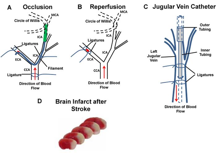
Figure 1: Diagram of the experimental stroke model, IV catheter installation, and representative infa...
Access restricted. Please log in or start a trial to view this content.
Materials
| Name | Company | Catalog Number | Comments |
|---|---|---|---|
| Heated pad | K&H Manufacturing Inc | 1060 | |
| Temperature monitor with rectal probe | Physitemp | 7029 | |
| Silk suture spool, 6-0 | Surgical Specialties Corporation | SP114 | |
| Silk suture on a needle, 3-0 | Ethicon | 1684G | |
| Nylon suture, 7-0 | Ethicon | 1696G | |
| Dental resin polysiloxane with hardener | Heraeus Kulzer | 65817930 | |
| Microinfusion IV pump | Kent Scietific | GT0897 | |
| Swivel 22GA | Instech | 375/22PS | |
| Laboratory tubing, 0.94 x 0.51 mm | Dow Corning | 508-002 | |
| Laboratory tubing, 3.18 x 1.98 mm | Dow Corning | 508-009 |
