The video demonstrates the setup and imaging process for in vivo two-photon microscopy of microglial dynamics in the hippocampal CA1 (Cornu ammonis 1) and dentate gyrus, ensuring optimal optical alignment and structural integrity assessment.
A subscription to JoVE is required to view this content. Sign in or start your free trial.
Method Article
In Vivo Two-Photon Imaging of Microglial Dynamics in the Mouse Hippocampus
1.2K views
⸱
June 17th, 2025
In This Article
Abstract
Protocol
All procedures involving animal models have been reviewed by the local institutional animal care committee and the JoVE veterinary review board.
1. In vivo, chronic two-photon imaging of microglia using CX3CR1-GFP mice
- After induction of anesthesia, wash the inside of the metal tube with sterile saline to remove debris. Ensure that the white alveus of the hippocampus is visible through the glass (Figure 1A) and that there are no complications, such as postoperative bleeding.
- Set the mouse under the two-photon microscope in the following steps:
- Set the mouse in the head-holding device under the objective lens of the two-photon microscope and on the motorized XY scanning stage. Use a heating pad for thermal support. Monitor and adjust the depth of anesthesia, as this quality check may take up to 30 mins. NOTE: The water-immersion objective lens with a working distance (WD) longer than 3 mm and a high numerical aperture (NA) is recommended. A correction collar of the objective lens can improve the resolution by compensating for the refractive index mismatch between the glass and the biomaterials.
- Fill the space between the glass and the objective lens with water, taking special care to avoid air bubbles. If necessary, expand the area of the metal plate using a plastic film, which holds more water, to cover the entire front lens of the objective.
- Turn on a femtosecond pulsed laser of 920 nm wavelength for the excitation of the fluorescent probes expressed in the brain and start image acquisition software.
- Adjust the focus to the CA1 (Cornu Ammonis 1) using the motorized stage and the motorized focus system. Position the target structure with the guidance of the fluorescence emitted from the brain parenchyma and the reflected light from the edge of the metal tube under continuous illumination by the pulsed laser.
- Measure the depths of the brain parenchyma touching the glass bottom by adjusting the focus with the motorized focus system. Compare the depths at different horizontal locations to judge the alignment of the glass bottom and the imaging plane.
- Adjust the angle of the mouse head by tilting the head-holding device until the glass bottom is set to be parallel to the imaging plane.
- Adjust the correction collar of the objective to achieve the highest resolution at the depth of the target structure in the CA1.
- Check the quality and intensity of images obtained by two-photon excitation of the hippocampus (Figure 1B).
NOTE: Image deterioration indicates the presence of tissue damage inside the brain. - Ensure that microglia have already recovered their ramified morphology (Figure 1C).
NOTE: The imaging data of microglia indicates that at least 3-4 weeks are required for microglia to return to their basal state. - Perform in vivo imaging in any layers of the CA1 for the particular purpose of the experiment.
Access restricted. Please log in or start a trial to view this content.
Results
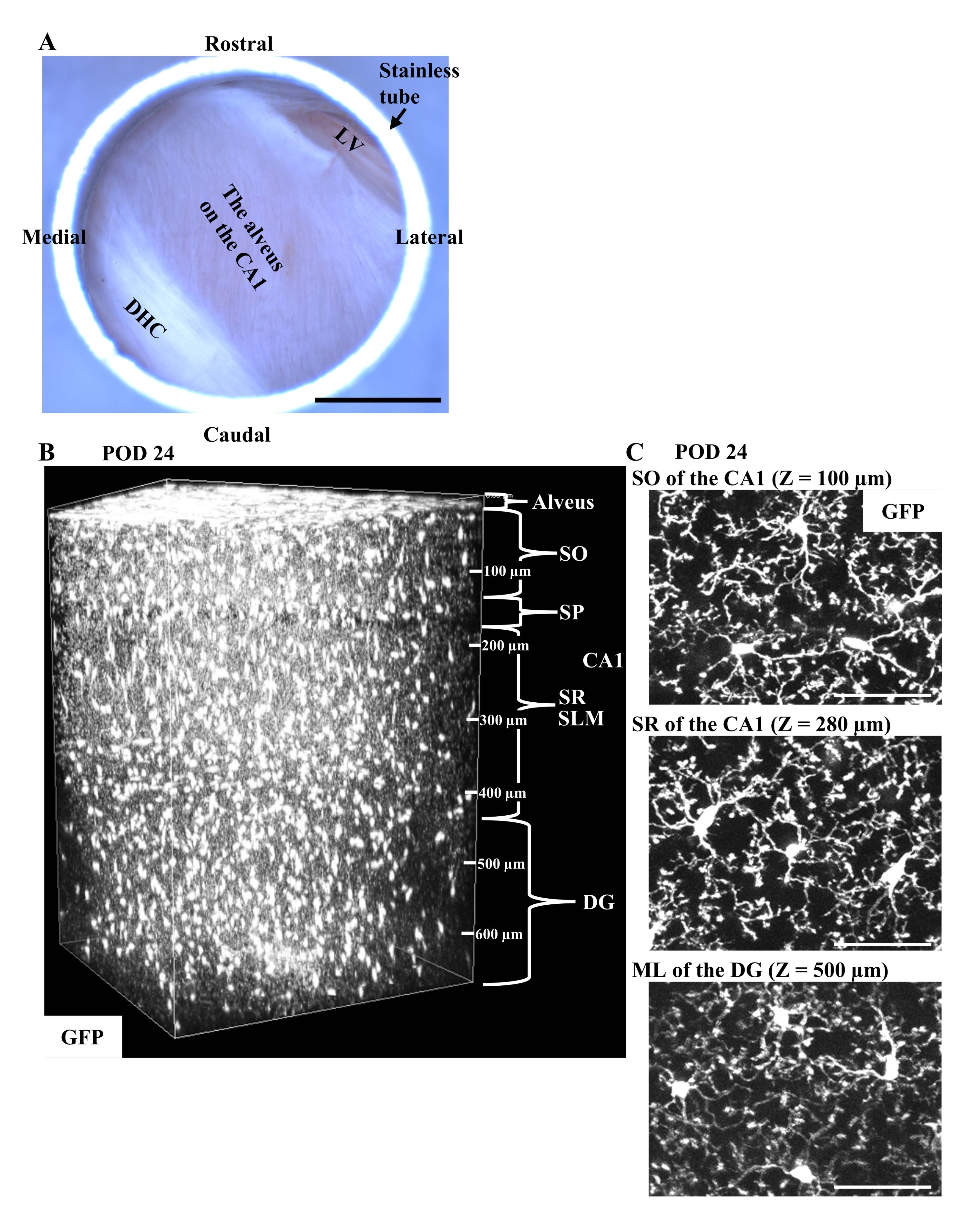
Figure 1: In vivo imaging of microglia in CX3CR1-GFP mice in the chro...
Access restricted. Please log in or start a trial to view this content.
Materials
| Name | Company | Catalog Number | Comments |
|---|---|---|---|
| A femtosecond pulsed laser | Spectra-Physics | MaiTai Deep See | A Ti:Sapphire laser used at 920 nm wavelength. |
| A two-photon microscope | Nikon | A1R MP+ | Microscope for the CA1 imaging. |
| An objective lens for two-photon imaging | Olympus | XLPLN25XSVMP | 25× objective with a long working distance, a high numerical aperture, and a correction collar |
| Aluminum plates | Narishige | CP-1 | Plates made of alminum for head fixation. |
| Circular glass coverslips | Matsunami Glass | 3φ No.1 | Coverslips bonded to stainless steel tubes. |
| CX3CR1-GFP mice | The Jackson Laboratory | 8451 | Transgenic mice for microglial imaging. |
| Cylindrical stainless steel tubes | MORISHITA | Custom-made | Metal tubes to be implanted. Outer diameter 3.0 mm, inner diameter 2.8 mm, height 1.7 mm. |
| Dental resin cement (Super-Bond C&B) | Sun Medical | 204610555 | Dental cement. |
| Head holding device | Narishige | MAG-2 | The head holding device of mice with angle adjusters |
| Heating pad | Bio Research Center | BWT-100A and HB-10 | Tools to provide thermal support for the mouse during surgery. |
| Heating pad | ALA Scientific | HEATINGPAD-1 and Hot-1 | Tools to provide thermal support for the mouse on the head holding device. |
| Ketamine (Ketalar) | Daiichi Sankyo Company | S9-001665 | Anesthesia during surgery. |
| Ophthalmic ointment | Sato Pharmaceutical | Used to prevent eye dryness during surgery. | |
| Povidone-iodine scrub solution | Meiji Seika Pharma | 2612701Q1137 | Used to disinfect the surgical site. |
| Stereo microscopes | Leica Microsystems | S8 APO | Microscopes for surgery. |
| Sterile saline | Otsuka Pharmaceutical Factory | 3311401A2026 | Washing solution during surgery. |
| Sterile waterproof pad | AS ONE | 8-5945-01 | Surgical platform. |
| Xylazine (Celactal 2%) | Bayer | Anesthesia during surgery. |
