The video describes the method for subretinal injection of viral vectors into the retinal pigment epithelium (RPE) in a mouse model. It details the procedure from anesthesia and eye preparation to precise lentiviral vector delivery, ensuring gene integration and expression in the RPE.
A subscription to JoVE is required to view this content. Sign in or start your free trial.
Method Article
Subretinal Injection of Viral Vectors into the Retinal Pigment Epithelium in a Mouse Model
1.3K views
⸱
August 7th, 2025
In This Article
Abstract
Protocol
All procedures involving animal models have been reviewed by the local institutional animal care committee and the JoVE veterinary review board.
1. Preparing Injection Kit and Viral Vectors
- Prepare the microliter syringe equipped with a 33 G blunt needle sterilized using ethylene oxide gas. Dilute the viral vectors in PBS (Phosphate buffered saline) for adequate titer in the microtube (i.e., 1 x 10⁶ TU/µl). Flush the syringe several times with viral vectors to remove any dead space in the syringe.
2. Subretinal Injection of Viral Vectors
- Anesthetize the adult mice (i.e., 6 - 8 weeks old) with an intraperitoneal injectio....
Access restricted. Please log in or start a trial to view this content.
Results
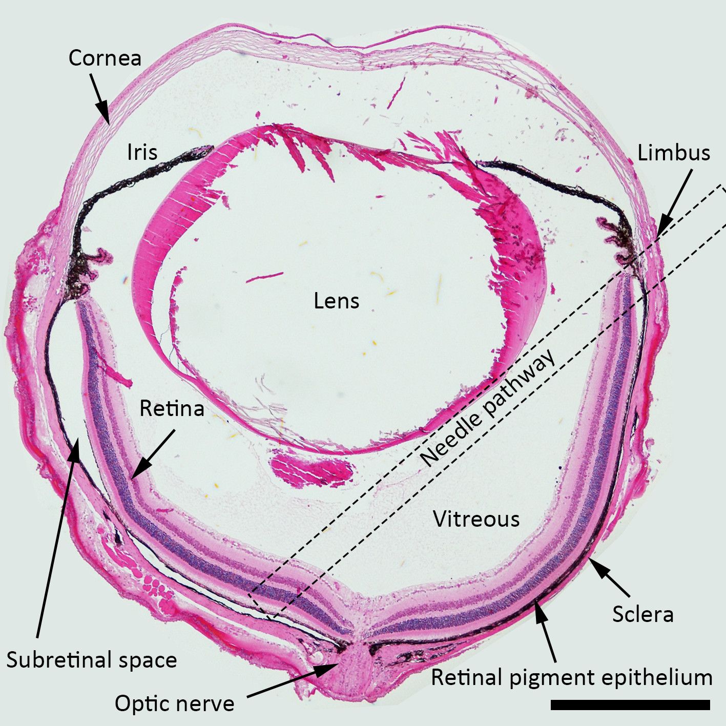
Figure 1. Schematic Diagram of the Subretinal Injection. H&E (Hematoxylin and Eosin.......
Access restricted. Please log in or start a trial to view this content.
Materials
| Name | Company | Catalog Number | Comments |
|---|---|---|---|
| Tweezers DUMONT #5 11cm DUMOSTAR 0.1 x 0.06 mm tips | WPI | 500233 | |
| VANNAS Scissors S/S, 105mm | WPI | 555583S | |
| 33G Blunt needle | WPI | NF33BL-2 | |
| NanoFil Syringe, 10 microliter | WPI | NANOFIL | |
| RPE-KIT | WPI | RPE-KIT | For easy one hand injection |
| 30Gx1/2 (0.3mmx 13mm) BD PrecisionGlideTM Needle | BD | 305107 | Initial puncture for subretinal injection |
| Microscope Cover Glasses (No. 1 3 mm diameter) | Warner Instruments | 64-0720 (CS-3R) | |
| Leica operating microscope | Leica | LM M80 | |
| Fluoresecein microscope | Nikon | Eclipse 80i | |
| Lentivirus | Thermo scientific | TMO.LV-Ctr | Used to dilute vectors |
| PBS | Gibco | 10010-015 | Used to dilute vectors |
| Troperin (Phenylephrin 0.5%-Tropicamide 0.5%) | Hanmi | For dilation | |
| Proparacaine Hydrochloride Ophthalmic Solution USP, 0.5% (Sterile) | Bausch&Lomb | For topical anesthesia | |
| Healon GV OVD | Abbott Medical Optics Inc. | ||
| Zoletil 50 (tiletamine hypochloride and zolazepam hypochloride) | Virbac | For general anesthesia | |
| Rompun® injection (Xylazine HCl) | Bayer | For general anesthesia | |
