This video demonstrates the application of repetitive transcranial magnetic stimulation (rTMS) in anesthetized rats to modulate cortical excitability. The procedure involves determining the motor threshold (MT) to establish baseline stimulation, followed by applying rTMS at an intensity above the MT to induce neuronal depolarization. This stimulation triggers calcium influx, promotes neurotrophic factor production, and enhances synaptic plasticity, ultimately influencing cortical excitability.
A subscription to JoVE is required to view this content. Sign in or start your free trial.
Method Article
Modulating Cortical Excitability in a Rat Using Repetitive Transcranial Magnetic Stimulation
750 views
⸱
August 7th, 2025
In This Article
Abstract
Protocol
All procedures involving animal models have been reviewed by the local institutional animal care committee and the JoVE veterinary review board.
1. Experimental Setup
- Animal preparation
- Allow male Sprague-Dawley rats 1 week to adapt to their new environment before starting the experiment.
NOTE: Although 8-week-old rats were used in the present study, a developing or adult brain can be chosen according to the research hypotheses.
- Allow male Sprague-Dawley rats 1 week to adapt to their new environment before starting the experiment.
- Inhalation anesthesia for induction
- Induce and maintain anesthesia with 5% and 2% of isoflurane dissolved in 40%/60% and 25%/75% oxygen/nitrogen via a chamber and nose cone, respectively. Adjust anesthesia depth to the level of abolishing the pedal withdrawal reflex to toe pinch to confirm proper anesthetization.
NOTE: Using awake animals can be a better choice in translational terms, but there are difficulties in restraining during rTMS, and they are prone to excessive stress. - Monitor body temperature with a rectal probe and maintain it at 37 °C by using a homeothermic blanket. Monitor anesthetic depth using pedal withdrawal reflex, temperature, respiratory rate, and heart rate.
- Induce and maintain anesthesia with 5% and 2% of isoflurane dissolved in 40%/60% and 25%/75% oxygen/nitrogen via a chamber and nose cone, respectively. Adjust anesthesia depth to the level of abolishing the pedal withdrawal reflex to toe pinch to confirm proper anesthetization.
- Switch over to intravenous anesthesia for maintenance
- Prepare the tail with an alcohol swab. Catheterize a lateral tail vein with a 24-gauge venous catheter for transition to intravenous (IV) anesthesia (Figure 1A). Load propofol intravenously (1 mg/[kg·min] over 10 min, using 10 mg/ml emulsion) to the animals. Discontinue isoflurane 5 min after starting propofol loading.
- Maintain propofol sedation at an infusion rate of 500 - 700 µg/(kg·min) throughout the experiment, as in a previous study. Supplement oxygen at 0.8 L/min via a nose cone.
NOTE: Anesthesia with propofol is to reduce potential suppression of cortical excitability by the inhalation agent. However, anesthesia is not mandatory in rTMS experiments, and awakened animals can also be used. Anesthesia methods should be decided in consideration of research hypotheses. - Use veterinary ointment on the eyes to prevent dryness while under anesthesia.
- Apply magnetic stimulation (see Section 2) 10 min after complete transition to IV anesthesia.
- Recovery conditions
- Monitor vital signs during the recovery phase. Do not leave the animal unattended until it has regained sufficient consciousness to maintain sternal recumbency. If an animal has undergone surgery, do not return it to the company of other animals until fully recovered.
NOTE: If surgery for a disease model is performed, post-surgical pain management is necessary. However, pain management is not needed for this rTMS experiment.
- Monitor vital signs during the recovery phase. Do not leave the animal unattended until it has regained sufficient consciousness to maintain sternal recumbency. If an animal has undergone surgery, do not return it to the company of other animals until fully recovered.
2. Repetitive Transcranial Magnetic Stimulation
- Stimulator and coil
- Apply stimulation by using a repetitive stimulator that delivers biphasic stimuli via a 25 mm figure-8 coil. Locate the center of the coil 0.5 cm lateral to the vertex on the biauricular line and angulate the coil 45° to the ground.
NOTE: The maximal magnetic field strength of the coil is 4.0 T. The magnetic coil is mounted firmly on a built-in holder.
- Apply stimulation by using a repetitive stimulator that delivers biphasic stimuli via a 25 mm figure-8 coil. Locate the center of the coil 0.5 cm lateral to the vertex on the biauricular line and angulate the coil 45° to the ground.
- Motor threshold
- Determine the motor threshold (MT) at the hot spot, with the center of the coil positioned 0.5 cm lateral to the vertex on the biauricular line and with the surface flat onto the calvaria. This is the same methodology used in a previous study.
NOTE: Define MT as the minimum stimulus intensity evoking 5 or more palpable contractions on the contralateral forepaw by 10 consecutive stimuli. Verify whether the stimulation is primarily causing contralateral muscle contraction to ensure unilateral stimulation.
- Determine the motor threshold (MT) at the hot spot, with the center of the coil positioned 0.5 cm lateral to the vertex on the biauricular line and with the surface flat onto the calvaria. This is the same methodology used in a previous study.
- Application of rTMS
- Apply rTMS 10 min after stabilization of deep anesthesia. Place the center of the coil at the target rTMS site, selected from the cerebral cortices depending on research questions. Then, tilt the coil to ensure direct contact between the coil center and the surface of the skull at the stimulation point.
NOTE: For example, angulate the coil 45° to the ground to minimize a potential direct effect of rTMS on the contralateral cortex (Figure 1B and 1C). - Subject the animals to a session of 20-min rTMS of the unilateral hemisphere. Using the software console, deliver rTMS with a low-frequency (1 Hz), high-frequency (20 Hz), or sham stimulation protocol, and set the stimulation intensity at 100 - 110% of the MT.
- Perform 1 Hz stimulation without rest. Using the software console, input "1,200" shots for "20" min. For 20 Hz stimulation, conduct 2 sec of stimulation followed by 28 sec of rest. Using the software console, input "1,600" shots for "20" min.
- For sham stimulation, tilt the coil perpendicular (90° rotation) to the calvaria and place the coil edge 2 cm apart from the head surface (Figure 1D). Fix the coil holder firmly to the main apparatus; there is no need to hold the coil by hand during the experiment.
NOTE: To compensate for acoustic and other nonspecific effects, distinct sham protocols should be used for distinct stimulation protocols. For example, 1-Hz sham stimulation can be used for 1-Hz rTMS experiments.
- Apply rTMS 10 min after stabilization of deep anesthesia. Place the center of the coil at the target rTMS site, selected from the cerebral cortices depending on research questions. Then, tilt the coil to ensure direct contact between the coil center and the surface of the skull at the stimulation point.
- Cooling the coil
- Use a water-cooling system to enable repetitive magnetic stimulation for more than 20 min at 1- and 20-Hz stimulation frequencies (Figure 2). Circulate icy water surrounding the whole length of the coil during the experiment to prevent overheating, although the temperature of the coil or stimulator is not monitored.
NOTE: Commercially available cooled rat coils can also be used. - If possible, monitor the coil temperature by viewing the heating gauge of the rTMS machine.
NOTE: There were no adverse consequences related to the rTMS stimulation. There is, however, a potential burn risk if metal ear identification tags are used near the stimulating coil.
- Use a water-cooling system to enable repetitive magnetic stimulation for more than 20 min at 1- and 20-Hz stimulation frequencies (Figure 2). Circulate icy water surrounding the whole length of the coil during the experiment to prevent overheating, although the temperature of the coil or stimulator is not monitored.
Access restricted. Please log in or start a trial to view this content.
Results
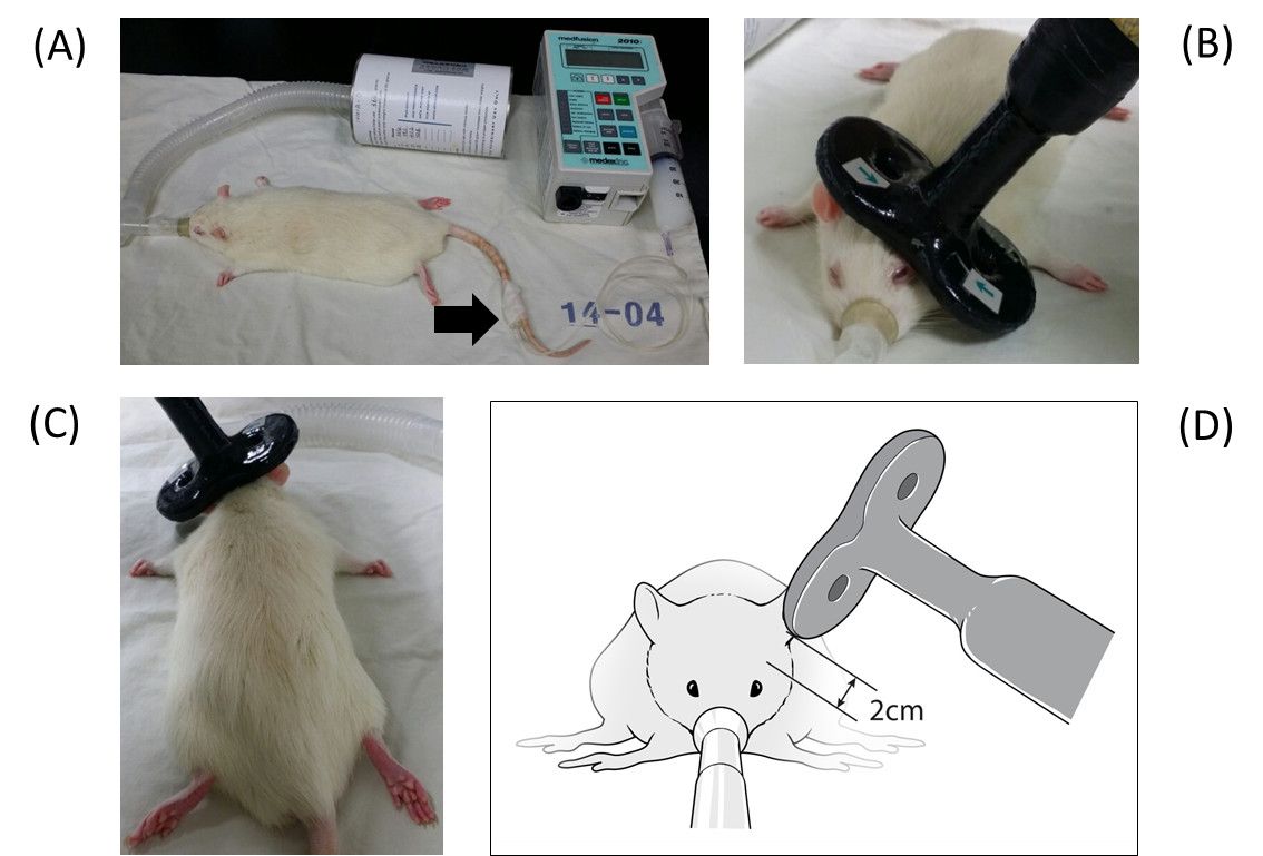
Figure 1. Experimental Settings. (A) An intravenous catheter is inserted...
Access restricted. Please log in or start a trial to view this content.
Materials
| Name | Company | Catalog Number | Comments |
|---|---|---|---|
| Homeothermic blanket with a rectal probe | Harvard apparatus | 507222F | |
| Isoflurane (Forane sol.) | Choongwae | ||
| Propofol (Provive Inj. 1% 20 ml) | Claris Lifesciences | ||
| Repetitive magnetic stimulator (Magstim Rapid²) | Magstim Company Ltd | ||
| 25 mm figure-of-8 coil | Magstim Company Ltd | 1165-00 |
