This video demonstrates a canine model for inferior alveolar nerve (IAN) regeneration using a biodegradable scaffold and ethanol-induced sympathetic blockade. The procedure involves surgically accessing the cervical sympathetic ganglion and injecting ethanol to induce vasodilation. The resulting increase in blood flow to the injury site, combined with Schwann cell-guided axonal regeneration, promotes nerve regeneration.
A subscription to JoVE is required to view this content. Sign in or start your free trial.
Method Article
Effects of Ethanol-Induced Sympathetic Ganglion Block on Nerve Regeneration in a Canine Model
499 views
⸱
August 7th, 2025
In This Article
Abstract
Protocol
All procedures involving animal models have been reviewed by the local institutional animal care committee and the JoVE veterinary review board.
1. Anesthesia and Skin Preparation
- Anesthetize the dog with a mixture of 5 mg/kg ketamine hydrochloride and 1 mg/kg xylazine by an intramuscular injection.
- Intubate with a tracheal tube of 7.5 mm diameter and 25 cm length.
- Place the dog in the right lateral position. Maintain general anesthesia with 3.2% sevoflurane with oxygen (1.0 L/min).
- Use a heating pad to maintain body temperature at 37 °C.
- Apply an ophthalmic gel over the anterior surface of the eyes to avoid corneal abrasion.
- Carefully shave the surgical field (left side chest area) using surgical clippers. Spray a substantial amount of alcoholic solution over the operative site. Wait at least 15 sec. Repeat the application 3 times.
- Record the heart rate and oxygen saturation during surgery.
2. Inferior Alveolar Nerve Reconstruction Using polyglycolic acid collagen (PGA-C) tube: Development of the Reconstruction-only Model
- Inject 3 mL of 1% lidocaine using a 27 G needle into the left mandibular gingiva as a local anesthetic and analgesic.
- Perform a 5-cm transverse incision with a number 15 scalpel blade in the left mandibular gingiva to expose the mandible of the animal.
- Use piezoelectric ultrasonic vibrations to grind the proximal aspect of the mandible into a 3-cm × 8-mm rectangle through the posterior mental foramen.
NOTE: The vibration frequency was 28 ‒ 32 kHz. - Remove the frontal part of the mandibular bone plate (dimensions, 3 cm × 8 mm) to expose the left IAN (Figure 1A).
NOTE: The reconstruction site corresponds to the root apex of the first molar - Transect the IAN with a scalpel to remove a 10-mm segment.
- Insert the proximal and distal stumps of the severed nerve into the nerve tube to a depth of 2 mm.
- Use 8-0 nylon sutures and a surgical microscope at 8X magnification to suture the tube to the proximal and distal nerve ends (Figure 1B).
- Return the bone plate to its original site in the mandible.
- Close the wound with 4-0 nylon sutures.
- One day after surgery, confirm that the mandibular bone plate is in its proper position.
- 4.10.1 Perform computed tomography (CT) imaging of the facial bone under anesthesia. Set CT parameters as follows: 120 kVp, 200 mAs, 0.5 mm/s, 0.5-mm slice thickness.
- Administer anesthesia using a mixture of 5 mg/kg ketamine hydrochloride and 1 mg/kg xylazine (Figure 2).
- 4.10.1 Perform computed tomography (CT) imaging of the facial bone under anesthesia. Set CT parameters as follows: 120 kVp, 200 mAs, 0.5 mm/s, 0.5-mm slice thickness.
- Administer ampicillin (100 mg/day) as an antibiotic and acetaminophen (100 mg/day) as an analgesic for a week after surgery.
3. Ethanol-induced cervical sympathetic ganglion block (CSGB): Development of the Reconstruction + CSGB Model
- Perform IAN reconstruction as described in section 2 and allow a week for recovery.
- Anesthetize the animal with 1.5% sevoflurane in oxygen (4 L/min) and air (6 L/min). Shave and clean the intended surgical field, as described in section 3.
- Mark the incision line with a surgical skin marker by drawing a line on the left side chest area (Figure 3, the incision line is 20 cm in length).
- Inject 5 mL of 1% lidocaine using a 21 G needle into the left side chest area as a local anesthetic and analgesic.
- Incise the left side chest skin with a number 10 scalpel blade.
- Incise the fat layer with an electric scalpel to expose the muscle fascia.
- Expose the serratus ventralis muscle and the scalenus muscle.
- Raise the serratus ventralis muscle and the scalenus muscle from ventral to dorsal to expose the second and third ribs (Figure 4).
- Perform a left lateral thoracotomy at the second and third intercostal space to expose the left cervical sympathetic ganglion (Figure 5).
- Inject 0.2 mL of 99.5% ethanol into the cervical sympathetic ganglion using a 30 G needle under direct visualization (Figure 6).
- Close the intercostal space with interrupted 1-0 absorbable stitches.
- Close the skin with interrupted 3-0 nylon stitches.
- Administer ampicillin (100 mg/day) as an antibiotic and acetaminophen (100 mg/day) as an analgesic for a week after surgery.
- At 1 week after CSGB, measure facial skin temperature with infrared thermography to confirm the CSGB.
Access restricted. Please log in or start a trial to view this content.
Results
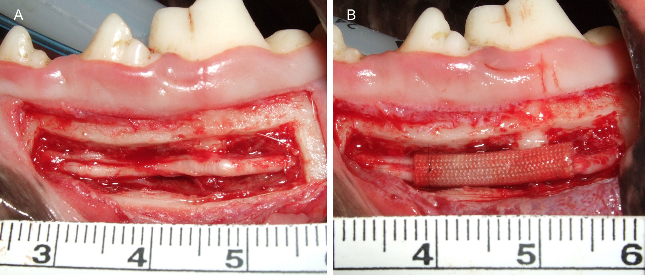
Figure 1: Left inferior alveolar nerve (IAN) pre- and post-reconstruction. A) Pre-recon...
Access restricted. Please log in or start a trial to view this content.
Materials
| Name | Company | Catalog Number | Comments |
|---|---|---|---|
| Surgical clippers | Roboz Surgical Instrument Company | RC-5903 | |
| Disposable scalpel (No.15) | Kai medical | 219ABBZX00073000 | |
| VarioSurg3 | Nakanishi | VS3-LED-HPSC, E1133 | Piezoelectric surgery for bone processing |
| 4-0 nylon sutures | Ethicon | 8881H | |
| 8-0 nylon sutures | Ethicon | 2775G | |
| Isepamicin sulfate | Nichi-Iko | 620005641 | |
| Disposable scalpel (No.10) | Kai medical | 219ABBZX00073000 | |
| 30-gauge needle | Nipro | 1134 | |
| 1-0 absorbable stitches | Ethicon | J347H | |
| 3-0 Nylon stitches | Ethicon | 8872H | |
| Neo Thermo | NEC Avio | TVS-700 | Infrared thermography |
