A method to monitor ubiquitin-proteasome activity in living cells is described. A degron-destabilized GFP- (GFP-dgn) and a stable GFP-dgnFS fusion protein are generated and transduced into the cell using a lentiviral expression vector. This technique allows to generate a stable GFP-dgn/GFP-dgnFS expressing cell line in which ubiquitin-proteasome activity can be easily assessed using epifluorescence or flow cytometry.
A subscription to JoVE is required to view this content. Sign in or start your free trial.
Method Article
Monitoring of Ubiquitin-proteasome Activity in Living Cells Using a Degron (dgn)-destabilized Green Fluorescent Protein (GFP)-based Reporter Protein
In This Article
Summary
Abstract
Proteasome is the main intracellular organelle involved in the proteolytic degradation of abnormal, misfolded, damaged or oxidized proteins 1, 2. Maintenance of proteasome activity was implicated in many key cellular processes, like cell's stress response 3, cell cycle regulation and cellular differentiation 4 or in immune system response 5. The dysfunction of the ubiquitin-proteasome system has been related to the development of tumors and neurodegenerative diseases 4, 6. Additionally, a decrease in proteasome activity was found as a feature of cellular senescence and organismal aging 7, 8, 9, 10. Here, we present a method to measure ubiquitin-proteasome activity in living cells using a GFP-dgn fusion protein. To be able to monitor ubiquitin-proteasome activity in living primary cells, complementary DNA constructs coding for a green fluorescent protein (GFP)–dgn fusion protein (GFP–dgn, unstable) and a variant carrying a frameshift mutation (GFP–dgnFS, stable 11) are inserted in lentiviral expression vectors. We prefer this technique over traditional transfection techniques because it guarantees a very high transfection efficiency independent of the cell type or the age of the donor. The difference between fluorescence displayed by the GFP–dgnFS (stable) protein and the destabilized protein (GFP-dgn) in the absence or presence of proteasome inhibitor can be used to estimate ubiquitin-proteasome activity in each particular cell strain. These differences can be monitored by epifluorescence microscopy or can be measured by flow cytometry.
Protocol
1. Plasmid Construction
- Order custom oligo-nucleotides encoding for dgn (ACKNWFSSLSHFVIHL11) and for dgnFS (HARTGSLACPTSSSICE) and ligate it into the pEGFP-C1 vector to obtain the fusion of the GFP with dgn/dgnFS (Figure 1).
- Amplify the coding sequence for GFP-dgn and GFP-dgnFS by PCR according to the protocol of the pENTR Directional TOPO Cloning Kit and continue with the pLenti6/V5 Directional TOPO Cloning Kit (Figure 6).
2. Virus Production
- The day before transfection (Day 1) seed out HEK 293FT cells in a T75 flasks so that they will be 90-95% confluent on the day of transfection.
- On the day of transfection dilute 36 μl of lipofectamine 2000 Reagent in 1.5 ml of DMEM without serum and antibiotics in a 15 ml falcon. Use another 15 ml falcon and dilute 3 μg pLenti6/V5 carrying the complementary DNA construct for either GFP-dgn or GFP-dgnFS, 2.5 μg envelope encoding plasmid pMD2.G and 7.5 μg vector backbone psPAX2 in 1.5 ml of DMEM without serum and antibiotics. After 5 min the diluted DNA is combined with the diluted lipofectamine 2000 Reagent.
- Incubate the mixture for 20 min at room temperature to allow the DNA-lipofectamine complexes to form.
- Remove the medium of HEK 293FT cells, replace it carefully by 7 ml of medium (without antibiotics) and add the DNA-lipofectamine mixture to the flask and rock it back and forth for mixing (Day 2). Incubate the flask overnight at 37 °C in a humidified 5% CO2 incubator.
- Change medium the next day (10 ml medium without antibiotics; Day 3).
- After 48 hr (Day 5) harvest supernatant, centrifuge at 300 x g for 5 min at room temperature and filter the supernatant through a 0.45 μm PVDF filter.
Culture medium - DMEM (HEK 293FT)
DMEM
10% FBS
0.1 mM MEM NEAA
6 mM L-glutamine
1 mM MEM Sodium Pyruvate
1% Pen-Strep (optional)
500 μg/ml Geneticin (optional)
3. Virus Concentration by Polyethylene Glycol (PEG) Precipitation
- Add one volume Polyethylene glycol solution (50 mM Polyethylene glycol, 41 mM NaCl, autoclave, pH =7.2; PEG) to four volumes of supernatant and incubate for 2 hr at 4 °C. Carefully mix it every 20-30 min by inverting.
- Spin down at 1500 x g for 30 min at 4 °C. A white pellet should be visible.
- Aspirate the supernatant and centrifuge again at 1500 x g for 5 min at 4 °C. Aspirate the remaining PEG solution.
- Resuspend the pellet in medium or phosphate-buffered saline (PBS) by pipetting up and down and vigorously vortex for 20 to 30 seconds. As a guideline use 500 μl for one T75 flask and aliquot it to 100 μl. Store the virus at -80 °C.
4. Titering of Virus
- Seed out 5×10⁴ U2-OS cells to each well of a 6-well plate the day before transfection.
- On the day of transfection remove the culture medium and replace it by 1 ml of DMEM containing 8 μg/ml polybrene (hexadimethrine bromide). Dilute the concentrated virus supernatant 1:10 and 1:100 and add 1 μl each to one well. For higher virus concentrations use 1 μl, 5 μl and 10 μl of concentrated virus supernatant for the remaining wells. One well is left as a positive control.
- The following day replace the medium by 2 ml of DMEM.
- Start with the selection the next day. Apply 10 μg/ml Blasticidin on each well. Replace the antibiotic containing medium every second day.
- Approximately 6-7 days after starting the selection the untransduced cells are dead. For the staining wash the cells three times with PBS and cover the cells with crystal violet (10 mg/l crystal violet in 20% ethanol) and incubate for 5-10 min at room temperature. Aspirate crystal violet (can be reused a few times) and rinse the wells twice with ddH2O and dry the plate.
- Count the colonies and multiply with 1,000 and the corresponding dilution (Figure 7). This procedure allows to calculate transfection units (TU) of the virus / ml.
Culture medium - DMEM (HFF-2/U2-OS)
DMEM
10% FBS
6mM L-glutamine
1% Pen-Strep
5. Transduction of Human Diploid Fibroblasts (This procedure can be used for any cell type.)
- Seed out 5×10⁴ human diploid fibroblasts (HDFs) to 6-well plates the day before transduction.
- Use a multiplicity of infection of two together with 8 μg/ml polybrene as transduction enhancer in a total of one milliliter.
- Change medium the next day.
- When the cells are at 70-80 % of confluency start the selection with 10 μg/ml blasticidin. After the selection is completed (no more untransfected cells die) the expansion of the cells can start and the cells are ready for experiments. Chronic selection with blasticidin allows to generate a stable cell line overexpressing GFP-dgn or GFP-dgnFS fusion proteins, ready to measure proteasome activity. This procedure guarantees a very high transfection efficiency independent of the cell type.
6. Measurement by Flow Cytometry
- Seed out 1×10⁵ cells (carrying either GFP-dgn or GFP-dgnFS) the day before the experiment.
- Treat control cells 3 hr prior the measurement with the proteasome inhibitor, e.g. 100 μg/ml LLnL.
- Wash twice with PBS and harvest the cells using 0.5 ml of trypsin. To stop the trypsinisation use 4.5 ml of culture medium and transfer the cells to a 15 ml falcon.
- Spin the cells at 300 x g for 5 min at room temperature.
- Aspirate the medium, resuspend the cells in PBS and spin down again at 300 x g for 5 min at 37 °C.
- Aspirate the PBS, resuspend in 400 μl of FACS buffer (10 mM Hepes, 140 mM NaCl, 2.5 mM CaCl2, pH=7.4) and transfer the cells into FACS tubes.
- Keep the cells on ice and measure samples by flow cytometry. The cells should not be stored longer than 30 min at 4 °C.
7. Representative Results
The GFP-dgn fusion protein carries a sequence which is targeted to the proteasome and therefore the protein is immediately degraded; it corresponds to the decrease in GFP fluorescence signal. The frameshift mutant (GFP-dgnFS) carries a mutated version of this sequence and is not degraded by proteasome; it leads to the higher green fluorescence. For these reasons, young HDFs with expected high proteasome activity and transduced with GFP-dgn show a low (6% positive cells) fluorescence signal in both flow cytometry measurement and in epifluorescence (Figure 2A and B, Figure 3). The same HDFs transduced with GFP-dgnFS display 39.7% of positive cells. The treatment of the cells with proteasome inhibitor LLnL (N-acetyl-L-leucyl-L-leucyl-L-norleucinal) raised the signal to maximum of 62.9% in both, GFP-dgn and GFP-dgnFS cells (Figure 2A and B).
To determine possible decline in proteasome activity in aged human skin samples, dermal fibroblasts isolated from young, middle-aged and old donors were infected with GFP-dgn and GFP-dgnFS, as described above and cultivated to the same passage number before analysis by flow cytometry. In these experiments, a clear-cut increase in GFP signal between young individuals (11.2±0.88% GFP-positive cells) and middle-aged donors (20.4±2.27% GFP-positive cells, P=0.003) has been observed, indicating a decrease in proteasome activity in samples obtained from aged donors7 (Figure 4). No further decrease in proteasome activity was observed in fibroblasts isolated from oldest individuals (Figure 4). Fluorescence intensity for GFP-dgnFS was in all cases ~90% (data not shown) which indicates a high transfection efficiency for the three different age groups.

Figure 1. The GFP-dgn and GFP-dgnFS sequence. Displayed is the 3'-end of GFP (green) the multiple cloning site of the pEGFP-CL1 vector (grey) and the dgn/dgnFS sequence (red).

Figure 2. Flow cytometry analysis of GFP-dgn and GFP-dgnFS human diploid fibroblasts. A. Young human foreskin fibroblasts (HFF-2) were infected with lentiviral vectors carrying a green fluorescent protein (GFP)–dgn gene or a GFP–dgnFS (frameshift) construct, as indicated and analyzed for GFP fluorescence using flow cytometry (FACS Canto II, Becton Dickinson). Where indicated, cells were also treated for 3h with proteasome inhibitor N-acetyl-L-leucyl-L-leucyl-L-norleucinal (LLnL). Uninfected cells were used as a control. Experiments were performed in triplicates. B. Data are representative of three independent experiments. The numbers reflect the amount of GFP positive cells. Click here to view larger figure.
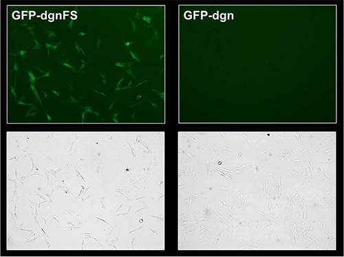
Figure 3. Fluorescence microscopy analysis of GFP-dgn and GFP-dgnFS HFF-2 cells.
Young HFF-2 were treated as in Figure 2. At 9 days after infection, cells were visualized by fluorescence and phase contrast microscopy.
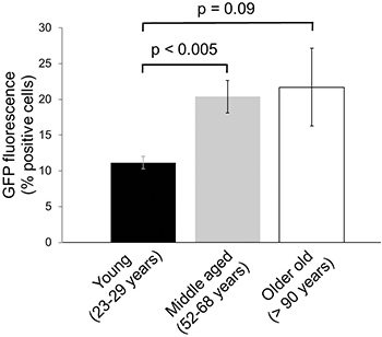
Figure 4. Changes in proteasome activity in human skin aging.
The human foreskin fibroblasts from nine different donors in the indicated age groups were minimally expanded and infected with lentiviral constructs coding for GFP–dgn. At 9 days after infection, the cells were analyzed by flow cytometry. Data were obtained on three samples per age group in duplicates (± SE).

Figure 5. Experimental approach. Custom-oligo nucleotides for dgn and dgnFS are cloned into the pEGFP-C1 vector and viruses are produced for each construct using HEK 293FT cells. The titer of the viruses is determined. Cells are transduced with the virus and expanded. After the favored treatment the cells are analyzed for fluorescence signal by flow cytometry.

Figure 6. Map of pLenti GFP-dgn. The map of the pLenti6/V5-DEST vector including the GFP-dgn sequence is displayed. Abbreviations: PCMV (CMV promoter), GFP-dgn (sequence of GFP-dgn), PSV40 (SV40 early promoter), EM7 (EM7 promoter), Blasticidin (Blasticidin resistance gene), ΔU3/3'LTR (3'LTR with deleted U3 region), SV40 pA (SV40 polyadenylation signal), Ampicillin (Ampicillin resistance gene), pUC ori (pUC origin), PRSV/5'LTR (RSV/5'LTR hybrid promoter), Ψ (HIV-1 Ψ packaging signal), RRE (HIV-1 Rev response element).

Figure 7. U2-OS titering plate. U2-OS were seeded out on a 6-well plate and transfected with different virus concentrations (dilution of 1/100 and 1/10, 1, 5 and 10 μl of concentrated viral supernatant). One well was used as untransfected control (NT). After 6-7 days, the cells were stained with crystal violet and air dried.
Access restricted. Please log in or start a trial to view this content.
Discussion
The first publication using green fluorescent protein (GFP) as a reporter substrate for ubiquitin-proteasome activity was published in 2000 12. Since then, GFP has become a common tool to visualize cellular activities, especially the ubiquitin-proteasome process. To monitor ubiquitin-proteasome activity in vivo a transgenic mouse model with a GFP-based reporter has been introduced 13. Additional in vivo research established another transgenic mouse model with a similar degron-desta...
Access restricted. Please log in or start a trial to view this content.
Disclosures
No conflicts of interest declared.
Acknowledgements
This study was funded by: National Research Network on Aging (NFN S93) by the Austrian Science Foundation (FWF), European Commission Integrated Projects MiMAGE and PROTEOMAGE, Netherlands Genomics Initiative/Netherlands Organization for Scientific Research (NGI/NWO; 05040202 and 050-060-810 NCHA), the EU funded Network of Excellence Lifespan (FP6 036894), and Innovation Oriented Research Program on Genomics (SenterNovem; IGE01014 and IGE5007).
Access restricted. Please log in or start a trial to view this content.
Materials
| Name | Company | Catalog Number | Comments |
|---|---|---|---|
| pEGFP-C1 Vector | BD Bioscience Clontech | 6084-1 | |
| pENTR Directional TOPO Cloning Kit | Invitrogen | K2400-20 | |
| pLenti6/V5 Directional TOPO Cloning Kit | Invitrogen | V496-10 | |
| Lipofectamine 2000 Reagent | Invitrogen | 11668019 | |
| DMEM | Sigma-Aldrich | D5546 | |
| PVDF filter (Rotilabo-Spritzenfilter) | Carl Roth Gmbh | P667.1 | |
| Polyethylene glycol | Sigma-Aldrich | P2139 | |
| NaCl | Merck & Co., Inc. | 1.06404.1000 | |
| Dulbecco's Phosphate Buffered Saline 1x (PBS) | Invitrogen | 14190 | |
| hexadimethrine bromide | Sigma-Aldrich | 10,768-9 | |
| Blasticidin | Invitrogen | R21001 | |
| Crystal violet | Sigma-Aldrich | C3886 | |
| FACS tubes | BD Biosciences | ||
| Penicillin Streptomycin (Pen-Strep) | Invitrogen | 15140130 | |
| L-glutamine 200 mM | Invitrogen | 25030024 | |
| Fetal Bovine Serum (FBS) | Biochrom AG | S0115 | |
| MEM Non-Essential Amino Acids (NEAA) 100x | Invitrogen | 11140035 | |
| MEM Sodium Pyruvate 100 mM | Invitrogen | 11360039 | |
| D-(+)-Glucose (45%) | Sigma-Aldrich | G8769 | |
| Geneticin | Invitrogen | 11811023 | |
| CaCl2 | Merck & Co., Inc. | C5080 | |
| Hepes | Sigma-Aldrich | H3375 | |
| Trypsin-EDTA (0.05%) | Invitrogen | 25300054 |
References
- Coux, O., Tanaka, K., Goldberg, A. L. Structure and functions of the 20S and 26S proteasomes. Annu. Rev. Biochem. 65, 801-847 (1996).
- Davies, K. J. Degradation of oxidized proteins by the 20S proteasome. Biochimi. 83, 301-310 (2001).
- Stangl, K., Stangl, V. The ubiquitin-proteasome pathway and endothelial (dys)function. Cardiovasc. Res. 85, 281-290 (2009).
- Tuoc, T. C., Stoykova, A. Roles of the ubiquitin-proteosome system in neurogenesis. Cell Cycle. 9, 3174-3180 (2010).
- Rock, K. L., et al. Inhibitors of the proteasome block the degradation of most cell proteins and the generation of peptides presented on MHC class I molecules. Cell. 78, 761-771 (1994).
- Lehman, N. L. The ubiquitin proteasome system in neuropathology. Acta Neuropathol. 118, 329-347 (2009).
- Koziel, R., Greussing, R., Maier, A. B., Declercq, L., Jansen-Durr, P. Functional Interplay between mitochondrial and proteasome activity in skin aging. J. Invest. Dermatol. 131, 594-603 (2010).
- Grillari, J., Grillari-Voglauer, R., Jansen-Durr, P. Post-translational modification of cellular proteins by ubiquitin and ubiquitin-like molecules: role in cellular senescence and aging. Adv. Exp. Med. Biol. 694, 172-196 (2010).
- Bulteau, A. L., Szweda, L. I., Friguet, B. Age-dependent declines in proteasome activity in the heart. Arch. Biochem. Biophys. 397, 298-304 (2002).
- Strucksberg, K. H., Tangavelou, K., Schroder, R., Clemen, C. S. Proteasomal activity in skeletal muscle: A matter of assay design, muscle type, and age. Anal. Biochem. , (2009).
- Bence, N. F., Sampat, R. M., Kopito, R. R. Impairment of the ubiquitin-proteasome system by protein aggregation. Science. 292, 1552-1555 (2001).
- Dantuma, N. P., Lindsten, K., Glas, R., Jellne, M., Masucci, M. G. Short-lived green fluorescent proteins for quantifying ubiquitin/proteasome-dependent proteolysis in living cells. Nat. Biotechnol. 18, 538-543 (2000).
- Lindsten, K., Menendez-Benito, V., Masucci, M. G., Dantuma, N. P. A transgenic mouse model of the ubiquitin/proteasome system. Nat. Biotechnol. 21, 897-902 (2003).
- Liu, J., et al. Impairment of the ubiquitin-proteasome system in desminopathy mouse hearts. FASEB J. 20, 362-364 (2006).
- Bowman, A. B., Yoo, S. Y., Dantuma, N. P., Zoghbi, H. Y. Neuronal dysfunction in a polyglutamine disease model occurs in the absence of ubiquitin-proteasome system impairment and inversely correlates with the degree of nuclear inclusion formation. Hum. Mol. Genet. 14, 679-691 (2005).
- Myung, J., Kim, K. B., Lindsten, K., Dantuma, N. P., Crews, C. M. Lack of proteasome active site allostery as revealed by subunit-specific inhibitors. Mol. Cell. 7, 411-420 (2001).
- Menendez-Benito, V., Heessen, S., Dantuma, N. P. Monitoring of ubiquitin-dependent proteolysis with green fluorescent protein substrates. Methods Enzymol. 399, 490-511 (2005).
- Lener, B., et al. The NADPH oxidase Nox4 restricts the replicative lifespan of human endothelial cells. Biochem. J. 423, 363-374 (2009).
Access restricted. Please log in or start a trial to view this content.
Reprints and Permissions
