The use of a model that mimics the condition of lung diseases in humans is critical for studying the pathophysiology and/or etiology of a particular disease and for developing therapeutic intervention. Here a noninvasive intratracheal intubation method that can directly deliver exogenous materials to mouse lungs is presented.
Method Article
Noninvasive Intratracheal Intubation to Study the Pathology and Physiology of Mouse Lung
In This Article
Summary
Abstract
The use of a model that mimics the condition of lung diseases in humans is critical for studying the pathophysiology and/or etiology of a particular disease and for developing therapeutic intervention. With the increasing availability of knockout and transgenic derivatives, together with a vast amount of genetic information, mice provide one of the best models to study the molecular mechanisms underlying the pathology and physiology of lung diseases. Inhalation, intranasal instillation, intratracheal instillation, and intratracheal intubation are the most widely used techniques by a number of investigators to administer materials of interest to mouse lungs. There are pros and cons for each technique depending on the goals of a study. Here a noninvasive intratracheal intubation method that can directly deliver exogenous materials to mouse lungs is presented. This technique was applied to administer bleomycin to mouse lungs as a model to study pulmonary fibrosis.
Introduction
Lung is an organ where many devastating diseases are commonly diagnosed. Among them, lung cancer is the second-most diagnosed cancer in both men and women, and the most common cause of cancer death. Chronic obstructive pulmonary disease, also known as emphysema and chronic bronchitis, is a very serious disease and the third leading cause of death in the United States. In 2011, it was estimated that 25.9 million Americans had asthma, including 7.1 million children under the age of 18. Asthma is the third leading cause of hospitalization among children under the age of 15 (American Lung Association, http://www.lung.org). In order to study the pathophysiology and/or etiology of these devastating diseases and their underlying mechanisms, the use of accurate models is critical in conjunction with convenient and noninvasive administration of various materials of interest to lung. Mice provide one of the best models to study the molecular mechanisms underlying the pathology and physiology of lung diseases because of the increasing availability of knockout and transgenic mice and a vast amount of available genetic information.
Various methods have been used by a number of investigators in different settings to deliver materials of interest to mouse lungs, including inhalation, intranasal instillation, intratracheal instillation, and intratracheal intubation1-4. The latter procedure has not been routinely used because it is considered rather difficult to perform. Intratracheal intubation described herein is a noninvasive, simple, and quick method to deliver materials of interest to mouse lungs in order to study the effect of the delivered materials on gene expression patterns, pathology and/or physiology of lung5. This technique assures the delivery of exogenous materials to a whole lung and does not involve any survival surgery and thus will likely be approved by any institutional animal care and use committees.
Protocol
The following protocol describes a noninvasive, simple, and quick method to deliver materials of interest to mouse lungs. This procedure was approved by the National Cancer Institute Animal Care and Use Committee.
1. Anesthesia
- First, anesthetize the mouse using a mixture of ketamine and xylazine (100 mg/kg body weight and 10 mg/kg body weight, respectively). This is the ACUC recommended anesthesia and dose. With this amount, mice are unconscious at least for ~20 min.
- Apply vet ointment to the eyes of the mouse in order to prevent drying of the eyes during anesthesia.
- After several minutes, pinch a foot of the mouse to check for consciousness. Once confirmed unconscious, place the mouse on an intubation stand angled at ~60° and hold it in place by hooking its upper incisors over a small rubber band located at the top of the stand.
2. Intratracheal Intubation
- Gently retract the mouse's tongue to one side using a Q-tip.
- When using the BioLITE Intubation Illumination System, carefully insert the intubation system until the larynx is visualized with the aid of the fiber-optic light guide.
- Once the epiglottis and the arytenoid cartilages are visualized, insert the fiber over the epiglottis and between the arytenoid cartilages, and advance until the proper length of catheter has been inserted.
Note: In order to obtain the proper length of catheter to be inserted, initially measure the length between the mouth and the bronchial bifurcation point by using a practice mouse of a similar size in advance (Figure 1). The length largely depends on the size of the mouse. Insertion of the catheter should stop above the bifurcation point (~1.5 cm for mouse with ~25 g of body weight). This ensures that the intubated material will go to all lobes. At least 50 practice mice may be required for a person performing the intubation to become proficient in the technique (Proficient means that the success rate of intubation is more than 95%).
- Once the catheter is inserted, quickly remove the fiber-optic light guide from the catheter to allow the animal to breathe normally. When the Intubation Illumination System is not used, directly insert a catheter as described.
- Add a solution containing materials of interest to a catheter. Make sure that the solution is sucked into the lung immediately after addition. Fifty microliters for ~25 g mouse body weight is routinely used.
3. Animal Recovery
- As soon as the solution is sucked into the lung, take down the mouse from the stand, and put it back into the original cage.
- Observe the mouse until it starts moving.
- Once confirmed that the mouse is in good condition, return the cage to a rack.
Results
Initially, green-dyed solution was used to intubate mice for practice. The lung was resected immediately after intubation to examine how evenly the color was distributed in the lung (Figure 2). This technique was applied to study bleomycin-induced pulmonary fibrosis using C57BL/6 mice. When the mice were intratracheally intubated with 1.2 U/kg of bleomycin or saline as control and necropsied 3 weeks later, the mice developed bleomycin-induced fibrosis throughout their lungs, supported by histology and the increased hydroxyproline content (Figure 3). The damaged area was evaluated using 20X objective created lung images and a 121-points lattice grid. The number of intersections (points) falling over the fibrous areas were counted and expressed as the percentage of total (121) points. The percentage of the damaged areas thus counted was proportional to the amount of bleomycin administered (Figure 4).

Figure 1. Illustration of mouse trachea. The red bars and arrows indicate the length where the measurement should be taken before intubation using a practice mouse. Click here to view larger image.
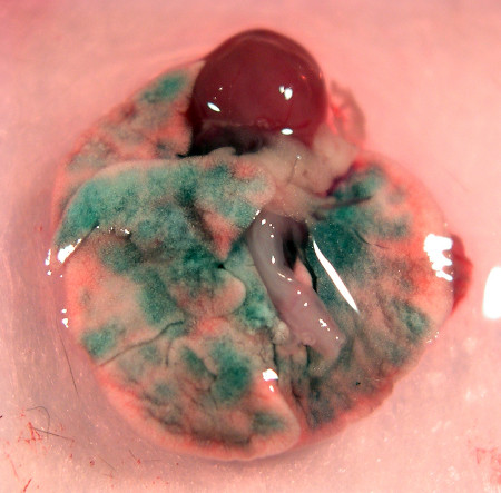
Figure 2. Appearance of lung right after being intubated with green dye. If the dye color can be seen in most parts of the lung, intubation is considered successful. Click here to view larger image.

Figure 3. Representative images of fibrotic lungs induced by intratracheal intubation of bleomycin. C57BL/6 mice were intubated with 1.2U/kg of bleomycin (B, D) or saline as control (A, C) and their lungs were histologically examined on day 21 post-bleomycin administration. Magnification: A, B: 40X; C, D: 100X. (E) Hydroxyproline content per lung measured on day 21 for control and bleomycin treated lungs. N=6, ***:P<0.001. Click here to view larger image.
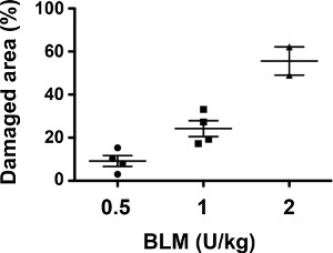
Figure 4. Relationship between bleomycin dose and damaged area of lung. Increased dose of bleomycin (0.5, 1, and 2 U/kg) proportionally increased the percentage of damaged areas. Click here to view larger image.
Discussion
Intratracheal intubation described here is a simple, yet excellent noninvasive method to evenly deliver materials of interest to mouse lung. This method allows study of the effect and/or role of the material administered on the physiology and/or pathology of lung. The materials administered can be endogenous molecules such as cytokines, or exogenous materials such as xenobiotic chemicals/drugs, carcinogens, pollutants, allergens, or viruses that result in various lung conditions that may represent a model for studying various human diseases6-10. This technique can also be used in conjunction with recombinant adenovirus or lentivirus to introduce overexpression or deletion of genes of interest in airway epithelial cells to study the role of these genes in homeostasis, physiology, pathology, and/or carcinogenesis of lung. The disruption of a gene can be achieved by transiently expressing Cre recombinase in epithelial cells to disrupt a floxed gene of interest in the epithelial cells11.
A catheter can easily be mistakenly inserted into the esophagus, which is juxtaposed to the trachea. In the method described here, the Intubation Illumination System provides guidance to the correct position where the catheter should be inserted5. The system consists of a fiber-optic fiber that is stably connected to a light source, and a specially engineered miniature optical lens system that allows the light to focus onto the fiber-optic fiber. The delivery end of the fiber-optic fiber fits into the disposable intravenous catheters that are used as intratracheal tubes. This system provides direct illumination of the oropharynx, enabling clear visualization of the larynx during intubation. Once one becomes proficient in carrying out this technique using the Illumination System, the use of the system is no longer necessary. The procedure can be efficiently performed with only a catheter, and the whole procedure requires only a few minutes. The exact same method can be used for rats with a larger-sized Illumination System or catheter.
Three points are critical for evenly distributing the material throughout the lung. First, always measure the length between the mouth and the bronchial bifurcation point before intubation in order to have an idea of how deep the catheter should be inserted using a practice mouse (Figure 1). Since this largely depends on the size of the mouse, the same insertion length can be used once it is determined for mice of a particular size. Second, make sure that the solution added to the intubation tube is sucked right after its addition. If the intubation tube is mistakenly inserted into the esophagus, the solution will not be sucked in immediately and thus stay in the tube. If this happens, repeat the whole process of intubation. This confirmation process assures that the solution is in the trachea, but not in the esophagus. The operator can try 3x at maximum to repeat the whole intubation procedure if the solution is not sucked into the lung. However, if this frequently happens, it is recommended to go back to practice to acquire higher successful rate. Lastly, mice should be unconscious during intubation, which assures that the mouse would not expectorate what was intubated. In this sense, the use of isoflurane with a nose cone, which may also physically interfere with intubation is not recommended.
Disclosures
The authors declare that they have no competing financial interests.
Acknowledgements
This work was supported by the Intramural Research Program of the National Cancer Institute, Center for Cancer Research.
Materials
| Name | Company | Catalog Number | Comments |
|---|---|---|---|
| KetaVed | Vedco | Ketamine | |
| AnaSed Injection 20 mg | LLOYD | Xylazine | |
| BioLite Stand | Braintree Scientific | RIS100 | For mice |
| BioLite Intubation Illumination System | Braintree Scientific | BIO MI-KIT | For mice |
| 22 G, 1 inch i.v. catheter | Terumo | SR-OX2225CA | For mice below 30 g. Catheter is used only for the case where the Intubation Illumination System is not used. |
| 20 G, 1 inch i.v. catheter | Terumo | SR-OX2025CA | For mice over 30 g. Catheter is used only for the case where the Intubation Illumination System is not used. |
References
- Das, S., Macdonald, K., Chang, H. Y., Mitzner, W. A simple method of mouse lung intubation. J. Vis. Exp. (73), e50318(2013).
- Miller, M. A., et al. Visualization of murine intranasal dosing efficiency using luminescent Francisella tularensis: effect of instillation volume and form of anesthesia. PLoS One. 7, e31359(2012).
- Rayamajhi, M., Redente, E. F., Condon, T. V., Gonzalez-Juarrero, M., Riches, D. W., Lenz, L. L. Non-surgical intratracheal instillation of mice with analysis of lungs and lung draining lymph nodes by flow cytometry. J. Vis. Exp. (51), e2702(2011).
- Riesenfeld, E. P., et al. Inhaled salmeterol and/or fluticasone alters structure/function in a murine model of allergic airways disease. Respir. Res. 11, 22(2010).
- Rivera, B., Miller, S., Brown, E., Price, R. A novel method for endotracheal intubation of mice and rats used in imaging studies. Contemp. Top. Lab Anim. Sci. 44, 52-55 (2005).
- Bonniaud, P., et al. Progressive transforming growth factor beta1-induced lung fibrosis is blocked by an orally active ALK5 kinase inhibitor. Am. J. Res. Crit. Care Med. 171, 889-898 (2005).
- Bivas-Benita, M., Zwier, R., Junginger, H. E., Borchard, G. Non-invasive pulmonary aerosol delivery in mice by the endotracheal route. Eur. J. Pharm. Biopharm. 61, 214-218 (2005).
- Haegens, A., et al. Myeloperoxidase deficiency attenuates lipopolysaccharide-induced acute lung inflammation and subsequent cytokine and chemokine production. J. Immunol. 182, 7990-7996 (2009).
- Jackson, P., et al. Exposure of pregnant mice to carbon black by intratracheal instillation: toxicogenomic effects in dams and offspring. Mutat. Res. 745, 73-83 (2012).
- Bodewes, R., et al. Pathogenesis of Influenza A/H5N1 virus infection in ferrets differs between intranasal and intratracheal routes of inoculation. Am. J. Pathol. 179, 30-36 (2011).
- Winslow, M. M., et al. Suppression of lung adenocarcinoma progression by Nkx2-1. Nature. 473, 101-104 (2011).
Reprints and Permissions
