"Freeze-cracking," a method for exposing the inner tissues of the nematode C. elegans to antibodies for protein localization, is demonstrated.
Method Article
Antibody Staining in C. Elegans Using "Freeze-Cracking"
In This Article
Summary
Abstract
To stain C. elegans with antibodies, the relatively impermeable cuticle must be bypassed by chemical or mechanical methods. "Freeze-cracking" is one method used to physically pull the cuticle from nematodes by compressing nematodes between two adherent slides, freezing them, and pulling the slides apart. Freeze-cracking provides a simple and rapid way to gain access to the tissues without chemical treatment and can be used with a variety of fixatives. However, it leads to the loss of many of the specimens and the required compression mechanically distorts the sample. Practice is required to maximize recovery of samples with good morphology. Freeze-cracking can be optimized for specific fixation conditions, recovery of samples, or low non-specific staining, but not for all parameters at once. For antibodies that require very hard fixation conditions and tolerate the chemical treatments needed to chemically permeabilize the cuticle, treatment of intact nematodes in solution may be preferred. If the antibody requires a lighter fix or if the optimum fixation conditions are unknown, freeze-cracking provides a very useful way to rapidly assay the antibody and can yield specific subcellular and cellular localization information for the antigen of interest.
Introduction
To determine the cellular and subcellular localization of proteins, scientists have traditionally labeled tissues with antibodies selected to specifically recognize particular proteins1. In some model organisms such as C. elegans, antibody staining has often been replaced by molecular genetic techniques, which yield results more quickly. These include transforming organisms with constructs consisting of gene fusions between the promoter and coding regions of the gene of interest and green fluorescent protein. However, molecular techniques are subject to a number of artifacts, including problems with knowing the true promoter and changes in expression of constructs at high copy number (the usual techniques in C. elegans)2,3. Therefore, staining with antibodies remains a goal for many scientists studying protein function in vivo.
Staining tissues with antibodies can be difficult, since antibodies may only recognize their antigen in a particular conformation1. For example, antibodies may recognize only denatured or intact antigen fixed in a particular way and may not recognize the antigen in situ. The problem of antibody staining in nematodes is exacerbated by the fact that the cuticle of the nematode forms a relatively impermeable barrier, blocking access of antibodies to tissues.
There are several different methods used for antibody staining in C. elegans (reviewed in our previous work4). To stain intact 'worms,' methods were developed to freeze and thaw in a relatively hard fixative (formaldehyde or glutaraldehyde); the freeze-thaw cycles help to crack the cuticle to allow rapid penetration of the fixative5. After fixation, the cuticle was permeabilized to allow penetration of the antibody; methods included treatment with reducing agents, collagenase, or both5-7. These treatments preserved morphology, but often reduced or destroyed antibody recognition. Alternative methods include dissection8 to allow antibody penetration.
"Freeze-cracking" is one way to gain access to the interior of the semi-intact worm to allow antibody staining9-10. Freeze-cracking can be performed with a variety of fixation conditions while avoiding collagenase and reduction treatments. The nematodes are placed between two adhesives slides, frozen, and then the slides are separated, leaving most of the nematode on the bottom slide and much of the cuticle on the top slide. The bottom slide can be placed in fixative and the entire slide with adhered nematodes is transferred through the antibody staining procedure. The method does present two major difficulties. First, it is difficult to apply the correct amount of pressure necessary to split the nematodes without seriously deforming them. Second, many of the worms will not stick to either slide, and will be lost in the fixative or rinses. However, with the proper slides and practice, the method will rapidly yield nematodes with reasonable morphology which can be used with a wide variety of fixatives and antibodies.
The method may be varied slightly, depending upon the goal of the experimenter. If staining of single nematodes is desired (e.g. to determine whether a single transformant has altered antibody staining), then slides with maximal adhesion (but higher background staining) can be used, such as laboratory-prepared slides with extra polylysine (see below). For formaldehyde or glutaraldehyde fixation, higher adhesion slides (laboratory-prepared polylysine slides) should be used, since nematodes adhere more poorly to polylysine after these fixations. If wild-type nematodes are being fixed with methanol and/or acetone, then slides with lower adhesion and lower background staining should be used (commercial or laboratory-prepared slides). If a variety of antibodies and fixatives are being used in the lab, a selection of slides may be prepared ahead of time and stored until needed.
After access to the nematode tissue has been gained, antibody staining procedures follow standard methods (with longer incubations characteristic of tissues rather than cells1,11).
Protocol
NOTE: Multiple protocol steps are presented with options for set-up and preparation depending on the specific conditions of the experiment. For these cases, alternative steps are presented as A, B, C, etc. The protocol with alternative steps is outlined in Figure 1.
1. Preparation of Polylysine Coated Slides
Three different types of slides may be used, depending upon the desired trade-off between ease of preparation, relative adhesion, and non-specific binding of the antibody. Details of slide preparation alternatives are given below in steps 1A-1C in order of increasing adhesion and complexity. Slides prepared by these different methods can be used together for a single experiment.
1A. No slide preparation
Commercially available polylysine coated slides provide low adhesion and low background.
1B. Slide preparation optimum for methanol-acetone fixation
Medium adhesion and low background.
- Prepare a polylysine solution for dipping the slides: Add 400 mg of very high molecular weight (150,000 - 300,000 D) poly-L-lysine to 200 ml double distilled water. Add 2 ml 10% sodium azide stock (final concentration 0.1%) as a preservative. CAUTION: dry sodium azide is reactive and all forms are toxic. Wear mask and gloves while weighing powder to make 10% stock in double distilled water.
- Wipe single end frosted glass slides with lint-free wipes, removing smudges and particles. This should be done even on slides purchased with the designation "precleaned."
- Place slides back-to-back in a slide holder, coplin jar or other slide staining container, keeping frosted edges up and not quite perfectly aligned (to make later separation easier). Place slice holder in beaker with 70% ethanol (reagent grade alcohol not needed) with 1% HCl in distilled water or fill staining jar to level of frosting. Rock slides in solution for a minimum of 5 min. CAUTION: This solution is corrosive, wear gloves. NOTE: This solution can be reused several times (up to several months).
- Rinse slides in running distilled water for 1 min.
- Dry slides completely in a dust free environment either by placing in a 40-60 °C oven for 30-60 min or by keeping overnight at room temperature.
- Incubate slides in polylysine solution (covering unfrosted parts) on rocker for 15 min.
- Repeat drying of slides.
- Repeat polylysine solution incubation and drying steps.
- Separate the slides using a slim metal object such as a thin weighing spatula. If they are properly adhered, they are difficult to separate. Wear appropriate safety gear (safety glasses) and work on bench top paper (to be discarded after use), since small bits of glass may fly loose when slides are separated.
- Store slides in clean (dust-free) slide box at 4 °C until needed.
1C. Slide preparation optimum for formaldehyde fixation
Highest adhesion but high background.
- Place slides prepared with method 1B flat in a dust-free, oven-safe dish (for drying in oven) or on a flat dust free surface (for air drying).
- Place a 20-60 μl drop of the polylysine solution on the middle of each slide.
- Dry slides completely in oven or at room temperature. The polylysine will leave behind pale ovals as it evaporates. These ovals are very adhesive. Unfortunately, they also bind to the primary and secondary antibodies, even after blocking.
- Store slides in clean (dust-free) slide box at 4 °C until needed.
2. Preparation of Frozen Nematode Slides
Prepare nematodes for staining by completely rinsing free of bacteria using method 2A (for plates of nematodes) or 2B (for individual nematodes).
2A. Preparation of slides from plates of nematodes
- Place a flat piece of metal on dry ice to prechill.
- Use distilled water and a glass pipette (decreases loss of worms) or micropipette to rinse worms from NGM plate with bacteria into a 1.5 ml microfuge tube. Plates can be synchronized or mixed ages; do not use plates with many dauers (difficult to crack) or starved worms (increased autofluorescence) unless necessary.
- Rinse the worms 3-4x with distilled water until free of bacteria. (To decrease loss of worms use the same glass pipette.) To pellet worms, either let them sit on the bench for 3-5 min (to collect mostly adults at bottom of tube) or spin 30 sec at ~1,000 rpm (~500 g) in a tabletop centrifuge (to collect adults, larva, and embryos).
- Remove most of water from tube with worms, leaving about twice the volume of water as worms (but at least 25 μl total volume).
- Label frosted side of the "bottom" (treated in step 1) polylysine slides with indelible ink and place slides on counter next to container with dry ice, label side up.
- Have an equal number of regular (wiped clean but not polylysine treated) "top" slides available.
- Place ~25 μl worms in water on the "bottom" adherent slide and spread the liquid containing the worms over the central portion of the slide using the side of pipette tip.
- Let the worms settle for 30 sec to 2 min. If the slide is appropriately sticky, the ends of the worms will stick as they contact the slide.
- Hold the bottom slide in one hand and place the top slide over the bottom slide so that all but the frosted parts are overlapping. The liquid should keep the slides slightly apart, so that the worms still move slightly. Do not let the slides slip over one another - this twists the worms.
- Carefully press straight down slightly on the top slide, using the thumbs and forefingers of each hand. With the ideal amount of pressure, a few of the largest hermaphrodites on the edge of the slides will rupture, while most of the adults and larvae will contact each slide but will stay intact.
- Immediately and carefully (without slipping) put the slides on the piece of metal on dry ice. The slides should appear frozen within one minute. Keep on dry ice for 5 min until thoroughly frozen.
- At this point, slides may be stored in a labeled box at -80 °C for several days. (If they are kept weeks at -80 °C, the water will start to sublime away and the worms will not stain as well). If slides are stored at -80 °C, let them 'warm up' on dry ice for 10 min before cracking.
- Proceed to step 3 (fixation).
2B. Preparation of slides of individual nematodes
- Place a flat piece of metal on dry ice to prechill.
- Use a worm pick to transfer individual nematodes to a drop of water on an NGM plate to free them from bacteria. Nematode(s) should be well-fed to decrease autofluorescence, if possible. Repeat transfer to new spots of water as needed until worm(s) are free of bacteria.
- Label frosted side of the "bottom" polylysine slides with indelible ink and place slides on counter next to container with dry ice, label side up. Have an equal number of less adherent "top" (untreated) slides available.
- Place a 5-10 μl drop of water on the region of the bottom slide double-treated with polylysine. Use the larger volume if transferring more worms, to prevent the water from evaporating before completion.
- Transfer the worm(s) to the drop of water with the worm pick, using the microscope to watch as the worms sink down to touch the sticky slide surface. Transfer as few as one worm to as many as 20+ worms per drop.
- Hold the bottom slide in one hand and place the top slide over the bottom slide so that all but the frosted parts are overlapping.
- Immediately and carefully (without slipping or compressing) put the slides on the piece of metal on dry ice. Keep on dry ice for 5-30 min. (Do not store these slides long term, as the slides easily dry out.)
- Proceed to step 3 (fixation).
3. Fixation
Fixatives are necessary to 'fix' the antigen in place in the cell, by either precipitating ("light fixation") or cross-linking ("hard fixation") the antigen. Fixation may disrupt antigenicity; most known antibodies work best with a particular fixation condition. For new antibodies, a range of fixation conditions should be tested.
For any fixation, the first step is to make phosphate buffered saline solution. Next fix slides with nematodes for antibody staining using method A (light fix using methanol and acetone) or B (harder fix using formaldehyde or glutaraldehyde).
- Prepare a sterile 10x PBS (phosphate buffered saline) stock solution by adding 80 g NaCl, 2.0 g KCl, 27.2 g Na2HPO4•H20, 2.4 g KH2PO4, 20 ml 10% sodium azide stock. To make this stock solution, weigh out sodium azide powder to make 10% solution in double distilled water. Stir saline with gentle heat until dissolved. CAUTION: dry sodium azide is reactive and all forms are toxic. Wear mask and gloves when working with dry sodium azide or solutions.
- Let saline cool (Tris pH is temperature sensitive), then pH to 7.2 with 1-10 M NaOH. Top to 1 L with double distilled water and autoclave to sterilize. Store at room temperature and dilute to 1x as needed with sterile double distilled water. CAUTION: NaOH is caustic - wear gloves during use.
3A. Light fix using methanol-acetone
- Put 100% methanol, 100% acetone, and 1x phosphate buffered saline (PBS) in staining jars on ice to prechill for at least 10 min. CAUTION: Methanol and acetone are toxic, wear gloves.
- Take a pair of bottom and top slides from the dry ice, swiftly twist them apart, and immediately immerse the "bottom" slide in ice cold methanol for 2 min. The "top" slide may be discarded.
- Transfer slides from methanol to ice cold acetone for 4 min. If the slides had many worms, most (90%) will fall off in the fix, even with the best prepared slides. If single worm slides were properly prepared, the worms should stay adhered.
- Place or dip the slides in the jar with PBS (to rinse off fixative) and proceed to step 4 (staining).
3B. Hard fix using formaldehyde or glutaraldehyde
- Dilute reagent grade formaldehyde or glutaraldehyde solution in 1x PBS to 0.5-4% final concentration. Aliquots (1.5 ml) may be stored tightly capped at -20 to -30 °C; defrost aliquots on ice just before using. NOTE: formaldehyde solutions degrade over time and with exposure to air. CAUTION: formaldehyde and glutaraldehyde are toxic, wear gloves and work in the hood.
- Take a pair of bottom and top slides from the dry ice, swiftly twist them apart, and immediately place the "bottom" slide with worms in the flat dish with lid. (The top slide can be discarded).
- Immediately pipette 200 μl toxic fixative over the center of the area of the slide with worms. The fixative will partially freeze in place, then melt to cover a larger region of the slide.
- If the worms are not totally covered with fixative, immediately and carefully add more fixative using a micropipette. Do not allow fixative to flow over the edge of the slide.
- Cover the dish with a lid and leave for 10 min to overnight at room temperature. Be sure the dish is humidified if longer incubations are used - this can be done by adding folded wet lint-free wipes to the dish. Do not let the wipes touch the slides.
- After fixation is complete, carefully pipette the fixative with loose worms into a 1.5 ml tube and dip the slide in a staining jar with PBS.
- Spin the tube with worms that came loose at high speed for 30 sec to pellet. Rinse the worms twice with PBS, then process in parallel with the worms on the slides (doing steps in microfuge tubes, rather than on slides).
- Proceed to step 4 (staining).
4. Antibody Staining
The antibody staining protocol is similar to standard protocols for any tissue on slides. This protocol is the same for all slides regardless of the preparation in steps 1-3.
- Prepare Antibody Buffer by mixing 700 ml double distilled water, 100 ml 10x PBS, 5 ml Triton X-100 (0.5% final), 2 ml 0.5 M EDTA pH 8 (1 mM final), 1 g BSA (0.1% final), 5 ml 10% sodium azide solution (0.05% final). pH to 7.2 with HCl, then add double distilled water to 1 L; store at 4 °C.
- Prepare Block by reconstituting donkey serum with double distilled water, then adding 5 ml serum to 45 ml antibody buffer (final 10% serum).
- Prepare primary and secondary Antibody Solutions by diluting antibody in antibody buffer plus 1% BSA. Recommended dilutions are 1:50-1:2,000 for primary antibodies and 1:1,000-1:5,000 for secondary antibodies (Cy3or OG488 conjugated donkey antibodies against species used to raise primary antibody).
- Prepare DAPI stock by mixing 2.5 mg/ml in double distilled water; store in 8 μl aliquots frozen at -30 °C. CAUTION: DAPI is a toxic DNA binding dye, wear gloves while weighing and dispensing into aliquots.
- Prepare 10 ml Mounting Medium by mixing 200 mg n-propyl gallate, 0.3 ml 1 M Tris pH 9, 2.7 ml double distilled water in a 15 ml conical tube. Heat at 65 °C for about 10 min until dissolved. Add 7 ml glycerol and an 8 μl aliquot of DAPI and vortex well. Aliquot medium into 1.5 ml tubes, wrap in foil, and store in dark at 4 °C (both air and light degrade the medium - if it becomes more yellow, discard in Biohazard waste). CAUTION: n-propyl gallate is a reactive anti-oxidant, wear gloves while weighing.
- Transfer slides to a staining jar with Block for 1 hr at room temperature or overnight at 4 °C (overnight preferable for hard fix). Avoid shaking to avoid losing worms.
- Transfer slides to staining jar with primary antibody or place slides flat in a humidified dish and top each with 100-500 μl primary antibody. Incubate slides overnight at 4 °C without shaking.
- After primary antibody incubation, transfer slides to staining jar of PBS.
- Filter Block through funnel lined with filter paper and store at 4 °C; if sterile filtered every 1-2 weeks, Block can be reused for 1 or more months. Similarly, filter and save primary antibody at 4 °C for reuse (for up to 10 or more rounds of staining).
- Rinse slides three times (~20 min each) in antibody buffer.
- Place slides in secondary antibody in a light-tight staining container and incubate 4 hr at room temperature or overnight at 4 °C.
- Transfer slides to staining jar of PBS. Filter and save secondary antibody at 4 °C for reuse (for up to 10 or more rounds of staining).
- Rinse slides three times (~20 min each) in antibody buffer.
- Transfer slides to PBS and leave in PBS (minutes to overnight at 4 °C) until ready to mount.
- Work rapidly on individual slides, so that the worms on front of slide stay wet. Remove one slide from the PBS, dry back of the slide with a lint-free wipe, and place on paper towel.
- Place ~20 μl of mounting medium over worms so that there are approximately equal parts medium and PBS on slide and tilt slide gently to mix the two solutions. CAUTION: Mounting medium is toxic.
- Tilt slide so frosted edge is lower and solution gathers near frosting, then place one edge of 24 x 60 mm coverslip on solution.
- Gently lower the coverslip to minimize air bubbles (which increase bleaching and disrupt viewing). If bubbles form, lift the edge of the coverslip slowly, then relower it without sliding the coverslip across the worms.
- Carefully dry off the edge of the slide without moving the coverslip, by compressing the edges with a lint-free wipe. The relatively dry edge of the slide must be visible all around the edge of the coverslip
- Seal the coverslip with 2-3 layers of nail polish. Slides can be placed in a flat slide holder during preparation and storage to protect from light. Once slide is well sealed and dry, clean the coverslip and back of the slide.
- Well-sealed slides can be kept in the dark for days at 4 °C or weeks at -20 °C. Over longer periods, DAPI staining of DNA and most green dyes (e.g. 488 nm excitation) fade. Reseal the coverslip with more nail polish as needed, e.g. after using any oil immersion lens.
Results
When worms are properly compressed, cracked, and lightly fixed, virtually the entire worm can be accessible to antibody staining (see Figure 2). The location of the nuclei as indicated by DAPI staining indicates which parts of the worm are intact When the worms are subjected to a harder fixative, such as formaldehyde, the morphology of the worm may become distorted (as seen by the unnaturally wavy appearance of the muscles in Figures 3A-D). Another common problem is uneven fixation or penetration of particular tissues. An example of this is shown in Figures 3E-H, where the muscle is stained only in a portion of the worm, despite the fact that the presence of other staining, including DNA staining, indicates that at least part of the body was present (i.e., not removed during freeze-cracking). The resulting pattern of partial staining can be readily identified with practice, so that the entire distribution of staining can be determined even if any single worm shows uneven staining.
Common problems encountered with freeze-cracking using any fixation condition are illustrated in the final figure (Figure 4). The most common problem is twisting of the sample due to relative motion of the two slides during compression. The fact that the body of the worm is twisted just posterior of the pharynx can be seen by the morphology of the stained tissues. This image also shows the problem with background staining, due to antibody sticking to the poly-lysine coating the slide. Note, however, that all of these images were collected using slides made during the first antibody staining attempts of undergraduates in a cell biology laboratory course. Even with practice, many individual worms will show the problems seen in Figure 4. However, on any one slide, worms like those seen in Figures 2 and 3 can be found and examined.
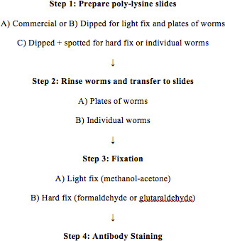
Figure 1. Outline of procedures. Flow chart indicating steps to perform the complete freeze-cracking procedure. Alternatives for varied fixation conditions and numbers of worms are indicated.
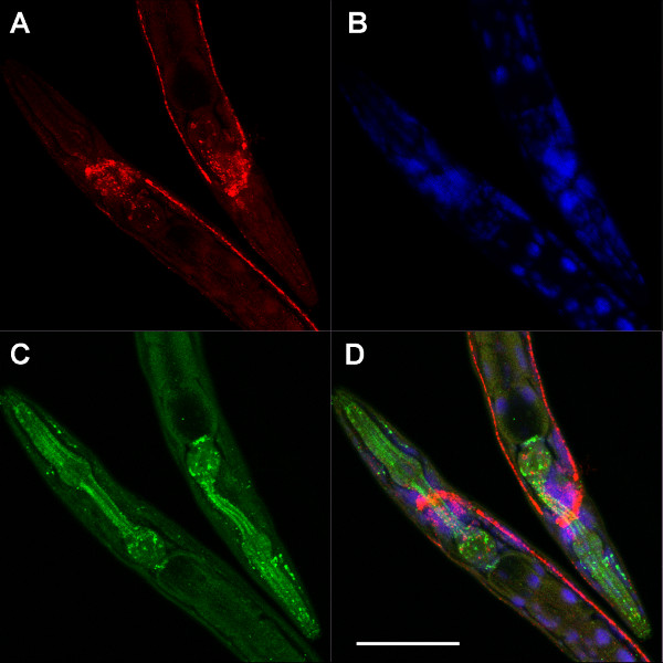
Figure 2. Lightly fixed, well-stained worms. Heads of two adult hermaphrodites fixed with methanol-acetone and labeled with multiple antibodies and dyes: A) Primary antibody to ChAT (choline acetyltransferase) and Cy3-conjugated secondary antibody, B) DAPI (blue DNA), C) Oregon Green 488-anti-GFP (green fluorescent protein), D) shows the overlay (with two cholinergic markers, Cy3-anti-ChAT and Cy5-anti-VAChT (vesicular acetylcholine transporter) shown as red). This transgenic strain (PS4657) expresses a translation fusion between GFP and AJM-1, found at pharyngeal apical junctions. The images are maximal projections of series of confocal images; scale bar is 50 μm.
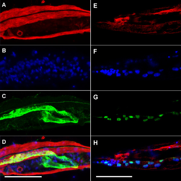
Figure 3. Formaldehyde-associated distortions. Formaldehyde fixed adults stained with Cy3-phalloidin (binds filamentous actin), DAPI (blue DNA), and Oregon Green 488-anti-GFP. A-D) Fours images of the body just posterior of the vulva (far left) in the same nematode, showing an unnaturally wavy morphology due to distortion induced by fixation. A) Red-phalloidin shows slightly irregular muscle morphology. B) Nuclei (blue) are evenly spread throughout the worm. C) Green shows the distribution of a CED-1::GFP fusion protein in the plasma membrane of the gonadal sheath cell, driven by the lim-7 promoter (strain MD701). D) Shows the overlay of these three dyes. E-H) Labeling can vary with different dyes on the same nematode. E) Phalloidin (red) labels only a portion of the muscle on one side of the worm. F) Nuclei (blue) are present throughout the worm (although stained with uneven intensity). G) The OG488-anti-GFP labels collagen-19::GFP in the cuticle on a side of the worm that lacks staining of the more central muscle tissue, indicating that muscle is present but not staining on one surface. H) Shows the overlay of the three dyes. The images are maximal projections of series of confocal images; scale bars are 50 μm.
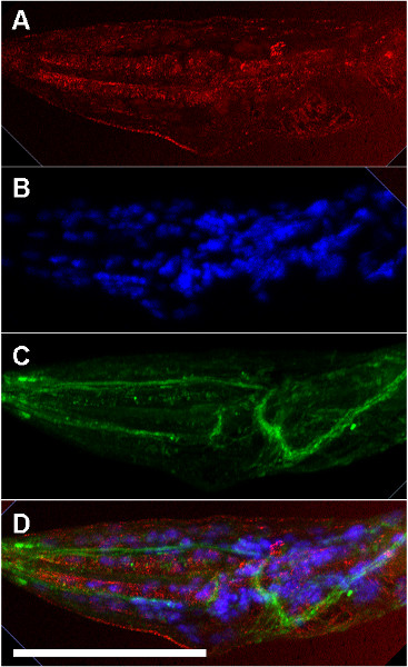
Figure 4. Compression-associated distortions. Formaldehyde fixed larva stained with A) Cy3-phalloidin (binds filamentous actin), B) DAPI (blue DNA), C) Oregon-Green 488-anti-GFP, D) overlay. The twist in the body just posterior of the pharynx is apparent, as the green excretory cell and ventral nerve cord cross over the rest of the body. High background staining is also apparent with Cy3-phallodin. This strain expresses a VHA-8::GFP fusion protein, located predominantly in the excretory cell. The images are maximal projections of series of confocal images that extended to the surface of the slide (leading to high background staining); scale bar is 50 μm.
Discussion
The freeze-cracking protocol is one of several methods for antibody staining in C. elegans. It provides a relatively simple way to stain worms, but does require specific reagents and practice for optimum results. Critical steps (outlined in Figure 1) include: 1) slide preparation (see steps 1 and 2) manual compression of the nematodes (see steps 2 and 3) rapid fixation (see step 3). First, to maximize adhesion of the nematodes to the slides, it is best to prepare slides with high molecular weight (long polymer) polylysine, instead of relying on commercially prepared slides. Commercial slides are coated with polylysine to allow cells to adhere, while freeze-cracking slides are designed to stick to the cuticle of entire nematodes. Second, the procedure for the correct compression of the worms between the slides before freezing is critical. It requires steady hands and practice to press the slides together using the correct amount of pressure so that individual worms contact both slides without destroying their morphology. Either too much or too little compression leads to increased loss of worms during fixation. Further care is needed to move the slides onto the dry ice chilled surface for freezing without moving the slides relative to one another. Any such motion will cause the worms to tear or twist. Third, as with any fixation technique, the frozen worms should be placed directly into fixative before defrosting. Otherwise, the antigen may diffuse, providing misleading information about subcellular localization. If these critical steps are all done properly, for best results it is still necessary to scan several stained slides to find worms with the best morphology.
One unavoidable aspect of this technique is that the worms will be compressed in one dimension. The compression is most noticeable in adults, where the apparent diameter of the round body in the z-axis may be only one-fourth that seen in the x- and y-axes. This compression tends to decrease in smaller larvae, and can be negligible in embryos. Due to the way the living nematodes move during slide preparation, the stained worms are often on their sides. This mimics the orientation of worms on agar dishes, with the lateral midline lying extending down the middle of the stained worm (see Figures 2-4). Thus, the compression is generally in the lateral dimension. While this actually makes standard fluorescent microscopy easier (more of the specimen will be in focus simultaneously), it means that 3-D reconstructions will not be accurate.
As with any antibody staining, it is important to check specificity of binding. In most species, this is done by controls such as affinity depleting your antibody or using no primary. In C. elegans, more specific means of checking for specificity are often available. Null mutants for the gene and protein of interest provide an excellent control for staining specificity. Both strains should be fixed under identical conditions before comparison, since staining is usually fixation dependent. If no null mutant is available, then it is relatively easy in C. elegans to decrease protein levels with RNAi (RNA inhibition12) and look for decreased staining in RNAi treated worms.
Polyclonal antibodies often show non-specific staining, even if affinity-purified with the antigen. Often, light fixation using methanol and acetone (which fix proteins by precipitating them rather than crosslinking them) gives lower non-specific staining than formaldehyde or glutaraldehyde (which generate more variant epitopes due to varied cross-linking)1,11. However, in C. elegans non-specific staining is very common under any fixation condition, especially in the gut. If your antigen is expressed in the gut, then this non-specific staining may mask your specific staining. If null mutants are available for your protein, then worms fixed with this method can be used to affinity deplete your serum. Since antigenicity depends upon fixation conditions, the worms that you use to deplete your antibody should be prepared in the same way as the wild-type worms you are staining. After one round of depletion of non-specific staining, mutant and wild-type worms can be stained in parallel with the depleted serum for an excellent control. Additional round of depletion with mutants can be done as necessary.
This technique is very useful for testing new antibodies for specific staining under a variety of conditions. These may be antibodies generated against C. elegans proteins or antibodies generated against conserved proteins from other species. While this method gives the best morphology with lightly fixed nematodes, it is relatively simple to try a range of fixatives to determine which conditions, if any, give specific results. Unlike other methods for hard fixation using formaldehyde or glutaraldehyde, enzymatic or reducing treatments are not needed for sample preparation. Since these treatments may decrease antigenicity, this increases the ability to identify specific antibodies and their optimum fixation conditions. If hard fixation conditions work best, the experimenter can then turn to methods performed on intact worms to obtain better morphology (summarized in our previous work 4). If light fixation preserves antigenicity, then this technique may be used for all further staining for light microscopy.
Disclosures
The author declares that she has no competing financial interests.
Acknowledgements
Funding was provided by NSF CCLI#0633618 and Ohio University. Some strains were provided by the Caenorhabditis Genetics Center. Graduate student Reetobrata Basu appears in the video.
Materials
| Name | Company | Catalog Number | Comments |
|---|---|---|---|
| poly-L-lysine slide (ESCO brand) | Fisher | 12-545-78 | Sigma brand slides also suitable |
| poly-L-lysine hydrobromide | Sigma | P1524 | IMPORTANT: Brand and high molecular weight of polylysine critical |
| single frosted edge slides | Fisher | 48312-002 | Other brands are suitable |
| sodium azide | Sigma | S2002-25G | TOXIC, wear gloves and mask while weighing out powder to make 10% stock. Wear gloves when pipeting stock solution |
| coplin staining jar | VWR | 47751-792 | Other brands are suitable |
| 5-slide mailer | Electron Microscopy Science | 71549-01 | One of many different brands of small slide holder, suitable for staining |
| Paraformaldehyde | VWR | MK262159 | IMPORTANT: Regular formalin solutions (37% formaldehyde) are NOT suitable. Either 1) Dissolve crysatlaline paraformaldehyde in phosphate buffer and store in small aliquots frozen. Or 2) buy electron microscopy grade paraformaldehyde in aliquots. TOXIC -wear gloves; dispose of excess down sink with water. |
| Glutaraldehyde solution, 25% in water | Sigma | G 5882 | IMPORTANT: purchase electron microscopy grade glutaraldehyde in ampules; TOXIC -wear gloves; dispose of excess down sink with water. |
| Triton X-100 | VWR | EM-9410 | Other brands are suitable |
| Bovine Serum Albumin | Fisher | ICN820451 | Other brands are suitable |
| Donkey Serum, lyophilized | Jackson Immunoresearch | 017-000-121 | Other brands are suitable |
| Cy3 Donkey anti-Mouse IgG multi-labeling | Jackson Immunoresearch | 715-165-150 | This company is recommended for high quality multi-label (affinity depleted) antibodies. Select appropriate dye and antigen (animal used to raise primary antibody). |
| n-propyl gallate | Fisher | ICN10274750 | Other brands are suitable |
| DAPI, 4’,6’-diamidino-2-phenylindole dihydrochloride | Sigma | D 9542 | TOXIC. Wear gloves and dispense into small aliquots and feeze to minimize exposure |
| Whatman #1 15 cm diameter filters | Fisher | 09805G | |
| Coverslip #1 1/2 rectangle 24 x 60 mm | VWR | 48393-252 | #1 1/2 thickness is optically best |
| Microscope slide folder | VWR | 48429-092 | Other brands are suitable |
References
- Harlow, E., Lane, D. Antibodies: A Laboratory Manual. , Cold Spring Harbor Laboratory Press. New York City, New York. (1988).
- Mello, C., Fire, A. DNA Transformation. Methods Cell Biol. 48, 451-482 (1995).
- Boulin, T., Etchberger, J. F., Hobert, O. Reporter gene fusions. WormBook. , WormBook. (2006).
- Duerr, J. S. Immunohistochemistry. WormBook. , WormBook. (2006).
- Finney, M., Ruvkun, G. The unc-86 gene product couples cell lineage and cell identity in C. elegans. Cell. 63, 895-905 (1990).
- Garriga, G., Desai, C., Horvitz, H. R. Cell interactions control the direction of outgrowth, branching and fasciculation of the HSN axons of Caenorhabditis elegans. Development. 117, 1071-1087 (1993).
- Miller, D. M., Shakes, D. C. Immunofluorescence microscopy. Caenorhabditis elegans: Modern Biological Analysis of an Organism. Epstein, H. F., Shakes, D. C. , Academic Press. San Diego, California. 365-394 (1995).
- Crittenden, S. L., Troemel, E. R., Evans, T. C., Kimble, J. GLP-1 is localized to the mitotic region of the C. elegans germ line. Development. 120, 2901-2911 (1994).
- Strome, S., Wood, W. B. Immunofluorescence visualization of germ-line-specific cytoplasmic granules in embryos, larvae, and adults of Caenorhabditis elegans. Proc. Natl. Acad. Sci. U.S.A. 79, 1558-1562 (1982).
- Albertson, D. G. Formation of the first cleavage spindle in nematode embryos. Dev. Biol. 101, 61-72 (1984).
- Harlow, E. Using Antibodies: A Laboratory Manual. , Cold Spring Harbor Laboratory Press. New York City, New York. (1999).
- Ahringer, J. Reverse Genetics. WormBook. , WormBook. (2006).
Reprints and Permissions
