Membrane trafficking involves transport of proteins from the plasma membrane to the cell interior (i.e. endocytosis) followed by trafficking to lysosomes for degradation or to the plasma membrane for recycling. Methods described in this article are designed to study endocytosis and recycling of plasma membrane proteins.
Method Article
The Cell-based L-Glutathione Protection Assays to Study Endocytosis and Recycling of Plasma Membrane Proteins
In This Article
Summary
Abstract
Membrane trafficking involves transport of proteins from the plasma membrane to the cell interior (i.e. endocytosis) followed by trafficking to lysosomes for degradation or to the plasma membrane for recycling. The cell based L-glutathione protection assays can be used to study endocytosis and recycling of protein receptors, channels, transporters, and adhesion molecules localized at the cell surface. The endocytic assay requires labeling of cell surface proteins with a cell membrane impermeable biotin containing a disulfide bond and the N-hydroxysuccinimide (NHS) ester at 4 ºC - a temperature at which membrane trafficking does not occur. Endocytosis of biotinylated plasma membrane proteins is induced by incubation at 37 ºC. Next, the temperature is decreased again to 4 ºC to stop endocytic trafficking and the disulfide bond in biotin covalently attached to proteins that have remained at the plasma membrane is reduced with L-glutathione. At this point, only proteins that were endocytosed remain protected from L-glutathione and thus remain biotinylated. After cell lysis, biotinylated proteins are isolated with streptavidin agarose, eluted from agarose, and the biotinylated protein of interest is detected by western blotting. During the recycling assay, after biotinylation cells are incubated at 37 °C to load endocytic vesicles with biotinylated proteins and the disulfide bond in biotin covalently attached to proteins remaining at the plasma membrane is reduced with L-glutathione at 4 ºC as in the endocytic assay. Next, cells are incubated again at 37 °C to allow biotinylated proteins from endocytic vesicles to recycle to the plasma membrane. Cells are then incubated at 4 ºC, and the disulfide bond in biotin attached to proteins that recycled to the plasma membranes is reduced with L-glutathione. The biotinylated proteins protected from L-glutathione are those that did not recycle to the plasma membrane.
Introduction
All living cells process information by trafficking cargo, such as extracellular ligands, microorganisms, nutrients, transmembrane proteins and lipids from the plasma membrane to endocytic vesicles (i.e. endocytosis). A reciprocal process called recycling balances endocytosis and returns much of the internalized membrane and cargo to the cell surface. The balance between endocytosis and recycling controls the plasma membrane composition and provides cells with information that has been resolved in time and space. Endocytosis and recycling are master regulators of diverse cellular functions such as nutrient uptake and metabolism, development, proliferation, differentiation and polarity, reprogramming, migration, cell adhesion and migration, cytokinesis, and neurotransmission1-3. Endocytic and recycling pathways are very dynamic and highly coordinated and allow cells to turn over the equivalent of the entire plasma membrane 1-5x per hour.
The cell-based L-glutahione protection assays are useful to study endocytosis and recycling of transmembrane proteins including receptors, channels, transporters, and adhesion molecules in epithelial and nonepithelial cells4-8. We have previously studied endocytosis and recycling of the Cystic Fibrosis Transmembrane Conductance Regulator (CFTR) in human airway epithelial cells and HEK293 cells9-15. The biotinylation-based assays described in the manuscript are optimized for examining endocytosis and recycling in epithelial cells cultured under polarizing conditions on semipermeable growth supports. These protocols can be modified to study endocytosis and recycling of proteins in epithelial cells cultured in plastic tissue culture dishes or in nonepithelial cells. Figures 1 and 2 contain examples of endocytic and recycling assays in epithelial and nonepithelial cells.
Endocytic assays are performed as previously described9-15. Cells are cultured on collagen coated semipermeable growth supports11,14. Alternatively, cells can be cultured in collagen coated plastic tissue culture dishes10,15. Cells are cooled rapidly to 4 ºC to stop membrane trafficking and the plasma membrane proteins are labeled at 4 °C with a cell membrane impermeable biotin. Biotin reacts with ε-amine of lysine residues and the disulfide bond is thiol-cleavable. After biotinylation cells are incubated at 37 °C to induce protein trafficking and load endocytic vesicles for 2.5, 5.0, 7.5, or 10 min. Subsequently, cells are cooled to 4 °C and the disulfide bond in biotin covalently attached to plasma membrane proteins is reduced with L-glutathione (GSH). At this point in the protocol, only proteins that were endocytosed from the plasma membrane are protected from GSH and thus, remain biotinylated. Cells remaining at 4 °C after biotinylation without incubation at 37 ºC or the GSH treatment would serve to determine the amount of CFTR biotinylated at time zero. Cells remaining at 4 °C after biotinylation without incubation at 37 ºC but with GSH treatment would serve to determine efficiency of the disulfide bonds reduction. Following the above described treatments, cells are lysed, biotinylated proteins are isolated by streptavidin agarose, eluted into SDS sample buffer, and separated by SDS-PAGE. The protein of interest is detected in the biotinylated samples by western blotting. The amount of biotinylated protein at 4 ºC at time zero (without the 37 ºC warming) is considered 100%. The amount of protein remaining biotinylated after GSH treatment at 4 ºC is considered background and is subtracted from the amount of protein remaining biotinylated after warming to 37 ºC at different time points. Protein endocytosis is calculated after subtracting the background and is expressed as the percent of biotinylated protein at each time point after warming to 37 ºC compared to the amount of biotinylated protein present at time zero.
Recycling assays are performed as previously described11,16. Cells are cultured on collagen coated semipermeable growth supports11. Alternatively, cells can be cultured on collagen coated plastic tissue culture dishes13. Cells are warmed to 37 °C after biotinylation to load endocytic vesicles with biotinylated proteins. The time of first incubation at 37 ºC is determined by the time when endocytosis of the protein of interest reaches maximum during the linear increase of the endocytic signal. The time is protein specific and may depend on the cell type and cell culture conditions. In our experience CFTR endocytosis reached maximum at 5.0 or 7.5 min13,14 (Figures 1 and 2). Subsequently, cells are cooled immediately to 4 °C and the disulfide bond in biotin attached to plasma membrane proteins is reduced with GSH. Next, cells are either lysed to determine the amount of endocytosed protein of interest or warmed again to 37 °C for different periods of time to allow endocytosed biotinylated protein of interest to recycle to the plasma membrane. Cells are then cooled again to 4 °C, and the disulfide bond on biotin attached to proteins recycled to the plasma membranes is reduced with GSH. Recycling of the protein of interest is determined from the difference between the amount of biotinylated protein after the first and second GSH treatment.
The feasibility of the endocytic and recycling assays depends on several factors. First, formation of cell monolayers is a prerequisite and cells that do not form monolayer or grow as multilayers are not suitable for assays described in this manuscript. Second, the abundance of the protein of interest at the cell surface and presence of an antibody to detect the protein by western blotting are critical. We recommend that the steady state abundance of the protein is first determined in whole cell lysates (WCL). Third, the ability to biotinylate the specific cell surface protein should be tested. Biotin attaches to lysine residues. Thus, the efficiency of biotinylation depends in part on the number of lysine residues in the protein’s extracellular domain. Accordingly, we recommend screening the protein sequence to determine whether lysine residues are present in the extracellular domain(s). Not all extracellular domain lysine residues may be equally accessible to biotin due to protein folding. Hence, protein biotinylation at steady state followed by western blotting should be performed to determine not only the steady state abundance of the protein at the cell surface but also to examine feasibility of the biotinylation-based assays for the protein of interest.
This protocol is optimized for examining endocytosis and recycling of wild type CFTR in human airway epithelial cells CFBE41o- cultured on 24 mm semipermeable growth supports in air-liquid interface9,10,13-15. CFTR polarizes to the apical membrane domain; thus, the protocol describes biotinylation of the apical membrane domain. Biotinylation of the basolateral membrane domain will be required to study endocytosis and recycling of proteins polarizing to the basolateral membrane. The endocytic assay protocol described in this manuscript has 6 conditions: Biotinylated only (BT = time zero; sample a); GSH control (GSH; sample b); and the 2.5, 5.0, 7.5, or 10 min endocytic time points (samples c; Table 1). The number and/or length of endocytic time points in the protocol can be modified as needed.
The recycling assay is performed after determining the time point when endocytosis of the protein of interest reaches maximum during the linear increase of the endocytic signal. This time point will be used to load endocytic vesicles with the protein of interest prior to inducing recycling. The time is protein dependent and may differ between cell types and culture conditions15. We have previously established that CFTR endocytosis reached plateau at the 7.5 min time point in human airway epithelial cells CFBE41o- stably expressing CFTR15. By contrast, CFTR endocytosis reached plateau at the 5.0 min time point in HEK293 cells stably expressing CFTR13. The recycling assay protocol described in this manuscript has 5 conditions: Biotinylated only (BT = time zero; sample a); GSH control (GSH; sample b); 5.0 min endocytosis (Endo; sample c), 5.0 min endocytosis followed by the 2.5 or 5.0 min recycling time points (Rec; samples d; Table 2). The number and/or length of recycling time points in the protocol can be modified as needed.
Protocol
1. Seeding Cells
- Pretreat 24 mm filters with 10% collagen I (prepare 10% collagen I in Minimal Essential Medium (MEM), cover the entire surface of the filter with the collagen solution, incubate under the UV light at room temperature for 30 min, and in a cell culture incubator at 37 ºC for 1 hr, suction off the excess collagen after incubation).
- Prepare cell culture medium (MEM gassed with CO2 for 20 min, 10% Fetal Bovine Serum (FBS), 50 U/ml penicillin, 50 U/ml streptomycin, 2 mM L-glutamine, 0.5 μg/ml puromycin)
- Seed CFBE41o- cells on six 24 mm filters for endocytosis and recycling, respectively, at 1 x 10⁶/filter.
- Remove the apical medium the day after seeding and feed daily from the basolateral side only.
- Feed with selection antibiotic negative medium 24 hr before the experiment. Perform experiment in CFBE41o- cells 6-10 days after seeding.
2. Preparations Before the Experiment (Similar for the Endocytosis and Recycling Assay)
- Set up a bench space in the cold room. Endocytic and recycling assays should be performed in the cold room except for the incubation at 37 °C. Plates containing the 24 mm filters should be placed directly on the bench top in the cold room.
- Turn on the 37 ºC incubator.
- Prepare 500 ml of PBS++, pH 8.2 (Dulbecco’s Phosphate Buffered saline (PBS), 1 mM magnesium chloride, and 0.1 mM calcium chloride, pH 8.2) and keep 250 ml at 37 °C in an incubator and 250 ml at 4 °C in the cold room.
- Fill wells in a 6 well plate with PBS++, pH 8.2, 37 °C and keep in the incubator at 37 °C.
- Fill wells in another 6 well plate with PBS++, pH 8.2, 4 °C and keep in the cold room at 4 °C.
- Prepare 100 ml of PBS++, pH 8.6 at 4 °C and keep in the cold room.
- Prepare biotin containing the disulfide bond and NHS ester at a concentration 0.8 mg/ml in PBS++, pH 8.2, 4 °C within 30 min of the biotinylation step (the volume of biotin buffer should cover completely the entire surface of the filter); we recommend 1.5 ml/24 mm filter.
- Prepare 100 ml of GSH buffer in water, pH 8.6 and cool to 4 ºC (75 mM sodium chloride, 1 mM magnesium chloride, and 0.1 mM calcium chloride, 50 mM GSH, 80 mM sodium hydroxide, and 10% FBS). GSH and sodium hydroxide should be added just before the experiment. Check the pH and adjust to 8.6; sodium hydroxide neutralizes the carboxyl groups and deprotonates half the cysteine residues in glutathione. It is strongly buffered at the pKa of this cysteine, which is 8.64. (Prepare the same volume of GSH buffer for the recycling assay).
- Prepare 50 ml of Lysis buffer, pH 8.2 and keep at 4 °C (25 mM HEPES, pH 8.2, 1% (v/v) Triton X-100, 10% (v/v) glycerol); add Complete protease inhibitor cocktail per 50 ml of lysis buffer and cool to 4 ºC; check the pH after adding Complete as a drop in pH may occur.
- Prepare Laemli sample buffer with 100 mM DTT.
- Prepare 1x Running buffer (100 ml of 10x Running buffer, 900 ml of water).
- Prepare 1x Transfer buffer and cool to 4 ºC (100 ml of 10x Transfer buffer without sodium dodecyl sulfate (SDS), 200 ml of methanol, 700 ml of water).
3. Endocytic Assay
CFTR polarizes to the apical membrane domain; thus, the protocol describes biotinylation of the apical membrane domain. Biotinylation of the basolateral membrane domain will be required to study endocytosis of proteins polarizing to the basolateral membrane.
Workflow: Biotinylation of cell surface proteins at 4 ºC → Warming to 37 ºC to load endocytic vesicles with biotinylated proteins → Cooling to 4 ºC to stop endocytic trafficking → Reduction of the disulfide bond in biotin attached to proteins that have remained at the cell surface → Cell lysis → Isolation of biotinylated (i.e. endocytosed) proteins with streptavidin agarose → Elution of biotinylated proteins from streptavidin agarose → Protein electrophoresis and western blotting.
- Bring the plate containing six 24 mm filters (Table 1: samples a-c) from the cell culture incubator and transfer quickly to the plate filled with cold (4 ºC) PBS++, pH 8.2 on ice.
- Let PBS++ overflow the apical surface to cover quickly the entire surface. Bring the plate on ice to the cold room and set on the bench top.
- Remove PBS++ quickly by turning the plate upside down holding filters in place to prevent falling out. Add ~2 ml of PBS++ to the apical and basolateral side.
- Wash filters with ~2 ml of cold PBS++, pH 8.2 3x for 2 min. Suction off the wash from one side at a time, apical or basolateral and add fresh PBS++, pH 8.2 to one side at a time.
- Suction off the last wash and add 1 ml of PBS++, pH 8.2 to the basolateral side. Add 1.5 ml of the biotin buffer to the apical side and incubate in the dark for 30 min. Avoid spilling buffers to the contralateral side.
- Suction off the biotin buffer from the apical side and wash with PBS++, pH 8.2 3x for 2 min.
- Set aside in a different plate two filters (Table 1: samples a and b), add ~2 ml of PBS++, pH 8.2 to the apical and basolateral side and keep in the cold room. Keep the remaining 4 filters in one plate (Table 1: samples c) and add ~2 ml of PBS++ pH 8.2 on the apical and basolateral side of each filter. Place the plate on ice and bring to the bench top next to the 37 ºC incubator.
- Transfer quickly filters from ice to the plate in the 37 ºC incubator filled with prewarmed PBS++ pH 8.2 and incubate one filter each precisely for 2.5, 5.0, 7.5, or 10 min covering the entire surface of each filter with warm PBS++ pH 8.2.
- During the incubation fill completely four wells in the plate on ice with cold (4 ºC) PBS++ pH 8.2.
- After the indicated time of incubation, turn each filter upside down to drain the warm (37 ºC) PBS++, pH 8.2 and transfer to the plate on ice. Add cold PBS++, pH 8.2 to overflow the apical side and cover the entire surface of each filter quickly.
- After transferring filters to ice bring the plate to the cold room and set on the bench top.
- Suction off PBS++ from both sides in all filters (samples a-c) and fill the basolateral side with 1 ml of PBS++ pH 8.6. Add 1.5 ml of PBS++, pH 8.6 to the apical side of the filter a (Table 1: sample a).
- Reduce the disulfide bond in biotin attached to the apical membrane proteins with the GSH buffer (pH 8.6) in filters b and c (Table 1: sample b and c).
- Add 1.5 ml of the GSH buffer to the apical side, incubate for 15 min, and repeat six times.
- Keep 1 ml of PBS++ pH 8.6 on the basolateral side. Avoid spilling the buffer to the basolateral side.
- After completing the procedure 3.15, wash all filters (Table 1: samples a, b, and c) briefly with PBS++ pH 8.2 2x, suction off the wash from the apical and basolateral side and set the plate with filters at a 45° angle to drain all wash and suction again.
- Place the plate flat on the bench top and add 500 μl of lysis buffer to the apical side of each filter and incubate in the cold room shaking for 15 min.
- Scrape cells with a rubber policeman and collect in a 1.5 ml Eppendorf tube.
- Spin at 14,000 x g for 10 min at 4 °C.
- Transfer the supernatants to fresh 1.5 ml tubes and discard the pellets. The supernatants are now called whole cell lysates (WCL).
- Add 100 μl of Laemli sample buffer with DTT to 50 μl of WCL (10% of the entire WCL) and incubate at 37 °C for 30 min (WCL samples).
- Prepare 200 μl aliquots of 50% streptavidin agarose and remove all wash buffer with a 27 G needle attached to a suction: wash 2x with 1 ml of PBS, pellet beads with pulse spin between the washes; wash once with 1 ml of lysis buffer to equilibrate before adding WCL; suction dry the agarose prior to adding WCL.
- Incubate the remaining WCL with washed streptavidin agarose (bring the volume up to 1.2 ml with lysis buffer and rotate in the dark for at least 2 hr or O/N in the cold room).
- Wash the streptavidin agarose–biotinylated protein complexes three times with 1 ml of lysis buffer (the lysis buffer does not need to contain Complete during washing), pulse spin the agarose between washes, and suction dry after the last wash.
- Incubate the streptavidin agarose–biotinylated protein complexes with 65 μl Laemli sample buffer with DTT at 85 °C for 5 min.
- Pulse spin and collect the eluted biotinylated protein complexes.
- Load 7.5% gels with 40 μl of the WCL samples and the entire volume of the biotinylated (BT) samples per well.
- Run gels at 120 V in SDS containing running buffer.
- Transfer proteins to a PVDF membrane at 90-95 V for 1.5 hours (as long as <400 A) in a transfer buffer without SDS.
- Block in 5% nonfat milk in TBS-T 0.1% O/N. Blot both membranes with anti-CFTR mouse monoclonal antibody CFF596 and a goat anti-mouse HRP-conjugated secondary antibody.
- Perform chemiluminescence. Reblot both membranes with either anti-ezrin or anti-actin antibody.
4. Recycling Assay
Workflow: Biotinylation of cell surface proteins at 4 ºC → Warming to 37 ºC to load endocytic vesicles with biotinylated proteins → Cooling to 4 ºC to stop endocytic trafficking → Reduction of the disulfide bond in biotin attached to proteins that have remained at the cell surface → Warming to 37 °C to allow biotinylated proteins from endocytic vesicles to recycle to the cell surface → Cooling to 4 ºC to stop endocytic trafficking → Reduction of the disulfide bond in biotin attached to proteins that have recycled to the cell surface → Cell lysis → Isolation of biotinylated proteins (i.e. those that have not recycled) with streptavidin agarose → Elution of biotinylated proteins from streptavidin agarose → Protein electrophoresis and western blotting.
- Biotinylate the apical surface proteins in five 24 mm filters (Table 2: samples a-d) following the procedure described in the Endocytic assay (section 3.1-3.6).
- In the cold room, set in a new plate two filters (Table 2: samples a and b), add ~2 ml of PBS++, pH 8.2 to the apical and basolateral side and keep in the cold room.
- Keep the remaining 3 filters in one plate (Table 2: samples c and d) with ~2 ml of PBS++ pH 8.2 on the apical and basolateral side of each filter. Place the plate with filters c and d on ice and bring to the bench outside of the 37 ºC incubator.
- Transfer filters quickly from the plate on ice to the plate filled with prewarmed PBS++ pH 8.2 in the incubator and keep for 5.0 min.
- During the incubation fill 3 wells of the plate on ice with cold (4 ºC) PBS++ pH 8.2.
- Transfer filters after the 5.0 min incubation at 37 ºC to the plate on ice. Let the cold PBS++ pH 8.2 to overflow the apical side and cover the entire surface of each filter quickly.
- Bring the plate on ice to the cold room and set on the bench top.
- Suction off PBS++ pH 8.2 from both sides of filters a, b, c, and d and add 1 ml of PBS++ pH 8.6 to the basolateral side.
- Keep filters a and b separately from filters c and d. Add 1 ml of PBS++ pH 8.6 to the apical side of filters a and b.
- Reduce the disulfide bond in biotin remaining at the cell surface in filters b, c and d following the procedure described in the endocytic assay (steps 3.13-3.15).
- Wash filters b, c, and d briefly with PBS++ pH 8.2 2x and replace with fresh PBS++, pH 8.2. Place filters d in a new plate and bring on ice to the bench top outside the 37 ºC incubator.
- Transfer quickly filters from the plate on ice to the plate in the incubator filled with prewarmed PBS++ pH 8.2 and incubate one filter each precisely for 2.5 or 5.0 min as described above in steps 4.4-4.9.
- Reduce the disulfide bond in biotin attached to the apical membrane proteins with the GSH buffer after the second incubation at 37 ºC in filters d as described in 4.4 with the exception that only three 15 min incubations with the GSH buffer will be done during this step. Keep filters a, b, and c in PBS++ pH 8.6 on the apical and basolateral side during this step.
- For the cell lysis, and Western blotting follow procedures described in the endocytic assay (steps 3.16-3.31).
Results
CFTR endocytosis was studied in CFBE41o- cells cultured on collagen-coated filters (Figure 1). Biotinylated CFTR was visualized by western blotting with mouse monoclonal antibody, clone 596 and an anti-mouse horseradish peroxidase antibody using the western blotting detection system followed by chemiluminesence. Quantification of biotinylated CFTR was performed by densitometry using exposures within the linear dynamic range of the film. CFTR endocytosis was calculated after subtracting the background and was expressed as the percent of biotinylated CFTR at each time point after warming to 37 ºC compared to the amount of biotinylated CFTR present at time zero (Figures 1A and 1B). CFTR endocytosis was linear between 0-7.5 min. Experiments in which the background CFTR was >10% were excluded due to inefficient GSH treatment (Figure 1D).
CFTR recycling was studied in HEK293 cells cultured in collagen-coated tissue culture dishes (Figure 2). CFTR endocytosis was linear between 0.0-5.0 min and reached maximum at the 5.0 min time point (Figure 2A), thus cells were incubated at 37 ºC for 5.0 min to load endocytic vesicles with biotinylated proteins including CFTR (Figures 2B and 2C). Recycling of endocytosed CFTR was calculated as the difference between the amount of biotinylated CFTR after the first and second GSH treatment.
Table 1. Endocytic assays.
| Endocytosis | BT | GSH | Endo-2.5 | Endo-5.0 | Endo-7.5 | Endo-10.0 |
| Sample | a | b | c2.5 | c5.0 | c7.5 | c10.0 |
| Biotin | + | + | + | + | + | + |
| 37 ºC | (-) | (-) | 2.5 min | 5.0 min | 7.5 min | 10 min |
| GSH | (-) | + | + | + | + | + |
Table 2. Recycling assay.
| Recycling | BT | GSH | Endo-5 | Rec-2.5 | Rec-5.0 |
| Sample | a | b | c | d2.5 | d5.0 |
| Biotin | + | + | + | + | + |
| 1st 37 ºC | (-) | (-) | 5 min | 5 min | 5 min |
| 1st GSH | (-) | (-) | + | + | + |
| 2nd 37 ºC | (-) | (-) | + | + | + |
| 2nd GSH | (-) | (-) | (-) | 2.5 min | 5 min |
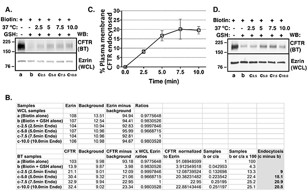
Figure 1. Summary of endocytic assays performed to determine CFTR endocytosis in CFBE41o- cells. Cells were cultured on collagen-coated filters. Representative western blots (A), representative densitometry values (B) and summary of experiments (C) demonstrating CFTR endocytosis as a function of time. Selective cell surface biotinylation and western blotting were used to determine the abundance of plasma membrane CFTR. Protein abundance was quantified by densitometry using exposures within the linear dynamic range of the film. At time zero, the amount of biotinylated (BT) CFTR was considered 100% (Table 1: sample a). At time zero, the amount of BT CFTR remaining after GSH treatment was considered a CFTR background (sample b; please, note this is a different background than the one subtracted from all samples as shown in Figure 1B). Background CFTR was 6.7 ± 0.9% (mean ± S.E.M.) in the experiments included for analysis. Background CFTR was subtracted from the BT CFTR after the 2.5, 5.0, 7.5, or 10 min warming at 37 ºC (samples c minus sample b). CFTR endocytosis was expressed as the percent of CFTR remaining biotinylated at the 2.5, 5.0, 7.5, or 10 min time points after subtracting background CFTR. CFTR endocytosis was linear between zero and 7.5 min. Ezrin abundance in the whole cell lysate (WCL) was used as a loading control. 4 experiments/group. Experiments in which the background CFTR was >10% were excluded due to inefficient GSH treatment (D). The amount of biotinylated CFTR in the GSH control (sample b) in the excluded experiment was 14.5%.
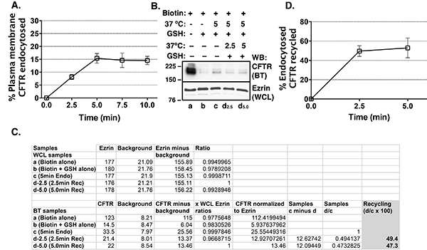
Figure 2. Summary of endocytic and recycling assays performed in HEK293 cells stably expressing CFTR. Cells were cultured in collagen-coated tissue culture plates. Summary of data demonstrating that CFTR endocytosis was linear between 0-5 min (A). Thus, in the recycling assays endocytic vesicles were loaded with biotinylated (BT) proteins including CFTR by warming at 37 ºC for 5 min. Protein abundance was quantified by densitometry using exposures within the linear dynamic range of the film. Representative western blot (B), representative densitometry values (C), and summary of experiments (D) demonstrating CFTR recycling as a function of time. At time zero, the amount of BT CFTR was considered 100% (Table 2: sample a). At time zero, the amount of BT CFTR remaining after GSH treatment was considered a CFTR background (sample b; please, note this is a different background than the one subtracted from all samples as shown in C). Experiments in which the background CFTR was >10% were excluded due to inefficient GSH treatment. Endocytic vesicles were loaded with BT proteins including CFTR by incubation at 37 ºC for 5 min followed by the GSH treatment to cleave biotin from proteins remaining at the plasma membrane (samples c and d). The amount of BT CFTR after the 5 min warming at 37 ºC followed by the GSH treatment represents endocytosed CFTR (sample c). Following the 5 min warming at 37 ºC and the first GSH treatment cells were warmed again at 37 ºC for 2.5 or 5.0 min to allow the endocytosed proteins to recycle to the plasma membrane and the biotin on recycled CFTR was reduced by the second GSH treatment (samples d). At this point only the CFTR that has not recycled from endosomes to the plasma membrane remained biotinylated (samples d). CFTR recycling was calculated as the difference between BT CFTR after the first GSH treatment (sample c) and second GSH treatment at 2.5 and 5.0 min (samples d) and was expressed as the percent of endocytosed CFTR. CFTR recycling was rapid and reached maximum by 2.5 min. Ezrin abundance in the whole cell lysate (WCL) was used as a loading control. 3 experiments/group.
Discussion
The success of endocytic and recycling assays depends on efficient biotinylation of CFTR at the plasma membrane. Biotin has to be prepared immediately before use because the NHS-ester moiety readily hydrolyzes and becomes nonreactive. Moreover, the biotinylation step requires strict temperature control at 4 ºC to stop protein trafficking. If the temperature is raised during the biotinylation step above 4 ºC protein trafficking may occur resulting in the biotinylation of variable amounts of CFTR.
The success of endocytic and recycling assays also depends on efficient reduction of the disulfide bond in the biotinylated CFTR remaining at the plasma membrane. The amount of CFTR remaining biotinylated after the GSH treatment should be <10% of the total biotinylated CFTR at time zero. Experiments where the amount of biotinylated CFTR is >10% in the GSH control sample should be excluded. Figure 1D demonstrates “failed” experiment where the amount of biotinylated CFTR in the GSH control was 14.5%. Insufficient reduction of the disulfide bond in the biotinylated CFTR may be secondary to poor control of temperature during the experiment. If the temperature is raised during the GSH treatment above 4 ºC protein trafficking may occur and the biotinylated CFTR endocytosed as a result of the unintentional increase in temperature will increased the background CFTR. Alternatively, insufficient reduction of the disulfide bond in the biotinylated CFTR may result from decreased half-life of GSH in aqueous solution caused by oxidation in air or from inappropriate pH. As previously determined GSH remains strongly buffered at pH 8.64. In our experience six 15 min treatments with the GSH buffer reduced the disulfide bond by more than 90%, more efficiently than one 90 min treatment.
The cell culture requires special attention. Biotin has to reach the plasma membrane domain in intact cells where the protein of interest is expressed. We have used the biotinylation based endocytic and recycling assays to study endocytosis and recycling of CFTR expressed in the apical membrane domain. These assays may be also used to study endocytic trafficking of proteins located in the basolateral membrane domain in cells cultured on semipermeable growth supports. Epithelial cells that form multilayers are not suitable for the these assays as biotin and GSH buffers will only reach the top layer of cells to detect the apical membrane proteins or the bottom layer to detect the basal membrane proteins. Epithelial cells cultured on plastic may be more prone to washing off during the assays. Random loss of cells during the experiment will compromise results. We recommend periodic examination of the monolayer integrity under a microscope in experiments where cells are cultured in plastic tissue culture dishes. Coating of tissue culture dishes with collagen may increased cell adherence.
The biotinylated protein samples should be routinely tested for contamination with intracellular proteins. Detection of intracellular proteins in the biotinylated protein samples may indicate insufficient washing of the streptavidin agarose–biotinylated protein complexes after incubation with WCL. Thus, the first step is to increase the washing efficiency. Moreover, presence of cells with compromised plasma membrane integrity will allow access of biotin to intracellular proteins. Western blotting for a protein expressed exclusively in an intracellular compartment such as the endoplasmic reticulum could be used to detect biotinylation of intracellular proteins. Alternatively, cytoskeletal proteins such as actin or ezrin may be used to test for contamination of the biotinylated protein samples with intracellular proteins. Cytoskeletal proteins may form complexes with transmembrane proteins and small amounts of the cytoskeletal proteins may be detected in the biotinylated protein samples. However, in our experience, the ratio of biotinylated/WCL ezrin or actin is <1:1,000 and thus, these proteins are suitable for determination of cell membrane integrity in the biotinylation-based assays.
The biotinylation reaction can be quenched with glycine or Tris to remove nonreacted biotinylation reagent as previously reported17. Moreover, GSH can be quenched with iodoacetamide6. In our experience the nonreacted biotinylation reagent and GSH can be efficiently removed by gentle washing with PBS++ without the use of quenching reagents.
The choice of streptavidin vs. neutravidin agarose to isolate the biotinylated protein complexes should be determined on case-by-case basis. In our experience streptavidin agarose provides efficient isolation of the biotinylated protein complexes with minimal binding of nonbiotinylated proteins, unlike the neutravidin agarose where the nonspecific binding occurs at levels that interfere with the assay results (unpublished observation).
Disclosures
The authors declare that they have no competing financial interests.
Acknowledgements
This study was supported by the U.S. National Institutes of Health (NIH) grants R01HL090767, R01HL090767-02S1, P30 DK06010, and The NepCure Foundation Established Investigator New Direction Grant, (to A.S.-U.).
Materials
| Name | Company | Catalog Number | Comments |
|---|---|---|---|
| CFBE41o- cells | reference10,11,13,14,15 | ||
| HEK293 cells | reference13 | ||
| Minimal Essential Medium (MEM) | Gibco | 11095 | |
| L-Glutamine | Cellgro | 25-005-CL | |
| Penicillin | Sigma | P0781 | |
| Streptomycin | |||
| Puromycin | InvivoGen | ant-pr-1 | |
| PureCol Bovine Collagen Solution Type I | Advanced Biomatrix | 5005-B | |
| Culture Reagents | |||
| Dulbecco’s Phosphate Buffered Saline (PBS) | Gibco | 14190 | |
| Calcium chloride | Sigma | C3306 | |
| Magnesium chloride | Sigma | M2670 | |
| Sodium chloride | Sigma | S3014 | |
| EZLink Sulfo-NHS-SS-Biotin | Pierce | 21331 | |
| L-Glutathione reduced (GSH) | Sigma | G6529 | |
| Streptavidin-agarose resin | Thermo Fisher | 20353 | |
| Sodium hydroxide | Sigma | 224165 | |
| Fetal bovine serum | Gemini Bioscience | 100-106 | |
| Milli-Q water | Millipore | Advantage System | |
| HEPES | Sigma | H3375 | |
| Triton X-100 | Bio Rad | 161-0407 | |
| Glycerol | FisherScientific | G33 | |
| Laemli Sample buffer | BioRad | 161-0737 | |
| Dithiothreitol (DTT) | Sigma | 646563 | |
| Complete Protease Inhibitor cocktail | Roche | 1697498 | |
| Streptavidin beads | Thermo Scientific | 20353 | |
| Chemiluminescence | Perkin Elmer | NEL104001EA | |
| Reagents | |||
| Nonfat dry milk | Carnation | N/A | |
| Tween-20 | BioRad | 170-6539 | |
| Tris-buffered Saline (TBS) | BioRad | 170-6435 | |
| Bovine Serum Albumin (BSA) | Gibco | 15120 | |
| Primary and secondary | |||
| Antibodies for Western Blotting | |||
| Mouse monoclonal Anti-CFTR 596 | Cystic Fibrosis Foundation Therapeutics, Inc., Chapel Hill, NC | ||
| Anti-ezrin | |||
| Running buffer 10x | BioRad | 161-0772 | |
| Transfer buffer 10x (without SDS) | BioRad | 161-0771 | |
| Methanol | Fisher | A4129 | |
| Coulter counter | Beckman Coulter | Z1Coulter Particle Counter | |
| Standard Cell Culture equipment | |||
| Cell culture hood with UV light | |||
| Stock flasks | |||
| Transwell filters-24mm | Corning | 3412 | |
| 4 cm cell culture dishes | |||
| pH meter | |||
| 15 ml and 50 ml conical tubes | |||
| 1.5 ml Eppendorf tubes | |||
| Rubber policeman | |||
| Cold room | |||
| Ice buckets and ice | |||
| 27 G needles | |||
| 37 ºC Incubator | |||
| Thermometer | |||
| Benchtop centrifuge | |||
| Cooled centrifuge | |||
| Electrophoresis equipment | |||
| Developer | |||
| Film | GE Amersham | 28-9068-36 | |
| PVDF membrane | Millipore | IPVH00010 | |
| ImageJ software | NIH |
References
- Sigismund, S., et al. Endocytosis and signaling: cell logistics shape the eukaryotic cell plan. Physiol. Rev. 92, 273(2012).
- Andersson, E. R. The role of endocytosis in activating and regulating signal transduction. Cell Mol. Life Sci. 69, 1755(2012).
- Grant, B. D., Donaldson, J. G. Pathways and mechanisms of endocytic recycling. Nat. Rev. Mol. Cell Biol. 10, 597(2009).
- Bretscher, M. S., Lutter, R. A new method for detecting endocytosed proteins. EMBO J. 7, 4087(1988).
- Watts, C., Davidson, H. W. Endocytosis and recycling of specific antigen by human B cell lines. EMBO J. 7, 1937(1988).
- Graeve, L., Drickamer, K., Rodriguez-Boulan, E. Polarized endocytosis by Madin-Darby canine kidney cells transfected with functional chicken liver glycoprotein receptor. J. Cell Biol. 109, 2809(1989).
- Garza, L. A., Birnbaum, M. J. Insulin-responsive aminopeptidase trafficking in 3T3-L1 adipocytes. J. Biol. Chem. 275, 2560(2000).
- Le, T. L., Joseph, S. R., Yap, A. S., Stow, J. L. Protein kinase C regulates endocytosis and recycling of E-cadherin. Am. J. Physiol. Cell Physiol. 283, C489(2002).
- Swiatecka-Urban, A., et al. PDZ-domain interaction controls the endocytic recycling of the cystic fibrosis transmembrane conductance regulator. J. Biol. Chem. 277, 40099(2002).
- Swiatecka-Urban, A., et al. Myosin VI Regulates Endocytosis of the Cystic Fibrosis Transmembrane Conductance Regulator. J. Biol. Chem. 279, 38025(2004).
- Swiatecka-Urban, A., et al. The short apical membrane half-life of rescued uF508-CFTR results from accelerated endocytosis uF508-CFTR in polarized human airway epithelial cells. J. Biol. Chem. 280, 36762(2005).
- Swiatecka-Urban, A., et al. Pseudomonas aeruginosa inhibits endocytic recycling of CFTR in polarized human airway epithelial cells. Am. J. Physiol. Cell. Physiol. 290, C862(2006).
- Swiatecka-Urban, A., et al. Myosin Vb is required for trafficking of the cystic fibrosis transmembrane conductance regulator in Rab11a-specific apical recycling endosomes in polarized human airway epithelial cells. J. Biol. Chem. 282, 23725(2007).
- Ye, S., et al. c-Cbl facilitates endocytosis and lysosomal degradation of cystic fibrosis transmembrane conductance regulator in human airway epithelial cells. J. Biol. Chem. 285, 27008(2010).
- Cihil, K. M., et al. Disabled-2 protein facilitates assembly polypeptide-2-independent recruitment of cystic fibrosis transmembrane conductance regulator to endocytic vesicles in polarized human airway epithelial cells. J. Biol. Chem. 287, 15087(2012).
- MacEachran, D. P., et al. The Pseudomonas aeruginosa secreted protein PA2934 decreases apical membrane expression of the cystic fibrosis transmembrane conductance regulator. Infect. Immun. 75, 3902(2007).
- Pagano, A., Crottet, P., Prescianotto-Baschong, C., Spiess, M. In vitro formation of recycling vesicles from endosomes requires adaptor protein-1/clathrin and is regulated by rab4 and the connector rabaptin-5. Mol. Biol. Cell. 15, 4990(2004).
Reprints and Permissions
