Various murine models of middle cerebral artery occlusion (MCAO) are widely used in experimental brain research. Here, we demonstrate the model of transcranial permanent distal MCAO which produces consistent cortical infarction of a size corresponding to damage imposed by the majority of human ischemic strokes.
Method Article
Modeling Stroke in Mice: Permanent Coagulation of the Distal Middle Cerebral Artery
* These authors contributed equally
Opens in a new tab
In This Article
Summary
Loading...
$$\rightleftharpoonup{xx}$$
$$\longleftharp{xx}$$,
$$\longrightharp{xx}$$,
Abstract
Loading...
$$\rightleftharpoonup{xx}$$
$$\longleftharp{xx}$$,
$$\longrightharp{xx}$$,
Stroke is the third most common cause of death and a main cause of acquired adult disability in developed countries. Only very limited therapeutical options are available for a small proportion of stroke patients in the acute phase. Current research is intensively searching for novel therapeutic strategies and is increasingly focusing on the sub-acute and chronic phase after stroke because more patients might be eligible for therapeutic interventions in a prolonged time window. These delayed mechanisms include important pathophysiological pathways such as post-stroke inflammation, angiogenesis, neuronal plasticity and regeneration. In order to analyze these mechanisms and to subsequently evaluate novel drug targets, experimental stroke models with clinical relevance, low mortality and high reproducibility are sought after. Moreover, mice are the smallest mammals in which a focal stroke lesion can be induced and for which a broad spectrum of transgenic models are available. Therefore, we describe here the mouse model of transcranial, permanent coagulation of the middle cerebral artery via electrocoagulation distal of the lenticulostriatal arteries, the so-called “coagulation model”. The resulting infarct in this model is located mainly in the cortex; the relative infarct volume in relation to brain size corresponds to the majority of human strokes. Moreover, the model fulfills the above-mentioned criteria of reproducibility and low mortality. In this video we demonstrate the surgical methods of stroke induction in the “coagulation model” and report histological and functional analysis tools.
Introduction
Loading...
$$\rightleftharpoonup{xx}$$
$$\longleftharp{xx}$$,
$$\longrightharp{xx}$$,
Stroke is the third most common cause of death and one the main reasons of acquired adult disability in developed countries1. Approximately 80% of this acute neurological disease is caused by brain ischemia resulting from an obstruction of cerebral blood flow while about 15% are caused by an intracerebral hemorrhage2. Despite ongoing research, intravenous administration of tissue plasminogen activator is the only approved pharmacological treatment for ischemic stroke so far and only available to a minority of stroke patients due to the short approved time window of 4.5 hr after stroke onset3,4. As there are no in vitro models, which can properly model the complex interactions between the brain, vasculature and systemic pathophysiological mechanisms during stroke, animal models are essential for preclinical stroke research.
Therefore, several ischemic stroke models have been developed in a variety of species. One of the most commonly used stroke models is the “filament model” where a suture filament is transiently introduced into the internal carotid artery and forwarded until the tip occludes the origin of the Middle Cerebral Artery (MCA), resulting in a stop of blood flow and subsequent brain infarction of subcortical and in case of prolonged occlusion also cortical regions5,6. In photothrombotic models of ischemic stroke, a photochemical occlusion of the irradiated cortical vessel is achieved after injection of a photosensitizer resulting in small, locally circumscribed lesions7. The permanent occlusion of the MCA distal of the lenticulostriatal arteries can be achieved by a ligation of the artery, its transient compression or by permanent coagulation8,9. The resulting infarct in this model predominantly affects the neocortex10 because the occlusion of the MCA in this model is distal to the lenticulostriatal arteries, which supply the basal ganglia.
Since the majority of human stroke lesions are located in the territory of the middle cerebral artery, all of the common stroke models resemble occlusions of the MCA or one of its branches11. The MCA is one of the major arteries that provides blood supply to the brain; it arises from the internal carotid artery, routes along the lateral sulcus where it then branches and projects to the basal ganglia and the lateral surfaces of the frontal, parietal and temporal lobes, including the primary motor and sensory cortex. The right and left MCA are connected to the anterior cerebral arteries and the posterior communicating arteries, which connect to the posterior cerebral arteries, creating the Circle of Willis (Figure 1).
As previously reported by Carmichael et al.11, infarcts modeled by the distal Middle Cerebral Artery Occlusion (MCAO) model in mice encompass about 10-15% of the hemisphere, thereby mimicking a majority of human stroke lesions which are located in the cortical MCA territory11,12. In 1981, Tamura et al. described a permanent, transcranial MCAO coagulation model in rats 8. However, the model described by Tamura involved a proximal occlusion of the MCA in order to circumvent the more distal bifurcation of the artery. Thus, the original “Tamura model” induces not only cortical but also striatal lesions, similar to the lesions obtained by the “filament model”6. Here, we describe the permanent distal MCAO model by transcranial electrocoagulation in mice. Additionally, we report related histological and functional methods to analyze the stroke outcome in this model. All methods are based on standard operating procedures developed and used in our laboratories.
Protocol
Loading...
$$\rightleftharpoonup{xx}$$
$$\longleftharp{xx}$$,
$$\longrightharp{xx}$$,
Ethics statement
The experiments reported in this video were conducted in accordance with national guidelines for the use of experimental animals and the protocols were approved by the German governmental committees (Regierung von Oberbayern, Munich, Germany). 10 week old, male C57Bl/6J mice are used in this study. The animals were housed under controlled temperature (22 ± 2 °C), with a 12 hr light-dark cycle period and access to pelleted food and water ad libitum. Analgesia and sedation protocols are described as approved by the local governmental committee but might differ from protocols used in other laboratories.
1. Preparation of the Material and Instruments
- Connect the heat blanket in order to maintain the operation area warm and maintain the mouse body temperature during anesthesia (37 °C).
- Prepare autoclaved scissors, forceps and cottons, dexpanthenol eye ointment and suture material. Prepare a syringe with saline solution (without needle) to maintain the operation area hydrated. Prepare the anesthesia gas (70% N2O + 30% O2 + isoflurane).
- Inject analgesics intraperitoneally: Metamizol 200 mg/kg, Carprofen 4 mg/kg and Buprenorphin 0.1 mg/kg.
- Place the mouse into the induction chamber with an isoflurane flow rate of 4% to anesthetize it until spontaneous movement of body and vibrissae stops.
- Transfer the mouse in lateral position with its nose into the anesthesia mask and maintain isoflurane concentration at 4% for approximately another minute, then reduce and it at approx.1.5% to maintain appropriate anesthesia.
- Apply dexpanthenol eye ointment on both eyes.
2. Distal MCAO Model
- Make a 1 cm skin incision between the ear and eye using little operation scissors after aseptical preparation of the surgical site using skin disinfectant.
- Separate the skin and localize the temporal muscle.
- Select in the high frequency generator the coagulation function, bipolar mode, select 12 W and connect the electrocoagulation forceps with the cable.
- Add a drop of saline and use the forceps to detach the temporal muscle from the skull in its apical and dorsal part, thereby, making a muscle flap without totally removing the muscle.
- Identify the MCA below the transparent skull, in the rostral part of the temporal area, dorsal to the retro-orbital sinus (Figure 2A). If the MCA bifurcation is not visible (due to an anatomical normal variation) identify the vessel most rostral.
- Add some saline on the skull and thin out the bone with the drill right above the MCA branch until it has a thin and translucent texture (Figure 2B).
- Carefully withdraw the bone above the artery with a very thin forceps.
- Select bipolar mode in the high frequency generator at 7 W. Coagulate the artery with the electrocoagulation forceps proximal and distal to the bifurcation (Figure 2C). When the bifurcation is not visible due to an anatomical variant, coagulate the correctly identified MCA branch (see above) at two sites of approx. 1mm distance. It is not necessary to grasp the artery with the forceps for coagulation, touching the artery carefully with the forceps on both sides from above is sufficient and induces less mechanical damage.
- Wait 30 sec and gently touch the artery with a blunted forceps to check for any blood flow due to spontaneous recanalization. In case of recanalization repeat the electrocoagulation once.
- Relocate the temporal muscle to its position, covering the burr hole.
- Suture the wound and place the animal in a nursing box at 32 °C to recover from anesthesia and return it to the cage. In general it takes 5-10 min for the animal to recover from anesthesia.
- Inject postoperative analgesia (i.p.) after 24 hr and then daily until the fifth postoperative day: Carprofen 4 mg/kg.
3. Sham Operation
Perform all procedures identically to the operation described above – including thinning of the skull and its removal – except for not coagulating the exposed artery.
4. Cylinder Test13
- Place the animal in a transparent acrylic glass cylinder (diameter: 8 cm; height: 25 cm) in front of two mirrors and videotape for 5 min. Adjust the camera centrally in front of the two mirrors and the cylinder to obtain an optimal video (Figure 3A).
- For assessment of independent forelimb use, score (1) contact of the cylinder wall with one forelimb during full rear and (2) landing with only one forelimb on floor after full rear. Count at least 20 contacts for one forelimb using slow motion or frame-by-frame function of the Video Lan Client (VLC) freeware software (http://www.videolan.org/vlc).
- For baseline analysis before surgery: perform the test twice per mouse, with a 1 hr break between trials. Forelimb use is expressed as a ratio of right/left-sided, independent forelimb use.
- After the MCA coagulation: perform the test again twice per mouse, with a 1 hr break between trials as indicated above.
5. Perfusion
- Anaesthetize the animals (e.g., by ketamine and xylazine 120/16 mg/kg body weight, respectively).
- Fix the animal in a supine position and open the abdominal cavity with a median cut. Remove rips and sternum. Make a small incision in the right atrium. Insert a perfusion cannula in the left ventricle and slowly perfuse with 20 ml of saline.
- Decapitate the animal, open the calvaria and gently detach the brain from the base of the skull.
- Freeze the brains in -40 °C cold isopentane for 10 min. Afterwards remove the brains from the isopentane and store until further analysis at -80 °C.
6. Infarct Volumetry
- Cryosectioning: Cut the brains serially on a cryostat to 20 µm thick sections every 400 µm on slides and store the slides at -20 °C.
- Cresyl violet (CV) staining:
- Prepare the staining solution: Mix 0.5 g of CV acetate in 500 ml H2O. Stir and heat (60 °C) until crystals are dissolved. Let the solution cool and store in a dark bottle. Reheat to 60 °C and filter before every use.
- Dry the slides at RT for 30 min. Then place them in 95% ethanol for 15 min and then in 70% ethanol for 1 min and afterwards in 50% ethanol for 1 min.
- Place the slides in distilled water for 2 min, refresh distilled water and place them in for 1 min. Afterwards place the slides in staining solution (60 °C) for 10 min and wash them twice in distilled water for 1 min.
- Place the slides in 95% ethanol for 2 min. Then place them into 100% ethanol for 5 min, refresh the 100% ethanol and place them in for 2 min. Afterwards cover the slides with mounting medium.
- Analysis:
Scan the slides and analyze the indirect infarct volume by the Swanson method14 to correct for edema:
(Ischemic area) = (Cortex area of contralateral side) – (Non-ischemic cortex area of ipsilateral side) (Figure 4A).
Results
Loading...
$$\rightleftharpoonup{xx}$$
$$\longleftharp{xx}$$,
$$\longrightharp{xx}$$,
Due to the short anesthesia time and moderate brain damage, approximately 10 min after transfer to their cages all the animals were awake, freely moving in the cage and interacting with littermates. The mortality during the MCAO surgery was less than 5%, mainly as a result of accidental subarachnoid hemorrhage or incorrect anesthesia. Mortality during the 7 day observation time after stroke induction occurs only very rarely in approximately 1-2% of animals. In the operation series of 10 animals for this report all of the animals survived the operation and the 7 day observation period, none of them had to be excluded due to exclusion criteria.
Behavioral deficits after MCA coagulation were assessed by the cylinder test13 analyzing forepaw use asymmetry. In this test, the ratio of independent left and right forepaw use is measured at the indicated time points after stroke induction and compared to baseline values obtained 24 hr before MCAO (Figure 3B). The animals presented a significant change in limb use asymmetry for combined wall exploration in the cylinder test 24 hr (1.72 ± 0.326; p < 0.05) and 3 days (1.36 ± 0.17; p < 0.05) after MCAO. Although the ratio improved during the 1 week observation time, motor asymmetry was still significant 7 days after MCAO (1.35 ± 0.29; p < 0.05) compared to baseline values.
We performed infarct volumetry using cresyl violet stained serial coronal brain sections 7 days after stroke induction (Figure 4B). Mean infarct volume was 15.4 mm3, thereby representing 12% of one brain hemisphere (Figure 4C). The variability of this stroke model is exceptionally low with a standard deviation of approximately 10%. The lesion area encompasses the somatosensory and motor cortex with only minor affection of subcortical structures. Moreover, localization of the infarct area is highly predictable with only minimal variability as shown in the schematic distribution diagram (Figure 4D).
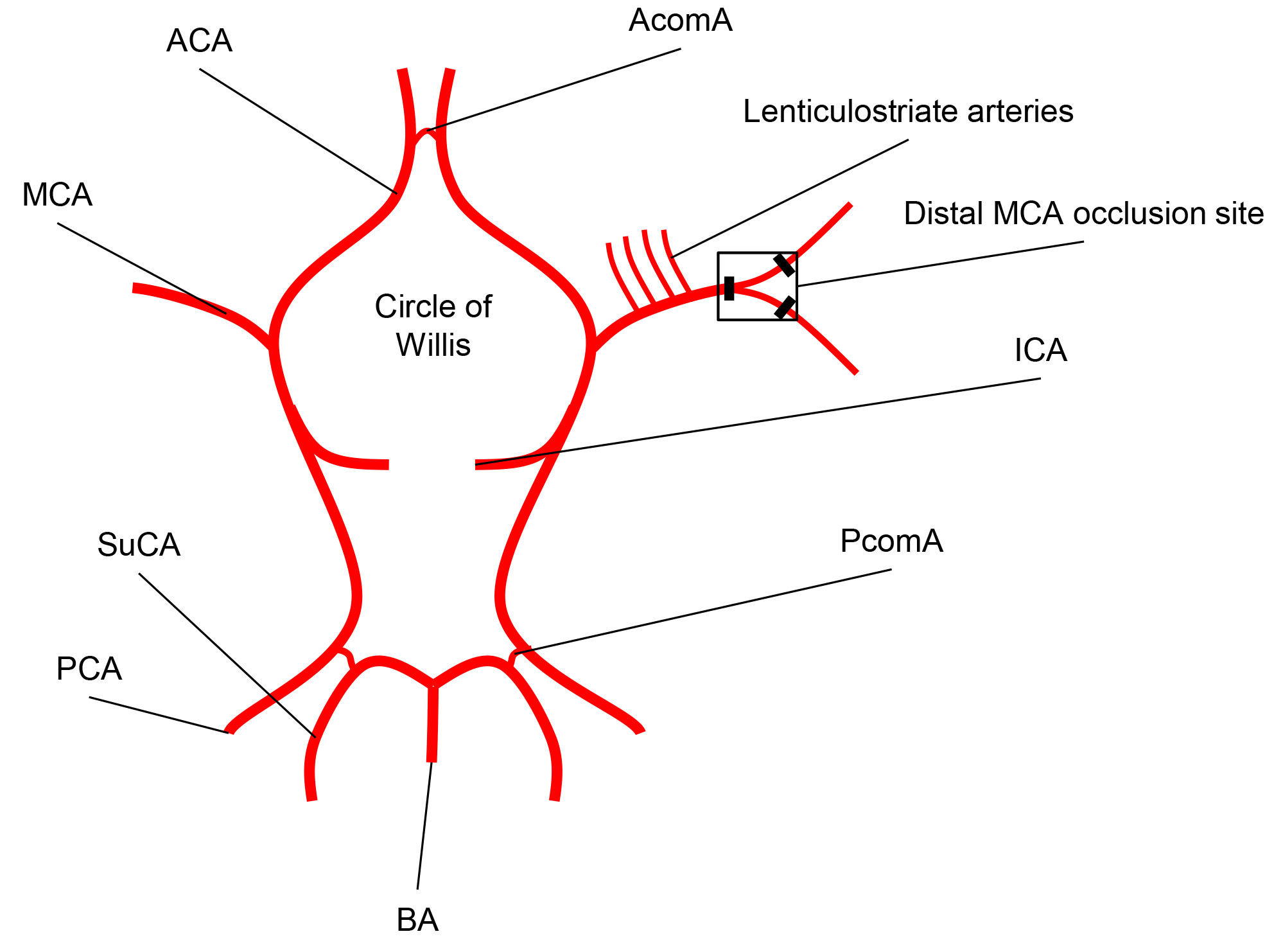
Figure 1. Schematic representation of the Circle of Willis. The arterial Circle of Willis is formed by the middle cerebral arteries (MCA) and the anterior cerebral arteries (ACA) which branch from the internal carotid artery (ICA), as well as by the posterior cerebral arteries (PCA) and the posterior communicating arteries (PComA). The MCA runs into the lateral sulcus where it branches to the cerebral cortex. The dominant MCA branch supplying the major part of the motor cortex and part of the somatosensory cortex is permanently occluded by the demonstrated model (AcomA = Anterior communicating artery; BA =Basilar artery; SuCA =Superior cerebellar artery).
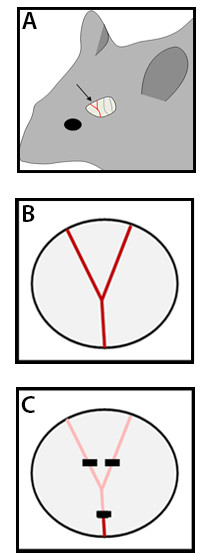
Figure 2. Transcranial view after removal of the temporal muscle and schematic view of MCA occlusion. (A) After removing the temporal muscle the cortical arteries can be viewed through the partially translucent mouse skull (in 8-12 week old mice). The dominant MCA branch can be identified in the rostral part of the temporal view as well as further cortical arteries branching from the MCA and the PCA in the caudal part. (B) Schematic view on the dominant MCA branch in its predominant variation with a bifurcation on the lateral temporal cortex after drilling a burr hole and removing the skull. (C) Black squares represent the MCA coagulation sites at the proximal and distal sides of the bifurcation.

Figure 3. Analysis of behavioral deficits. (A) Cylinder test set-up: the mouse is placed into a vertical cylinder and mirrors are placed behind in order to register all of the movements using a video camera. (B) Forelimb use asymmetry was analyzed using the cylinder test. Left/right ratios of independent forelimb use were calculated 24 hr before MCAO and at the indicated time points after stroke induction. N = 10, *p < 0.05 between indicated time point and baseline (control) value.
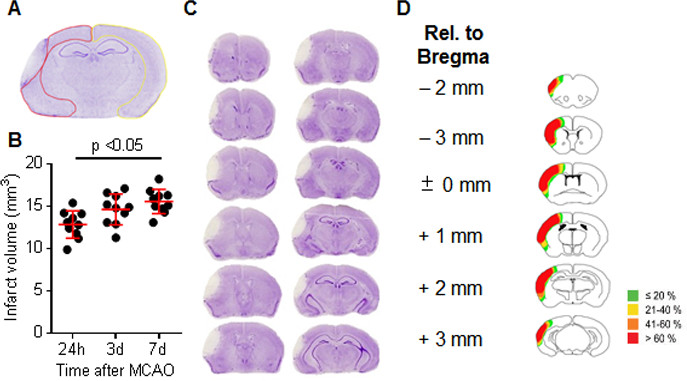
Figure 4. Volumetric infarct analysis and infarct outcome after distal MCAO. (A) Representative image of a cresyl violet stained coronal brain section 7 days after MCAO. The yellow line is indicating selection of the contralateral (right) cortex and the red line is selecting the non-infarcted (stained) cortex of the ipsilateral brain. The pale area in the left hemisphere depicts the infarcted tissue area. (B) Infarct volume analysis of 10 brains (each dot representing one individual brain) 24 hr, 3 days and 7 days after distal MCA coagulation. The horizontal red line represents the mean, error bars indicate standard deviation. (C) Representative cresyl violet stained coronal brain sections every 400 µm at 7 days after MCA coagulation. (D) Schematic distribution of the infarcted brain tissue 7 days after MCAO. Each slide depicts the accumulated information of infarct distribution (color coded as indicated) at the given section in relation to the bregma (modified image from: Liesz A et al., Brain, 2011; by permission of Oxford University Press). Please click here to view a larger version of this figure.
Discussion
Loading...
$$\rightleftharpoonup{xx}$$
$$\longleftharp{xx}$$,
$$\longrightharp{xx}$$,
The present protocol describes the experimental stroke model of distal, permanent MCAO by transcranial electrocoagulation – the so-called “coagulation model”. This model has meanwhile become one of the most frequently used animal models in experimental stroke research12. Compared to other focal brain ischemia models, the coagulation model as presented in this video has the advantage of a very short operation time of approximately 10 min when performed by a trained scientist. Hence, brief anesthesia times can be achieved in this model, which is a favorable feature of an experimental stroke model because the impact of anesthetics on neuroprotection and stroke outcome is well-known15. Moreover, as previously described by Carmichael et al.11, we corroborate that the resulting infarct volume and localization after distal permanent MCA coagulation corresponds to ischemic brain lesions in the majority of human strokes in proportion to brain size. Human stroke is mainly small in size with lesions of approximately 5-15% of the hemisphere, based on previous population studies and clinical imaging trials16-18, in contrast to extensive stroke lesions with compressive brain edema which occur in less than 10% of clinical strokes19. Thereby, stroke lesions in the MCA territory of around 12% of the hemisphere achieved by the presented model can be regarded as a translationally relevant stroke volume. However, it has to be taken into account that different mouse strains or used anesthesia protocols might affect the resulting lesion volume20.
Mortality during the observation period after stroke induction in this model is virtually absent. The overall mortality of less than 5% consists mainly of deaths during operation because of anesthesiological complications or sacrifice because reaching of exclusion criteria. In order to warrant the low variability of this model and its excellent reproducibility, we suggest the following exclusion criteria: 1) Any subarachnoid hemorrhage during the operation. 2) Operation time longer than 15 min. 3) Recanalization of the MCA after two attempts for electrocoagulation with only transient MCAO. Moreover, animals need to be examined daily after MCAO (basic physiological behavior, fur appearance and body weight) to control for pain, discomfort or sickness behavior.
Several measures might be implemented for analysis of stroke outcome like laser speckle measurement, magnetic resonance imaging, behavioral tests or histological analysis. In this protocol we provide exemplary methods for behavior analysis and infarct volume analysis. Several test for behavioral analysis after focal brain ischemia have been developed and used in experimental stroke research. Suitable tests for sensorimotor dysfunction previously used by our group in this stroke model21,22 were the Rotarod test23, Sticky label test24, Corner test25 and the Cylinder test13, which is demonstrated in this video. The cylinder test consistently depicts motor asymmetry in the acute phase after distal permanent MCAO and also detects consecutive regain of motor function.
Despite the obvious advantages, some limitations of this stroke model have to be taken into account. First, a trepanation of the skull is needed in order to coagulate the artery, thereby producing a potential access for peri-operational infections of the brain, although bacterial infections of the surgical wound, temporal muscle or the brain itself have never been detected by ourselves or documented by others using this model. Moreover, mechanical damage to the cortex during preparation and coagulation cannot be excluded but can be limited by careful drilling and removal of the skull, constant humidification of the surgical site and minimal necessary electrocoagulation (see exclusion criteria). Although the course of the MCA as depicted in Figure 2 is found in the majority of C57Bl/6 mice, we describe in the protocol how to proceed in normal variations of the vessel course to minimize model variability. Furthermore, we suggest the use of multiple (3 in case of bifurcation; 2 without bifurcation of the MCA) occlusion sites to minimize the risk of partial recanalization of the MCA, which in our experience is an important factor for the variability of this model.
In terms of behavioral tests, only minor behavioral deficits can be detected in the above-mentioned behavioral tests and functional regeneration can be observed within the first week after stroke. Thus, more advanced test systems with higher sensitivity and qualitative test parameters like the skilled reaching test27 might be more suitable to detect long-term functional outcome in this model.
Finally, due to the permanent coagulation of the MCA no reperfusion can be obtained, which is a feature observed in a substantial percentage of stroke patients due to spontaneous clot lysis or therapy28. However, a previously described thromboembolic stroke model26 provides the option for a complimentary stroke model with reperfusion of cortical brain ischemia. Taken together, the high reproducibility, the possible long-term observation due to minimal mortality and the comparable relative infarct volume and localization in respect to human stroke distinguish the “coagulation model” as a valuable model for basic and translational stroke research.
Disclosures
Loading...
$$\rightleftharpoonup{xx}$$
$$\longleftharp{xx}$$,
$$\longrightharp{xx}$$,
The authors have no competing interests to disclose.
Acknowledgements
Loading...
$$\rightleftharpoonup{xx}$$
$$\longleftharp{xx}$$,
$$\longrightharp{xx}$$,
This work was funded by the excellence cluster of the German research foundation “Munich Cluster for Systems Neurology (SyNergy)” and by the Daimler-Benz foundation to A.L.
Materials
| Name | Company | Catalog Number | Comments |
|---|---|---|---|
| Camera Canon Eos 500D | Canon | Optics: 18-55 mm; 30 fps; 640 x 480 video function | |
| Mirror | Kristallform | 2677089 | 30 x 30 cm |
| Transparent acrylic glass cylinder | H&S Kunststofftechnik | Diameter: 8 cm, height: 25 cm | |
| Heating blanket | FHC DC Temperature Controller | ||
| Fine Scissors | FST | 15000-00 | |
| Mayo Scissors | FST | 1410-15 | |
| Forceps | FST | 11616-15 | |
| Cottons | NOBA Verbondmitel Danz | 974116 | |
| Saline solution | Braun | 131321 | |
| Bepanthen pommade | Bayer | ||
| Isoflurane | Abbot | B506 | |
| Anesthesia system for isoflurane | Drager | ||
| Stereomikroskop | Zeiss | Stemi DV4 | |
| Electrosurgical device | ERBETOM ICC 80/50 HF-Chirurgiegerät | ||
| Drill | Proxxon | D-34343 | |
| Ketamine | Inresa Arzneimittel GmbH | ||
| Xylacine | Albrecht | ||
| 5ml Syringe | Braun | ||
| Phosphate Buffered Saline PH: 7.4 | Apotheke Innestadt Uni Munchen | P32799 | |
| Isopentane | Fluka | 59070 | |
| Cryostat | Thermo Scientific CryoStarNX70 | ||
| Superfrost Plus Slides | Thermo Scientific | J1800AMNZ | |
| Cresyl violet | Sigma Life Science | C5042-10G | |
| Acetic acid | Sigma Life Science | 695092 | |
| Ethanol 70% | CLN Chemikalien Laborbedorf | 521005 | |
| Ethanol 96% | CLN Chemikalien Laborbedorf | 522078 | |
| Ethanol 99% | CLN Chemikalien Laborbedorf | ETO-5000-99-1 | |
| Roti-Histokit mounting medium | Roth | 6638.1 | |
| C57Bl/6J mice | Charles River | 000664 |
References
Loading...
$$\rightleftharpoonup{xx}$$
$$\longleftharp{xx}$$,
$$\longrightharp{xx}$$,
- Donnan, G. A., et al. Stroke. Lancet. 371, 1612-1623 (2008).
- Di Carlo, A., et al. Frequency of stroke in Europe: A collaborative study of population-based cohorts. ILSA Working Group and the Neurologic Diseases in the Elderly Research Group. Italian Longitudinal Study on Aging. Neurology. 54, 28-33 (2000).
- Hacke, W., et al. Thrombolysis with alteplase 3 to 4.5 hours after acute ischemic stroke. N. Engl. J. Med. 359, 1317-1329 (2008).
- Jauch, E. C., et al. Guidelines for the early management of patients with acute ischemic stroke: a guideline for healthcare professionals from the American Heart Association/American Stroke Association. Stroke. 44, 870-947 (2013).
- Longa, E. Z., et al. Reversible middle cerebral artery occlusion without craniectomy in rats. Stroke. 20, 84-91 (1989).
- Engel, O., et al. Modeling stroke in mice - middle cerebral artery occlusion with the filament model. J. Vis. Exp. (47), e2423(2011).
- Zhang, Z., et al. A new rat model of thrombotic focal cerebral ischemia. J. Cereb. Blood Flow Metab. 17, 123-135 (1997).
- Tamura, A., et al. Focal cerebral ischaemia in the rat: 1. Description of technique and early neuropathological consequences following middle cerebral artery occlusion. J. Cereb. Blood Flow Metab. 1, 53-60 (1981).
- Chen, S. T., et al. A model of focal ischemic stroke in the rat: reproducible extensive cortical infarction. Stroke. 17, 738-743 (1986).
- Tureyen, K., et al. Infarct volume quantification in mouse focal cerebral ischemia: a comparison of triphenyltetrazolium chloride and cresyl violet staining techniques. J. Neurosci. Methods. 139, 203-207 (2004).
- Carmichael, S. T. Rodent models of focal stroke: size, mechanism, and purpose. NeuroRx. 2, 396-409 (2005).
- Howells, D. W., et al. Different strokes for different folks: the rich diversity of animal models of focal cerebral ischemia. J. Cereb. Blood Flow Metab. 30, 1412-1431 (2010).
- Schallert, T., et al. CNS plasticity and assessment of forelimb sensorimotor outcome in unilateral rat models of stroke, cortical ablation, parkinsonism and spinal cord injury. Neuropharmacology. 39, 777-787 (2000).
- Swanson, R. A., et al. A semiautomated method for measuring brain infarct volume. J. Cereb. Blood Flow Metab. 10, 290-293 (1990).
- Kitano, H., et al. Inhalational anesthetics as neuroprotectants or chemical preconditioning agents in ischemic brain. J. Cereb. Blood Flow Metab. 27, 1108-1128 (2007).
- Effect of intravenous recombinant tissue plasminogen activator on ischemic stroke lesion size measured by computed tomography. NINDS; The National Institute of Neurological Disorders and Stroke (NINDS) rt-PA Stroke Study Group. Stroke. 31, 2912-2919 (2000).
- Sowell, E. R., et al. Mapping cortical change across the human life span. Nat. Neurosci. 6, 309-315 (2003).
- Brott, T., et al. Measurements of acute cerebral infarction: lesion size by computed tomography. Stroke. 20, 871-875 (1989).
- Hacke, W., et al. Malignant' middle cerebral artery territory infarction: clinical course and prognostic signs. Arch. Neurol. 53, 309-315 (1996).
- Majid, A., et al. Differences in vulnerability to permanent focal cerebral ischemia among 3 common mouse strains. Stroke. 31, 2707-2714 (2000).
- Liesz, A., et al. Boosting regulatory T cells limits neuroinflammation in permanent cortical stroke. J. Neurosci. 33 (44), 17350-17362 (2013).
- Liesz, A., et al. Inhibition of lymphocyte trafficking shields the brain against deleterious neuroinflammation after stroke. Brain. 134, 704-720 (2011).
- Jones, B. J., Roberts, D. J. A rotarod suitable for quantitative measurements of motor incoordination in naive mice. Naunyn Schmiedebergs Arch. Exp. Pathol. Pharmakol. 259, 211(1968).
- Bouet, V., et al. The adhesive removal test: a sensitive method to assess sensorimotor deficits in mice. Nat. Protoc. 4, 1560-1564 (2009).
- Zhang, L., et al. A test for detecting long-term sensorimotor dysfunction in the mouse after focal cerebral ischemia. J. Neurosci. Methods. 117, 207-214 (2002).
- Orset, C., et al. Mouse model of in situ thromboembolic stroke and reperfusion. Stroke. 38, 2771-2778 (2007).
- Farr, T. D., Whishaw, I. Q. Quantitative and qualitative impairments in skilled reaching in the mouse (Mus musculus) after a focal motor cortex stroke. Stroke. 33, 1869-1875 (2002).
- Kassem-Moussa, H., Graffagnino, C. Nonocclusion and spontaneous recanalization rates in acute ischemic stroke: a review of cerebral angiography studies. Arch. Neurol. 59, 1870-1873 (2002).
Reprints and Permissions
Request permission to reuse the text or figures of this JoVE article
Request Permission