This method evaluates cancer cell invasion from spheroids into a surrounding 3D matrix. Spheroids are generated via the hanging drop culture method and then embedded in a matrix comprised of basement membrane materials and type I collagen. Invasion out of the spheroids is subsequently monitored.
Method Article
A Cancer Cell Spheroid Assay to Assess Invasion in a 3D Setting
In This Article
Summary
Abstract
The invasive nature of cancer cell lines is thought to correlate with their metastatic potential. Most traditional assays, however, do not examine these invasive features in a three-dimensional environment and the resulting data suffer from reduced biological applicability. Here an approach is presented to visualize the invasive ability of cell lines in a physiologically relevant setting. The cancer cell spheroid invasion assay first utilizes gravity to generate spheroids within drops of media that hang from the lid of a cell culture dish. Next, these spheroids are embedded in a 3D matrix consisting of a mixture of basement membrane materials and type I collagen. Cancer cell egression from the spheroids into the surrounding matrix is then monitored over time. The method described here can be modified to examine invasion after coculture of different cell types, inclusion of drugs/inhibitors, or alterations in extracellular matrix (ECM) constituents.
Introduction
It is established that cancer cell motility is predictive of metastatic potential, given that such behavior facilitates invasion through the basement membrane and entry into the circulatory system1. Most research on motility has focused on how cells behave in a two-dimensional (2D) setting, though it is becoming widely recognized that the movement of cells in a three-dimensional (3D) matrix is more representative of how these same cells will actually behave in vivo2. 3D culture systems are increasingly used to study cellular behaviors that range from cell morphology, to growth kinetics and drug sensitivity3. The desire to monitor cancer cell invasion within the context of a 3D milieu has led to the synthesis of previously established techniques involving the generation of 3D cell aggregates (spheroids) via the hanging drop culture method4, followed by embedding these spheroids into a 3D extracellular matrix (ECM) composed of collagen and basement membrane materials5. This method seeks to improve upon these previously established techniques by providing a streamlined approach that can be easily utilized to compare invasion under a variety of experimental conditions.
More traditional ways to assess cell motility in vitro are the scratch-wound assay and the transwell assay6. The former assay depicts cell motility in a 2D setting, and is therefore independent of a variety of features critical for in vivo invasion, e.g. protease activity7. The transwell assay can better model cell invasion when the well inserts are coated with ECM substrates but, only a single parameter, i.e. the appearance of cells on the opposite membrane surface is measured, and many nuances of cell invasion are thus not readily observable. In contrast to these techniques, the cancer cell spheroid invasion assay (Figure 1) allows for the real-time monitoring of cell invasion in a setting that is not only physiologically relevant, but also permits important cell line-specific features to be visualized, such as individual vs. collective cell migration8. This method also affords advantages over standard 3D culture growth assays. The generation of cellular aggregates via the hanging drop method initially constrains cell movement, so that cells will be incentivized to invade after this constraint is lifted. Furthermore, once that constraint is lifted, cell egress will proceed in a uniform direction that can then be conveniently quantitated.
The most popular ECM materials used for cancer cell spheroids assays are Matrigel and type I collagen, where each of these components has important and distinct roles in influencing metastatic behavior. Matrigel is a secreted mixture of proteins produced by Engelbreth-Holm Swarm mouse sarcoma cells, and is enriched in basement membrane proteins such as laminin, entactin, and type IV collagen9. For this reason Matrigel is henceforth referred to as “basement membrane materials.” These basement membrane materials provide essential ligands needed for integrin adhesion during cancer cell invasion10 in addition to many other proteins that exert a range of effects on cell behavior11. In comparison, type I collagen, commonly prepared from acid digests of tendons and other dense collagenous structures12, is a much simpler matrix material that serves as a major structural element of the connective tissue and stroma supporting tissues and organs of the body. It has been demonstrated that the physical characteristics of collagen can regulate a number of features of cell motility; for example, the alignment of collagen fibrils at the tumor-stromal interface permits cancer cells to subsequently migrate along those fibrils when invading into the stroma13. In the assay presented here, both type I collagen and basement membrane materials are utilized as tools to study 3D cancer cell-stroma interactions.
The effect of inhibition or stimulation of pathways that control invasion can be monitored after the cells have been embedded in the 3D matrix. Cells can be pretreated during growth in the hanging drops or upon transfer to the 3D culture, depending on whether a lengthy treatment will be required to modulate invasion. For shorter treatments, it is recommended that the drug be mixed with the spheroid suspension after collection, as well as the media that will surround the 3D cultures, to facilitate adequate drug exposure to the cells. Next, normal or tumor-associated stromal cells can be admixed with the matrix material to evaluate their role in modulating tumor cell invasion, or to determine how paracrine and autocrine signaling influences cell behavior. This idea was shown in a study where the coculture of colon cancer and endothelial cells in hanging drops led to a vascular network within the spheroids14. Finally, the ECM constituents can also be altered, as cancer cell invasion is impacted by different substrates15. The method presented below will thus provide a framework for assessing cancer cell invasion under a variety of conditions. In general it was found that not all cell lines will create spheroids in the hanging drops and epithelial-looking cell lines typically form regular spheres.
Protocol
1. Generation of Spheroids
- Prepare single-cell suspension for hanging drop cultures by detaching adherent cancer cell cultures of ~70% confluence using a PBS wash followed by exposure to 0.05% trypsin-EDTA solution.
- Neutralize the trypsin solution with cell culture media and count the cells using an aliquot of the cell suspension. Note: The specific cell culture media will depend on the cell line being tested. Follow ATCC media recommendations, where the cell culture media is typically DMEM + 10% FBS. More information can be found in the Materials Table.
- Perform a dilution to allow for the seeding of 500-1,000 cells per 20 µl drop of cell culture media.
- Acquire 10 cm dish and add 5 ml of sterile PBS to the bottom. Note: This step protects the hanging drops from evaporation. Lower-cost “bacteriological grade” dishes can be used in place of cell culture grade dishes, as cells will not contact the culture surface.
- Use a multi-channel pipet to transfer 20 µl droplets of the diluted cell suspension to the inner surface of the lid.
- Pipet 40 drops (5 rows of 8 drops) onto the lid of the 10 cm dish.
- Invert the lid and place over the culture dish. Incubate the hanging drop cultures at 37 °C for 72 hr to generate spheroids.
- Flip the lid in a confident, yet controlled manner. Note: Inverting the lid too fast or too slow may cause the droplets to shift. Some cell lines may form spheroids in 48 hr or less, while others may require more than 72 hr to produce compact aggregates.
2. Embedding of Spheroids into 3D Matrix
- Thaw an aliquot of growth factor-reduced basement membrane materials at 4 °C overnight before embedding the spheroids.
- Optional: Layer wells with ECM in advance to prevent potential spheroid interaction with the tissue culture surface.
- Pipet 200 µl of ECM per well on a 24-well plate.
- Tilt the plate to ensure that the ECM covers the entire well surface.
- Carefully remove excess ECM with a pipet.
- Incubate the plate until the wells are dry: 3 hr at room temperature or overnight at 4 °C.
- Optional: Layer wells with ECM in advance to prevent potential spheroid interaction with the tissue culture surface.
- Collect the spheroids by tilting the lid of the tissue culture dish and pooling the media. Transfer the media with the spheroids into a 1.5 ml microcentrifuge tube.
- Allow 10 min for the spheroids to settle at the bottom of the microcentrifuge tube. Spheroids should be visible by eye.
- Mix 100 µl of the basement membrane materials with 100 µl of cold (4 °C) type I collagen in a separate pre-chilled tube. Keep the mixture at 4 °C to prevent either gel from solidifying prematurely.
- Use pre-chilled pipet tips if transferring small volumes.
- Give care when mixing the basement membrane materials and collagen type I to prevent air bubble formation. Note: Air bubbles may hinder imaging later in the protocol if they become embedded in the gel.
- Aspirate the spheroids from the 40 µl bottom portion of the microcentrifuge tube, and combine with the basement membrane materials/type I collagen mixture. Be careful to prevent air bubble formation when mixing. Note: The solution containing of 100 µl basement membrane materials, 100 µl collagen type I, and 40 µl of the spheroids will create enough material for 4 independent 3D cultures. This can be scaled up or down.
- Pipet 40 µl drops of the viscous mixture into the centers of wells on a 24-well plate. Keep the plate level to prevent the mixture from running into the side of the well. Note: A single column on the 24-well plate – 4 wells – is recommended for each condition to be tested.
- Place the plate into a 37 °C incubator and leave undisturbed for 30 min. The 3D culture will polymerize during this period.
- Slowly submerge the 3D cultures in 1 ml of cell culture media.
- Ensure that the media is warm when adding it into the wells to further promote gel polymerization.
- CRITICAL STEP: Gently add the media to the 3D cultures. Pipetting the media too quickly into the wells can lead to detachment of the 3D cultures from the tissue culture surface.
3. Monitoring and Analyzing Spheroid Invasion
- Image invasion from the spheroids into the surrounding 3D matrix at time-points decided by the investigator. Acquire photographs using an inverted microscope with the 20x objective lens. Note: Ideal time-points will differ depending on the cell line being tested. More invasive cell lines will begin their egress from the spheroids shortly after plating and, therefore, take initial photographs within a couple of hours after plating. Generally, photographs are taken at 0 hr (after plating), 24 hr, and 48 hr. Invasion typically concludes 24 to 48 hr after plating.
- Quantitate invasive ability using image analysis software like Image J.
- Analyze invasion as the cell distance from the edge of the spheroid.
- Use the Straight Draw Tool to mark the radius and/or maximal invasive distance.
- Click “Analyze” in the top menu, and then click “Measure” to display the length measurement.
- Analyze invasion as the total invasive area outside the spheroid.
- Use the Freehand Draw Tool to trace the border of the spheroid and/or total invasive area.
- Click “Analyze” in the top menu, and then click “Measure” to display the area measurement.
- Use a plugin like ROI Manager Tools to create measurements for a stack of images.
- Analyze invasion as the cell distance from the edge of the spheroid.
Results
Using the spheroid invasion assay (Figure 1), a panel of cancer cell lines was tested for their ability to form spheroids, as well as for the amount of cell egress exhibited after implantation in a 3D matrix consisting of basement membrane materials and type I collagen (Table 1). These results demonstrate that not every cancer cell line will create well-formed spheroids, where cell lines possessing an epithelial morphology in vitro tended to produce regular and smooth aggregates. The ability of different cell lines to invade in the assay was also variable. Cell lines that are established as being invasive like 4T1, E0771, and U-87 MG exhibited a high degree of spheroid egress in the assay. Less aggressive cell lines, like the MCF-7, BT474, and MCF10DICS.com did not invade, as was anticipated. Unexpected, however, was the lack of invasion displayed by A-431 and COLO 357 PL cells. An example of the data for some of these lines is shown (Figure 2). For 4T1 and E0771 cells, invasion was already pronounced 24 hours after embedding the spheroids into the 3D matrix and, for U-87 MG, invasion commenced directly after implantation into the matrix.
The qualitative assessment of cellular invasion can be further supported by quantitation in image analysis software. Two different strategies for quantitating the extent of invasion are included (Figure 3), and the strategy selected depends on the type of invasion witnessed. In ImageJ, invasive distance or invasive area is first demarcated using the software’s draw tool, after which comparative values are generated as pixel measurements. Microscopy images can be imported as a stack to enable more efficient data acquisition but, if doing so, a plugin like ROI Manager Tools is recommended to facilitate efficient measuring.
| Cell Line | Cancer Type | Species | Sphere-Forming Ability | Assay Invasion |
| 4T1 | Mammary | Mouse | +++ | ++ |
| A-431 | Epidermoid | Human | +++ | - |
| BT-474 | Breast | Human | ++ | - |
| COLO 357 PL | Pancreas | Human | ++ | - |
| E0771 | Mammary | Mouse | +++ | +++ |
| LNCaP | Prostate | Human | + | - |
| MCF-7 | Breast | Human | +++ | - |
| MCF10DCIS.com | Breast | Human | ++ | - |
| MDA-MB-231 | Breast | Human | - | ? |
| PANC-1 | Pancreas | Human | + | ++ |
| PC-3 | Prostate | Human | - | ? |
| SK-BR-3 | Breast | Human | - | ? |
| U-87 MG | Glioblastoma | Human | +++ | +++ |
Table 1: Sphere-formation and invasion by different cell lines. Cell lines tested are scored according to their sphere-forming ability and invasion.

Figure 1: Visual workflow of the spheroid invasion assay. Spheroids are generated in drops of media that hang from the lid of a tissue culture dish for 72 hr. Next, the drops are pooled, and the spheroids are transferred to a 4 °C mixture of basement membrane materials and type I collagen. Following spheroid resuspension, the viscous mixture is pipetted into the wells of a 24-well plate, after which it is given 30 min at 37 °C to solidify into a 3D culture. Warm media is then added to the wells. Cell exit from the spheroids is then monitored over time.
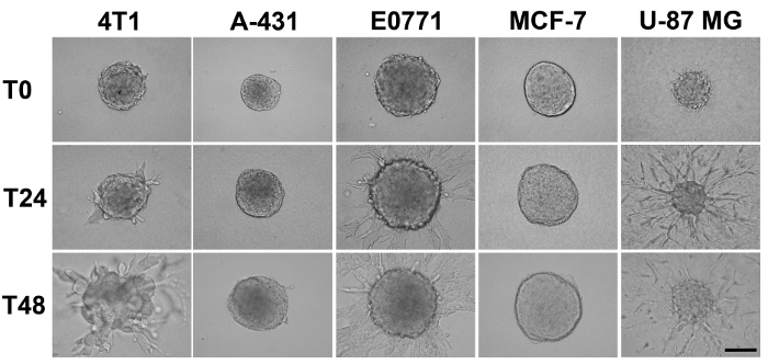
Figure 2: Example of assay data. The 4T1, E0771, and U-87 MG cells show invasion from the spheroids whereas the A-431 and MCF-7 cells do not. Invasion of U-87 cells is most pronounced after 24 hr, while maximal invasion by the 4T1 and E0771 cells occurs later. For the most invasive E0771 and U-87 MG cell lines, cell egress appears to offset the increase in spheroid size otherwise observed with the less invasive lines. Scale bar, 200 µm. Please click here to view a larger version of this figure.

Figure 3: Quantitation of invasion. Invasion can be quantitated by image analysis, e.g. using Image J software. (a) Calculation of invasion as a function of the longest invasive distance emanating from the spheroid. (b) Total area invaded by cells leaving the spheroid. Please click here to view a larger version of this figure.
Discussion
This study evaluated the performance of a panel of cancer cell lines in a spheroid invasion assay (Table 1). Generally, we find that spheroid formation is enhanced in the more epithelial-looking cell lines, where the presence of cell-cell junctions promotes the formation of a spheroid-like architecture. Known cell lines that have undergone an epithelial-mesenchymal transition, like MDA-MB-231 cells do not form spheroids in the hanging drop culture most likely due to their reduced E-, N-, and P-cadherin expression16.
Some of the steps in the spheroid invasion assay require close adherence to the protocol. It is absolutely critical to work quickly when pipetting the basement membrane and collagen mixture, and it is crucial that one keeps the mixture near 4 °C while working as it will begin to solidify at room temperature. Also, soluble collagen preparations are acidic, and are induced to gel by the addition of a small amount of sodium hydroxide and salt, followed by warming to 37 °C. In our hands, simply mixing equal volumes of collagen with the basement membrane materials renders it gel-competent upon warming because the basement membrane component is able to neutralize the collagen. If smaller ratios of basement membrane:collagen are planned, the collagen should be neutralized prior to adding the basement membrane mixture to facilitate polymerization. Once formed, most spheroids are resistant to dissociation and will survive resuspension in this viscous mixture that will comprise the 3D culture. During the resuspension steps, one must also exert care to restrict the formation of air bubbles, as these can hinder imaging. When adding culture media to the wells after the gel has solidified, it is important that one pipets slowly to prevent the 3D cultures from detaching from the dish.
The egress of cancer cells into the surrounding 3D matrix is generally correlated with the invasive ability of the lines, where the E0771 and U-87 MG cells demonstrated the most invasion in the panel of cell lines examined here (Figure 2). Interestingly, these two lines also exhibit different modes of invasion. Whereas the E0771 cells invade in a gradual and collective manner, the U-87 MG cells display individual and rapid exit from the spheroids. The spheroid invasion assay thus allows one to compare the different modes used by lines to invade the substrate.
Different methods of analyzing invasion are presented in Figure 3 and the preferred manner of analysis may depend on the mode of invasion. Regardless of the type of analysis employed, this assay was designed to allow for the rapid quantitation of invasion out of a large number of spheroids. The pooling of spheroids into a 3D culture mixture, and then aliquoting this mixture to create 4 independent replicate cultures, leads to the establishment of multiple spheroids that can be monitored for a given experimental condition (Figure 1). The assay is hence easily amendable to statistical comparison due to the large potential “n,” and the phenotype shared by spheroids of the same type seems to exhibit minimal variability. Note that less aggressive cell lines like BT-474 and MCFDCIS cells form smooth, compact spheres that have a much smaller size than more aggressive lines. As a consequence, during the initial cell seeding into the hanging drops, the starting cell number needs to be adjusted accordingly.
Some metastatic cell lines performed against our expectations and did not exhibit invasive behavior in this assay. Notable examples include the A-431 and the COLO 357 PL cells. It is possible that the lack of invasion displayed by these lines is due to the type of ECM substrate that the spheroids were embedded into. Indeed, others have shown that basement membrane materials (e.g. Matrigel) and collagen can have disparate effects on the invasion of cells17 perhaps due to different matrix metalloprotease requirements for migration18. Thus, tweaking the ratio of collagen:basement membrane materials is another way to modify assay outcomes, where reducing the basement membrane material component to a third of the total mixture, thereby allowing for an increase in the amount of collagen, could further enhance invasion by some cell types. This idea is exemplified in a recent study that found a positive correlation between the invasive ability of mammary organoids and the number of type I collagen fibrils present, where collagen fibril formation was promoted by extending the 4 °C preincubation period of pH neutralized collagen19.
The discovery that some cell lines acted contrary to expectation shows some of the limitations of this assay. Although monitoring cancer cell invasion out of a spheroid into a 3D mixture consisting of collagen and basement membrane materials is more physiologically relevant than a 2D motility assay, it is still a model system that omits some biological complexities otherwise found in vivo. For example, metastatic ability can be modulated by the distinct microenvironments that support orthotopic vs. ectopic tumor growth20 and, as such, it is important to acknowledge that a variety of stromal factors can profoundly influence the metastatic process. A number of these factors are absent in the invasion assay presented here and could explain some of the observed inconsistencies.
In summary, the cancer cell spheroid invasion assay presented here provides a flexible framework for monitoring invasion in a biologically relevant setting, supports the discovery of mechanisms of cell invasion, and can potentially aid in the development of novel anti-metastatic therapies.
Disclosures
The authors declare that they have no competing financial interests.
Acknowledgements
Supported by NIH grants P30 CA051008 and T32 CA009686 (ATR).
Materials
| Name | Company | Catalog Number | Comments |
|---|---|---|---|
| Matrigel Growth Factor Reduced | Corning | CB-40234 | |
| Collagen Type I, Rat Tail, 100 mg | Millipore | 08-115 | |
| DMEM | Life Technologies | 11995-065 | |
| RPMI 1640 Medium | Life Technologies | 11875-093 | |
| PBS | Life Technologies | 10010-023 | |
| 0.05% Trypsin-EDTA | Life Technologies | 25300-054 | |
| Fetal Bovine Serum, Heat Inactivated | Omega Scientific | FB-12 | |
| 100 mm TC-treated Dishes | Corning Incorporated | 430167 | |
| 24-well TC-treated Plates | NEST Biotechnlology | 702001 | |
| Olympus IX-71 Inverted Microscope | |||
| Cell lines were maintained in DMEM + 10% FBS, with the expection of BT-474 and LNCaP cells, which were mantained in RPMI + 10% FBS. |
References
- Chambers, A. F., Groom, A. C., MacDonald, I. C. Metastasis: Dissemination and growth of cancer cells in metastatic sites. Nat Rev. Cancer. 2 (8), (2002).
- Schmeichel, K. L., Bissell, M. J. Modeling tissue-specific signaling and organ function in three dimensions. J Cell Sci. 116 (12), 2377-2388 (2003).
- Yamada, K. M., Cukierman, E. Modeling tissue morphogenesis and cancer in 3D. Cell. 130 (4), (2007).
- Foty, R. A simple hanging drop cell culture protocol for generation of 3D spheroids. J Vis Exp. 51, (2011).
- Del Duca,, Werbowetski, D., T,, Del Maestro,, F, R. Spheroid preparation from hanging drops: characterization of a model of brain tumor invasion. J Neuro-Oncol. 67 (3), (2004).
- Justus, C. R., Leffler, N., Ruiz-Echevarria, M., Yang, L. V. In vitro cell migration and invasion assays. J Vis Exp. 88, (2014).
- Ramos-DeSimone, N., Hahn-Dantona, E., Sipley, J., Nagase, H., French, D. L., Quigley, J. P. Activation of matrix metalloproteinase-9 (MMP-9) via a converging plasmin/stromelysin-1 cascade enhances tumor cell invasion. J Bio Chem. 274 (19), Forthcoming.
- Friedl, P., Wolf, K. Tumour-cell invasion and migration: diversity and escape mechanisms. Nature Rev. Cancer. 3 (5), 362-374 (2003).
- Kleinman, H. K., Martin, G. R. Matrigel: Basement membrane matrix with biological activity. Semin Cancer Biol. 15 (5), 378-386 (2005).
- Shaw, L. M., Chao, C., Wewer, U. M., Mercurio, A. M. Function of the integrin α6β1 in metastatic breast carcinoma cells assessed by expression of a dominant-negative receptor. Cancer Res. 56 (5), 959-963 (1996).
- Hughes, C. S., Postovit, L. M., Lajoie, G. A. Matrigel: a complex protein mixture required for optimal growth of cell culture. Proteomics. 10 (9), (2010).
- Rajan, N., Habermehl, J., Coté, M. -F., Doillon, C. J., Mantovani, D. Preparation of ready-to-use, storable and reconstituted type I collagen from rat tail tendon for tissue engineering applications. Nat Protoc. 1 (6), (2006).
- Provenzano, P. P., Eliceiri, K. W., Campbell, J. M. Collagen reorganization at the tumor-stromal interface facilitates local invasion. BMC Med. 4 (1), (2006).
- Timmins, N. E., Nielsen, L. K. Generation of multicellular tumor spheroids by the hanging-drop method. Methods Mol Med. 140, 141-151 (2007).
- Brenner, W., Groß, S., Steinbach, F., Horn, S., Hohenfellner, R., Thüroff, J. W. Differential inhibition of renal cancer cell invasion mediated by fibronectin, collagen IV and laminin. Cancer Lett. 155, 199-205 (2000).
- Nieman, M. T., Prudoff, R. S., Johnson, K. R., Wheelock, M. J. N-cadherin promotes motility in human breast cancer cells regardless of their E-cadherin expression. J Cell Biol. 147 (3), Forthcoming.
- Cheung, K. J., Gabrielson, E., Werb, Z., Ewald, A. J. Collective invasion in breast cancer requires a conserved basal epithelial program. Cell. 155 (7), (2013).
- Sodek, K. L., Brown, T. J., Ringuette, M. J. Collagen I but not Matrigel matrices provide an MMP-dependent barrier to ovarian cancer cell penetration. BMC Cancer. 8 (1), 223-2210 (2008).
- Nguyen-Ngoc, K. V., Ewald, A. J. Mammary ductal elongation and myoepithelial migration are regulated by the composition of the extracellular matrix. J Microsc. 251 (3), 212-223 (2013).
- Glinskii, A. B., Smith, B. A., Jiang, P., Li, X., Yang, M., Hoffman, R. M., Glinsky, G. V. Viable circulating metastatic cells produced in orthotopic but not ectopic prostate cancer models. Cancer Res. 63 (14), 4239-4243 (2003).
Reprints and Permissions
