We describe here a reproducible protocol for isolating the mouse circle of Willis.
Method Article
Protocol for Isolating the Mouse Circle of Willis
In This Article
Summary
Abstract
The cerebral arterial circle (circulus arteriosus cerebri) or circle of Willis (CoW) is a circulatory anastomosis surrounding the optic chiasma and hypothalamus that supplies blood to the brain and surrounding structures. It has been implicated in several cerebrovascular disorders, including cerebral amyloid angiopathy (CAA)-associated vasculopathies, intracranial atherosclerosis and intracranial aneurysms. Studies of the molecular mechanisms underlying these diseases for the identification of novel drug targets for their prevention require animal models. Some of these models may be transgenic, whereas others will involve isolation of the cerebro-vasculature, including the CoW.The method described here is suitable for CoW isolation in any mouse lineage and has considerable potential for screening (expression of genes, protein production, posttranslational protein modifications, secretome analysis, etc.) studies on the large vessels of the mouse cerebro-vasculature. It can also be used for ex vivo studies, by adapting the organ bath system developed for isolated mouse olfactory arteries.
Introduction
The cerebral arterial circle (circulus arteriosus cerebri), also known as the circle of Willis (CoW), loop of Willisor Willis polygon) was first described by Thomas Willis in 1664. It is a circulatory anastomosis located around the optic chiasma and hypothalamus that can be considered as a central hub supplying blood to the brain and surrounding structures. Blood enters this structure via the internal carotid and vertebral arteries and it flows out of the circle via the interior middle and posterior cerebral arteries. Each of these arteries has left and right branches on either side of the circle. The basilar, post communicating, and anterior communicating arteries complete the circle (Figure 1 and Figure 2). The risk of impaired blood flow in any of the outflow arteries is minimized by the merging of blood entering the circle from the carotid and cerebral arteries, thereby guaranteeing that sufficient blood is supplied to the brain. This structure also serves as the main route for collateral blood flow in severe occlusive diseases of the internal carotid artery.
Several types of cerebrovascular disorders have their origin in the CoW. The most common are cerebral amyloid angiopathy (CAA)-associated vasculopathies, intracranial atherosclerosis and intracranial aneurysms. 1,2,3 These disorders may lead to hypoperfusion due to vasodilation, and intracerebral and/or subarachnoid hemorrhages ultimately translating into ischemic or hemorrhagic strokes or, at best, a transient ischemic attack. Recent advances in diagnostic procedures, including neuroimaging, possibly combined with angiography, have made it possible to diagnose these major cerebrovascular diseases clinically, without the need for a brain biopsy. Nevertheless, effective and specific treatments (pharmacological or endovascular) are currently lacking and there is therefore a need to define new molecular targets.
The identification of novel drug targets for the prevention of these diseases in humans will require animal models and ways of isolating the cerebro-vasculature including the CoW. Such models should provide evidence of and clues to the specific changes, including inflammatory changes, occurring in the walls of the large vessels in animal models of intracranial artery aneurysm, CAA or intracranial atherosclerosis. 4,5,6
We have established a method for mouse CoW isolation to facilitate studies of vessel inflammation in Alzheimer's disease (AD) and related diseases, such as CAA. This method for isolating the mouse CoW was developed for the assessment of inflammatory cerebrovascular gene expression during disease progression. Together with the detection of amyloid beta deposition within the walls of the leptomeningeal and pial arteries, this method could make it easier to determine the possible relationship between inflammatory gene expression in the cerebro-vasculature wall and Aβ-peptide accumulation. The vascular network of the brain, including the leptomeningeal and pial in the subarachnoid space, is an extension of the large arteries forming the circle of Willis. The method described here could be used to isolate the CoW of any mouse lineage and could be used for all types of screening (e.g., gene expression, protein production and posttranslational protein modifications) on the large vessels of the mouse cerebro-vasculature.
Protocol
All procedures were performed in accordance with European Community standards for the care and use of laboratory animals, with the approval of the local ethics committee for animal experimentation (Ile de France-Paris-Committee, Authorization 4270).
1. Anesthesia
- Infuse a lethal dose of pentobarbital (up to 1 mg/10 g body weight) intraperitoneally (27-gauge needle and 1-ml syringe) into adult mice before surgery.
2. Vessel Perfusion
NOTE: There is no need to apply vet ointment to the eyes during vessel perfusion. This procedure is rapid (5-10 minutes) and ends in the death of the animal. Confirm the lack of response with a toe pinch.
- Using iris scissors, make an incision, about 4 cm long, into the abdominal wall and peritoneum, just beneath the rib cage.
- Make a small incision (a few millimeters long) in the diaphragm and then continue the incision of the diaphragm along the entire length of the rib cage to expose the pleural cavity.
- Lift the sternum away and clamp the tip of the sternum with the hemostat; place the hemostat on the neck. Carefully trim the adipose tissue connecting the sternum to the heart.
- Pass the 15-gauge perfusion needle through the left ventricle into the apex of the heart.
- Finally, use scissors to cut one of the liver lobes to create an outlet.
NOTE: An alternative outlet can be created by using iris scissors to create an incision to the right atrium. - Perfuse the animal with 25 to 50 ml of phosphate-buffered saline (PBS) with a pump operating at a rate of 2.5 ml/min. The liver should blanch as the blood is replaced with PBS.
- After approximately five minutes, once the fluid from the liver is completely clear, stop the perfusion.
- If immunostaining or regular staining is planned, perfuse the animal with 50 ml of paraformaldehyde (PFA; 4% in PBS) for 15 min.
NOTE: Caution, PFA fumes are toxic. Perfusion of the animal with PFA should be carried out in a ventilated fume hood.
3. Isolation of the Brain and the Circle of Willis
- Isolation of the brain
- Remove the head with a pair of surgical scissors.
- Make a midline incision with iris scissors, along the skin from the neck to the nose.
- Trim off the skin to expose the skull and remove any residual muscles and adipose tissues with iris scissors.
- Place the sharp end of the iris scissors into the foramen magnum on one side and carefully slide them along the inner surface of the skull to the external auditory meatus (also known as the ear canal).
- Reproduce the incision described in 3.1.4 on the contralateral side and make a midline cut along the inner surface of the inter-parietal bone to the start of the sagittal suture.
- Plant the iris scissors in the frontal bone, right between the eyes, in the sagittal suture and then open them to split the skull in two.
- Lift out the brain, grabbing the olfactory bulbs and using the iris scissors to cut off the nerve connections on its ventral surface.
- Remove the brain and place it in a 60-mm Petri dish containing ice-cold PBS for CoW isolation. Completely immerse the brain in the PBS. If the brain was fixed with 4% PFA (for subsequent sectioning and immunostaining or regular staining), keep it in a bath of 4% PFA at 4 °C for 24 hr.
- Isolation of the circle of Willis
NOTE: A dissecting microscope is required for CoW isolation. The brain should be kept at 4 °C throughout the entire procedure.- Put the brain upside down (i.e., on its dorsal surface) to visualize the CoW.
- Use a small forceps to grab the anterior cerebral arteries (ACA) at the base of the olfactory lobes (a, Figure 1) and exert pressure to dissociate them from the vessel continuum. Use the same procedure to cut the middle cerebral arteries (MCA) in b (Figure 1).
- Use the sharp ends of the forceps to lift and remove the major arteries forming the CoW from the cortex.
- Lift up the start of the posterior communicating arteries (PCA) to disconnect them from the brain, by gripping the middle cerebral arteries (MCA) with the forceps. Pick up the anterior arteries (ACA and MCA) and pull them gently over the optic chiasm in an anterior-dorsal direction. To prevent disruption of the CoW, interrupt the the procedure to deal with the other arteries.
- Repeat steps 3.2.2 and 3.2.3 for the superior and posterior cerebellar arteries (SCA)/(PCA) (c, Figure 1) and for the basilar artery (BA) (d, Figure 1), pulling them in a dorsal-anterior direction. Stop at the end of the procedure described in 3.2.4.
- Remove the entire CoW by pulling gently with the forceps. Place the CoW in a 60-mm Petri dish filled with ice-cold PBS and remove any remaining attached brain tissue with two forceps, holding the CoW in place with small pins.
- Keep the harvested CoW at -80 °C for subsequent processing for RNA purification (RNA extraction yields large amounts of RNA — approximately 500 ng) or protein extraction.
Note: The CoW can be maintained ex vivo for 24 hr, by adapting the organ bath system developed for isolated mouse olfactory artery. 7
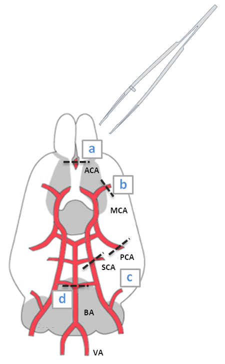
Figure 1: Schematic Diagram of a Ventral View of the Mouse Brain Highlighting the CoW. The CoW is formed from the two internal carotid arteries (MCA), which are derived from the two anterior cerebral arteries (ACA); the basilar artery (BA) branches into the posterior (PCA) and superior (SCA) cerebral arteries, and two vertebral arteries (VA).
Results
The PBS-perfused mouse is killed and the CoW is isolated as described in section 3.2 of the protocol. When the dissection is performed correctly, the CoW should come out in one piece and should be slightly transparent due to the absence of residual blood in the vasculature.
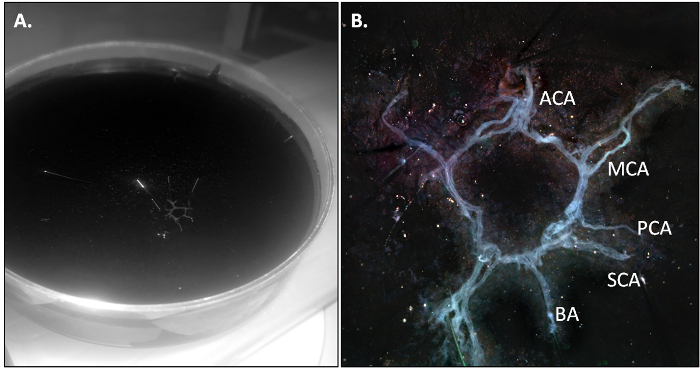
Figure 2: The Mouse CoW after Isolation. (A) Overview of the CoW in a 10-cm Petri dish. (B) Details of the various branches of the CoW. MCA for middle cerebral arteries, ACA for anterior cerebral arteries, BA for basilar artery, PCA for posterior cerebral arteries and SCA for superior cerebral arteries. Please click here to view a larger version of this figure.
The purity of the CoW preparation can be checked by ensuring that specific vascular genes are strongly expressed whereas the expression of neuronal genes is undetectable. More specifically, the cleaned CoW should specifically express vascular smooth muscle cell markers, such as smooth muscle actin (SMA), smooth muscle-myosin heavy chain (SM-MHC) and smoothelin. These markers are barely expressed in other parts of the brain. An opposite pattern of expression should be obtained for neuronal genes, with neuronal markers (Mog, Map2) only barely detectable in the CoW.
Levels of mRNA can be assessed by RT-qPCR with normalization relative to a reference gene transcript (here, that of GAPDH). Typical results are shown in Figure 3A and B.

Figure 3: Expression of Neuronal and Contractile Genes in the Mouse Circle of Willis and Brain. The RT-qPCR results were normalized against those for a reference gene (GAPDH). They are expressed as the mean ± SD of 6 to 12 independent experiments; Comparison of the CoW with the brain: n.s, not significant, *, P < 0.05, **, P < 0.005, and ***, P < 0.001. (A) Expression of contractile genes: SMA (smooth muscle actin), smoothelin, SM-MHC (smooth muscle-myosin heavy chain 11) and E-selectin. (B) Expression of neuronal genes: Mog (myelin oligodendrocyte glycoprotein) and Map2 (microtubule associated protein 2).
The expression pattern of the endothelial marker E-selectin in brain lacking the CoW is very similar to that obtained for CoW samples, possibly reflecting the existence of a brain capillary network. Table 1 lists the mean Cp numbers for each transcript.
| A. | |||||||||||||||
| Mean Cp | GAPDH | SMA | Smoothelin | SM-MHC | E-Sel | ||||||||||
| Mean | SD | N | Mean | SD | N | Mean | SD | N | Mean | SD | N | Mean | SD | N | |
| CoW | 20.86 | 0.93 | 12 | 22.02 | 0.67 | 12 | 25.29 | 1.89 | 12 | 23.37 | 0.80 | 12 | 30.56 | 1.66 | 12 |
| Brain | 18.71 | 0.72 | 6 | 33.91 | 2.06 | 6 | 31.08 | 3.17 | 6 | 26.05 | 0.73 | 6 | 28.45 | 0.63 | 6 |
| B. | |||||||||||||||
| Mean Cp | GAPDH | Mog | Map2 | ||||||||||||
| Mean | SD | N | Mean | SD | N | Mean | SD | N | |||||||
| CoW | 21.04 | 0.41 | 6 | 32.13 | 1.63 | 6 | 30.21 | 0.85 | 6 | ||||||
| Brain | 19.09 | 0.49 | 6 | 26.43 | 0.88 | 6 | 23.30 | 0.99 | 6 | ||||||
Table 1: Mean Crossing Point for the Expression of Neuronal and Contractile Genes in the Mouse Circle of Willis and Brain. Calculations are based on comparison of the precise cycle determined by crossing points (Cp) at a constant level of fluorescence. The crossing point is the cycle number corresponding to the maximum second derivative of the amplification curve. Higher Cp values are associated with lower levels of expression. For a given gene, expression is considered to be undetectable if the Cp exceeds 35. (A) Cp of the reference gene: GAPDH; and of contractile genes: SMA, smoothelin, SM-MHC and E-selectin. (B) Cp of the reference gene: GAPDH; and neuronal genes: Mog and Map2.
Discussion
We describe here a reproducible protocol for the isolation of the circle of Willis. The most common cerebrovascular disorders involving the CoW are CAA-associated vasculopathies, intracranial atherosclerosis and intracranial aneurysm, all of which affect the walls of arterial vessels. The risk factors are well known, but the molecular pathogenesis of these cerebral disorders remains poorly understood and specific biological markers for predicting their occurrence are lacking. There is considerable interest in methods for isolating the CoW from transgenic mice to link macroscopic observations with molecular changes. For example, by inducing consistent aneurysms in a mouse model in which a particular gene is knocked out(as described by Hosaka et al.8) and analyzing the CoW, it should be possible to determine whether treatments targeting the protein encoded by this gene could prevent intracranial aneurysms and/or subarachnoid hemorrhages. Obviously, this method could also be used to analyze the effect of a particular drug on the progression of CAA-associated vasculopathies in the various transgenic mouse models of AD.
It is easier to isolate mouse arteries than to purify entire mouse microvessels, but such sampling is nevertheless more difficult in mice than in rats. Indeed, the rat CoW is embedded in much stiffer meninges, making it possible to isolate the whole structure in one go. In addition to the problem of the small size of the vessels in mice, this procedure is also tricky due to the transparency of the vessels following their perfusion with PBS before the removal of the brain. We therefore recommend practicing without the infusion, starting from step 3 after anesthesia to make it easier to distinguish the blood vessels. Finally, as mouse cerebral vessels are very fine and break easily, the dissection should be carried out very carefully, without rushing, to ensure that the entire structure is isolated in one piece, by first grabbing the ACA. The purity of the CoW preparation can be evaluated by comparing the levels of expression of the neuronal and vessel genes. With practice, a single CoW provides only sufficient RNA for the study of 5 - 10 gene expressions. For large-scale screening, several mouse CoWs should be pooled.
Gauthier et al.9 described a method for isolating small pieces of microvessels (based on the use of collagenase/dispase to digest fragments of brain and of glass beads to trap them) that yields smooth muscle or endothelial cells that can be maintained in culture when placed in an appropriate medium. The advantage of our method is that it isolates large vessels, without altering vessel structure. The dissection of specific branches of the CoW may also be of interest, to determine the correlation between the gene expression profile of a particular branch and possible susceptibility to cerebrovascular diseases, such as intracranial aneurysm. Such approaches may provide an explanation as to why some ACA anatomical and genetic variations are correlated with a higher prevalence of ACA aneurysms. This dissection requires pressure to be exerted with forceps at the base of each specific branch, to dissociate it from the other branches, at the start of the CoW isolation procedure.
Thus, in addition to making it possible to derive cells from the CoW 10 or to screen for gene expression 11 and protein production 12 or posttranslational protein modifications, this method can be used for ex vivo studies, by simply adapting the organ bath system developed for isolated mouse olfactory arteries. 7 Following incubation of the entire CoW in culture medium containing antibiotics, the secretome released by this specific structure can be obtained and analyzed. An analysis of this secretome in mouse models of CAA would make it possible, for example, to determine the CAA-related inflammatory status of these cerebral arteries. The resulting conditioned medium could also be used to determine the effects of the secretome on each vessel wall cell type phenotype. Pathological CoW preparations could also be used to identify possible differences in biochemical signals, such as cyclic guanosine monophosphate (cGMP), cyclic adenosine monophosphate (cAMP), or Ca2+ concentrations, detectable with fluorescence resonance energy transfer (FRET)-based biosensors. Virus-delivered biosensors have been used on brain slice preparations containing experimentally accessible mature living neurons with a preserved morphology, leading to the detection of profound differences in D1 response between the pyramidal cortical neurons and striatal medium spiny neurons. 13
The modeling of cerebrovascular diseases in transgenic mice expressing FRET-based second messenger sensor proteins 14 for the imaging of biochemical signals in the isolated CoW should further provide further insight into the biochemistry, pathophysiology, and pharmacotherapy of these diseases.
Disclosures
The authors have nothing to disclose.
Acknowledgements
This work was supported by Paris VI University and a Pierre Fabre Innovation grant.
Materials
| Name | Company | Catalog Number | Comments |
|---|---|---|---|
| Dulbecco’s Phosphate Buffered Saline | Sigma-Aldrich | D8537 | |
| Dumont #55 Forceps | Fine Science Tools | 11295-51 | |
| Hardened Fine Iris Scissors | Fine Science Tools | 14090-11 | |
| Scissors - Straight / Sharp / Sharp 16.5 cm | Fine Science Tools | 14002-16 | |
| Dumont #7b Forceps | Fine Science Tools | 11270-20 | |
| Stereoscopic Zoom Microscope | Nikon | SMZ745T | |
| CellBIND Surface 60mm Culture Dish | Corning | #3295 | |
| Peristaltic Pump - MINIPULS 3 | Gilson | M312 | |
| Pentobarbital Sodique | Ceva Santé Animale | FR/V/2770465 3/1992 |
References
- Beckmann, N., et al. Age-dependent cerebrovascular abnormalities and blood flow disturbances in APP23 mice modeling Alzheimer's disease. J Neurosci. 23 (24), 8453-8459 (2003).
- Sadasivan, C., Fiorella, D. J., Woo, H. H., Lieber, B. B. Physical factors effecting cerebral aneurysm pathophysiology. Ann Biomed Eng. 41 (7), 1347-1365 (2013).
- Ritz, K., Denswil, N., Stam, O., van Lieshout, J., Daemen, M. Cause and mechanisms of intracranial atherosclerosis. Circulation. 130 (16), 1407-1414 (2014).
- Tulamo, R., Frösen, J., Hernesniemi, J., Niemelä, M. Inflammatory changes in the aneurysm wall: a review. J Neurointerv Surg. 2 (2), 120-130 (2009).
- Yamada, M. Cerebral amyloid angiopathy: emerging concepts. J Stroke. 17 (1), 17-30 (2015).
- Oy, B. Intracranial atherosclerotic stroke: specific focus on the metabolic syndrome and inflammation. Curr Atheroscler Rep. 8 (4), 330-336 (2006).
- Lee, H. J., Dietrich, H. H., Han, B. H., Zipfel, G. J. Development of an ex vivo model for the study of cerebrovascular function utilizing isolated mouse olfactory artery. J Korean Neurosurg Soc. 57 (1), 1-5 (2015).
- Hosaka, K., Downes, D. P., Nowicki, K. W., Hoh, B. L. Modified murine intracranial aneurysm model: aneurysm formation and rupture by elastase and hypertension. J Neurointerv Surg. 6 (6), 474-479 (2013).
- Gauthier, S. A., Sahoo, S., Jung, S. S., Levy, E. Murine cerebrovascular cells as a cell culture model for cerebral amyloid angiopathy: isolation of smooth muscle and endothelial cells from mouse brain. Methods Mol Biol. 849, 261-274 (2012).
- Choi, S., Kim, J., Kim, K., Suh, S. Isolation and in vitro culture of vascular endothelial cells from mice. Korean J Physiol Pharmacol. 19 (1), 35-42 (2015).
- Peters, D. G., Kassam, A. B., Yonas, H., O'Hare, E. H., Ferrell, R. E., Brufsky, A. M. Comprehensive transcript analysis in small quantitiesof mRNA by SAGE-Lite. Nucleic Acids Res. 27 (24), (1999).
- Badhwar, A. Stanimirovic, Hamel, & Haqqani The proteome of mouse cerebral arteries. J Cereb Blood Flow Metab. 34 (6), 1033-1046 (2014).
- Castro, L., Brito, M., et al. Striatal neurones have a specific ability to respond to phasic dopamine release. J Physiol. 591 (13), 3197-3214 (2013).
- Hübscher, D., Nikolaev, V. Generation of transgenic mice expressing FRET biosensors. Methods Mol Biol. 1294, 117-129 (2015).
Reprints and Permissions
