Here we describe protocols to disrupt mammalian cells by solid-state milling at a cryogenic temperature, produce a cell extract from the resulting cell powder, and isolate protein complexes of interest by affinity capture upon antibody-coupled micron-scale paramagnetic beads.
Method Article
Protein Complex Affinity Capture from Cryomilled Mammalian Cells
In This Article
Erratum Notice
Important: There has been an erratum issued for this article. View Erratum Notice
Summary
Abstract
Affinity capture is an effective technique for isolating endogenous protein complexes for further study. When used in conjunction with an antibody, this technique is also frequently referred to as immunoprecipitation. Affinity capture can be applied in a bench-scale and in a high-throughput context. When coupled with protein mass spectrometry, affinity capture has proven to be a workhorse of interactome analysis. Although there are potentially many ways to execute the numerous steps involved, the following protocols implement our favored methods. Two features are distinctive: the use of cryomilled cell powder to produce cell extracts, and antibody-coupled paramagnetic beads as the affinity medium. In many cases, we have obtained superior results to those obtained with more conventional affinity capture practices. Cryomilling avoids numerous problems associated with other forms of cell breakage. It provides efficient breakage of the material, while avoiding denaturation issues associated with heating or foaming. It retains the native protein concentration up to the point of extraction, mitigating macromolecular dissociation. It reduces the time extracted proteins spend in solution, limiting deleterious enzymatic activities, and it may reduce the non-specific adsorption of proteins by the affinity medium. Micron-scale magnetic affinity media have become more commonplace over the last several years, increasingly replacing the traditional agarose- and Sepharose-based media. Primary benefits of magnetic media include typically lower non-specific protein adsorption; no size exclusion limit because protein complex binding occurs on the bead surface rather than within pores; and ease of manipulation and handling using magnets.
Introduction
A typical application of the presented procedures is to stabilize and obtain a high yield and purity of endogenous protein complexes of interest for interactomic characterization1. It is understood that dynamic networks of both stably and transiently associated macromolecular complexes, chiefly comprised of proteins, orchestrate cellular processes2,3. While there are many experimental approaches for identifying protein-protein interactions, affinity capture is among the most widely used methods for isolating and dissecting physiological protein complexes4-6. Moreover, this technique has the advantage of yielding the macromolecular complexes as physical entities, not just as data points in a read-out; advantageously, the obtained complexes may thus be used in a host of additional downstream biochemical, enzymatic, and structural assays7-9. The presented protocols have been developed in response to the need to map protein-protein interaction networks and characterize macromolecular complexes in their roles as the effector molecules of cell biology. They are detailed with respect to their application to mammalian cells grown in tissue culture, but are equally applicable to a wide range of biological samples given appropriate system-specific tweaks.
The history of affinity capture stretches back to the early twentieth century with the first immunoaffinity chromatography experiments — resembling what is commonly referred to nowadays as immunoprecipitation (IP) — appearing in the literature by the early 1950s. Mainstream use of the technique further developed through the turn of this century to the present10-13. Diverse cell and molecular biological studies have utilized mechanical breakage of cells by milling at cryogenic temperatures for at least the last forty years14-19; and molecular separations utilizing (super)paramagnetic beads have become increasingly common over the last two decades20. Combining these different technologies has served to synergistically improve the results that can be obtained in protein complex affinity capture experiments1, as evidenced by an extensive body of work produced by ourselves and the wider research community7,9,11,18,19,21-23. Supporting results are presented in Figure 1.
A collection of caveats and considerations applicable to executing effective affinity capture experiments can be found in reference1. Typically this approach is most appropriate to: (I) catalog the interactors of a protein of interest, i.e., use protein mass spectrometry (MS) to identify hitherto unknown copurifying proteins (exploratory analysis); (II) assay for the presence of a particular interacting partner, i.e., use MS or western blot to detect a particular protein or limited set of proteins suspected to interact with the protein of interest (hypothesis testing); or (III) prepare endogenously assembled protein complexes containing the protein of interest for further study by additional techniques (preparative workup). Before embarking on an affinity capture experimental regime it is absolutely essential to have a high quality affinity reagent that binds to the target protein, typically an IP-competent antibody against the native target protein of interest or against a tag appended to a fusion protein. It is also critical to have appropriate methods of experimental readout in place: general protein staining (such as Coomassie blue, Sypro Ruby, or silver, following SDS-PAGE), western blotting, and protein MS are all commonly used in conjunction with affinity capture1. The presented protocols utilize antibody conjugated magnetic beads as the affinity medium. While the function of the affinity medium can initially be validated in tests that utilize few experimental parameters, to obtain the best results each experiment should be empirically optimized1,11,24. The protocols are separated into three distinctive phases: (1) preparation of frozen cell material; (2) cell breakage by solid state milling at cryogenic temperature; and (3) protein extraction and affinity capture using antibody-coupled paramagnetic beads.
Protocol
1. Cell Harvesting and Freezing
- Grow 1-8 g of cell material using the appropriate culturing conditions for the cell line of interest25,26. This protocol is optimized for up to 8 grams of cells (~109 cells), modified from references19,27,28. Typically, ~5 g of HEK-293 or HeLa cells can be obtained from eight 500 cm2 culture plates grown to ~90% confluency.
CAUTION: These protocols use liquid nitrogen (LN2), capable of causing severe cryogenic burns. Don protective clothing and exercise appropriate handling precautions. - Pour off the growth medium (waste) into a large beaker.
- Place the culture dish on ice in a large rectangular ice pan.
- Add 20 ml of ice-cold 1x Phosphate Buffered Saline (PBS) to the culture dish and release the cells from the dish using a large cell scraper; transfer the cells to a 50 ml tube, pre-chilled on ice; hold the tube on ice.
NOTE: For all cell handling steps use an electro-pipettor set to "low" and 25 ml pipettes to avoid excessive shearing of cells during transfer manipulations. Arrange 50 ml collection tubes and 1x PBS in an ice bucket prior to initiating the procedure. - Add an additional 10 ml of ice-cold 1x PBS to the same dish. Collect the remaining cells and transfer them to the 50 ml tube.
- Repeat for each dish; cell suspensions from different dishes may be combined to reduce sample number and plastic waste.
NOTE: Because the cells themselves will not constitute a large proportion of the suspension volume, three plates worth of cell suspension can be combined into two 50 ml tubes. Because 50 ml tubes actually hold more than the nominal volume, eight plates can typically be spread across five such tubes. - Centrifuge for 5 min at 1,000 x g, 4 °C.
- Carefully pour off the supernatant. Resuspend each pellet in 10 ml ice-cold 1x PBS. Consolidate the resuspended pellets, up to 5 per 50 ml tube, to reduce sample number.
- Centrifuge 5 min at 1,000 x g, 4 °C.
- Carefully pour off the supernatant. Resuspend the pellet in 10 ml ice-cold 1x PBS
- Remove the plunger from a 20 ml syringe and set it aside. Cap the nozzle of the syringe and transfer the cell suspension to it.
- Place the syringe inside a 50 ml tube and centrifuge 5 min at 1,000 x g, 4 °C.
- Aspirate the supernatant with a fine tip pipette attached to a vacuum trap system until just the top layer of cells begins to be sucked up. This results in a wet cell pellet.
- Uncap the syringe, insert the plunger and drip the cells directly into a large plastic beaker filled with LN2, held in an LN2 bath in a Styrofoam box. Forcibly plunge the remaining cells from the syringe.
- Transfer the frozen cells to 50 ml tubes by pouring. Loosely cap the tubes to allow excess LN2 to evaporate; hold them overnight at -80 °C. Tighten caps fully the next day. The frozen cell may be stored in this way at -80 °C until cryomilling.
NOTE: Do not tightly close the tubes before all the LN2 has visibly evaporated, otherwise excessive pressure may cause the tube to explode. Not closing the tubes after the LN2 has dissipated may result in the accumulation of frost on the cells material, adding excess water weight and effectively reducing the protein concentration upon milling.
2. Cryogenic Disruption of Frozen Cells
NOTE: All tools for milling and manipulations should be pre-cooled with LN2. A small decanter will be required for adding LN2 within the jar and for pouring LN2 over the top of the closed milling jar. A homemade tool is shown in reference19. Cryogenic gloves will be needed to handle the LN2 cooled milling apparatus. The milling jar lids used here (see Table of Materials) typically ship with a rubber gasket installed; this will need to be replaced with a commercially available Teflon lid gasket to reliably execute the following protocol. Furthermore, because pressure from gaseous N2 can build up to high levels within the jar during milling, we recommend installing a commercially available 5 bar/500 kPa pressure valve as a safety release precaution28.
- Remove cell beads from -80 °C storage and hold in a 50 ml tube holder in an LN2 bath.
- Pre-cool the 50 ml jar, two 20 mm balls, and lid in a clean rectangular ice bucket containing LN2. Pre-cool the Polytetrafluoroethylene (PTFE) insulator if using one; either a set of pucks (see reference27), or a sleeve-and-puck (see Figure 2). Pre-cooling is complete when the violent boiling of LN2 calms.
- Set appropriate counterbalance on the mill.
NOTE: This will be equal to the mass of the jar, jar lid, the milling balls being used, and the quantity of cells to be added to the jar. If any PTFE insulators are used, their mass should also be included. We suggest recording the masses of the jar, lid, and balls (and their combined mass, including any insulator used) in advance and recording them on an informational sheet stored near the mill. - Using forceps, place the two pre-cooled 20 mm milling balls and the frozen cells inside the pre-cooled milling jar.
Note: Due to small losses of material on the jar and ball surfaces during milling, the percentage of recovered material increases as the mass of cells milled increases. By the method described, material losses are very modest, being on the order of ~0.3 g wet cell weight (WCW). The manufacturer’s guidelines indicate the maximum volume of sample loaded should be ~1/3 of jar volume.29 The total volume of the balls should be ~1/3 and remaining ~1/3 free space is for free movement of the balls. - Add LN2 to the jar up to ~½ full, place the lid on the jar and transfer the assembly to the mill.
NOTE: First install the chosen insulator if using one. - Clamp the assembly in place and perform three cycles of milling using following program: 400 rpm, 3 min, reverse-rotation each minute, no interval break. Cool the milling jar with LN2 in between each cycle.
NOTE: During milling a distinct clunking noise is generated as the balls collide within the jar in planetary motion. If these sounds are not heard, milling is not occurring. Terminate the milling cycle, do not count this cycle, move the jar assembly back to LN2 and examine the contents of the jar. Ensure no ice has formed and if it has, chip it away from the jar walls with a pre-cooled spatula. Start again from step 2.5.
If using no insulator or insulator pucks, move the jar assembly back to LN2 between cycles to re-cool and add LN2 to ~½ full each time. The LN2 added will evaporate within the jar as the temperature of the jar increases during milling. This results in pressure build-up within the jar. Therefore, when disengaging the jar from the mill, release the clamp very slowly. If the clamp is released rapidly, cell powder may escape with rapid depressurization. A slow release of the clamp allows the pressure to escape from the jar in a gentle, controlled fashion, and prevents loss of cell material. The gentle escape of gaseous N2 can often be heard hissing as the clamp is slowly released — this is normal.
If using a sleeve-and-puck insulator, the jar assembly can be left in the mill and LN2 added to the insulator/jar assembly in situ. - Move the jar back to LN2, and let rest momentarily to cool. If using a sleeve-and-puck insulator, the jar can be removed from the sleeve with the aid of two spatulas, providing leverage on either side. Once the jar is resting in LN2, carefully remove the lid, remove the balls using forceps, and transfer the powder to a pre-cooled 50 ml tube using a pre-cooled spatula. Adding a little LN2 to the open jar / powder can help to dislodge powder that is caked onto the balls, before removing them.
NOTE: Once the jar has been opened, add a small amount of LN2 to it before removing the balls. This will help to recover powder caked on the surfaces. Cell powder should be held at -80 °C or below until use. In our experience, cell powder can be stored in this way, essentially indefinitely, without affecting performance.
3. Affinity Capture of Protein Complexes from Cell Extracts
NOTE: The following protocol implements affinity media comprised of an affinity ligand, bait, or antibody that interacts with the protein of interest, coupled to micron-scale paramagnetic beads. These can be prepared in-house19,30, using commercially available kits, or purchased as ready-made reagents.
- Cell Extract Preparation
NOTE: When handling cell powder, remember to use utensils and tubes pre-cooled with LN2. Tubes containing the cell powder should always be held on LN2 when not at -80 °C. Place the 50 ml tube(s) containing cell powder in a tube rack, in a Styrofoam container with LN2.- Weigh out 100 mg of cell powder into a 1.5 or 2 ml microfuge tube.
NOTE: We have observed that this quantity of cells is a good starting point that will typically yield the target protein in the tens to hundreds of nanograms range after affinity capture for a moderately expressed protein (~50 kDa mass, present at thousands of copies per cell), presuming the extraction and capture efficiencies of the target are ≳70%. Such yields provide for direct visualization of the purified fraction by SDS-PAGE and protein staining. - Tare analytical balance with the empty microfuge tube. Dispense cell powder into a tube using an LN2 cooled spoon or spatula. Check the mass of the powder dispensed within tube on the analytical balance.
NOTE: To ease the weighing out of cell powders, small volumetric measuring spoons may be used. These have been found to give reproducible results (see reference19). We have found best results using screw-cap or 'safe-lock' microcentrifuge tubes. Pressure from evaporating LN2 that may enter the tubes can cause standard microcentrifuge tubes to pop open during subsequent warming just prior to the addition of the extraction solution - potentially resulting in loss of the sample. - Open the tube (or loosen screw-cap) with cell powder and let stand at RT for 1 min. This will release pressure within the tube and prevent the immediate freezing of the extraction solution when added to the powder. No thawing is observed during this 1 min incubation.
- Add 400 µl of extractant supplemented with protease inhibitors and vortex briefly, then hold on ice while proceeding to step 3.1.5. Samples should be held on ice between all subsequent manipulations until elution.
NOTE: The best composition of the extractant will depend upon the protein complex(es) to be purified. Some general guidance is provided in Table 1 and the supporting references. - Use a microtip ultrasonicator to give the sample a brief low energy pulse to disperse any aggregates. (Ultra)sonicate with 4 pulses (2 sec each, 2 A; approximately 15-20 J of total energy).
NOTE: Vortexing dispenses the cell powder into the extractant, but depending on the solution character, some aggregates may be observed. We have found that dispersing these aggregates provides for the best yield during subsequent affinity capture. A brief microtip sonication easily disperses these aggregates. The solution should appear semi-translucent but homogenous, without obvious aggregates. Water bath sonicators tend to be too low power to efficiently disperse such aggregates without significantly longer sample handling times, but may be suitable with appropriate settings. - Clarify the extract by centrifugation (e.g., 20,000 x g, 10 min, 4 °C).
NOTE: An aliquot of the clarified cell extract may be saved for comparison to the contents of the pellet, the post-affinity capture supernatant, and the fractions obtained after elution from the affinity medium to determine the efficiency of extraction and capture of the target protein, e.g., by western blot (see Discussion). - Remove the supernatant (clarified extract) and proceed to the affinity capture.
NOTE: The pellet may be re-extracted in SDS-PAGE sample loading buffer at 70 °C to determine the degree of release of the target protein during the initial non-denaturing extraction by comparison to the extract obtained in step 3.1.6.
- Weigh out 100 mg of cell powder into a 1.5 or 2 ml microfuge tube.
- Affinity Capture
- Prewash the magnetic affinity medium. This can be done while cell extracts are being centrifuged.
- Place the tube(s) on the magnet. The beads will accumulate on the side of the tube within seconds, permitting the storage solution to be removed.
- Add 500 µl of extraction solution to beads. Briefly vortex mix at medium speed (sufficient to resuspend). Pulse-spin the tubes in a mini-centrifuge to collect all contents to the bottom. Doing so ensures the minimum carryover of solutions. Place tubes on the magnet and aspirate the solution.
NOTE: Magnetic media with distinctive characteristics are available from a wide variety of commercial suppliers. Results may vary depending upon these characteristics, including bead size, uniformity, surface coating, and antibody coupling chemistry. Side-by-side trials in your application are recommended before settling on a choice reagent. - Initiate the affinity capture by transferring the clarified cell extract to a 1.5-2.0 ml tube containing pre-washed affinity medium and vortex briefly to resuspend.
- Incubate for 30 min at 4 °C with continuous gentle mixing on a rotator wheel; the beads should remain suspended throughout the incubation.
- Pulse-spin the tubes in a mini-centrifuge to collect all contents to the bottom of the tube. Aspirate the supernatant and wash the beads three times with 1 ml of cold extraction solution as described in step 3.2.3. Place the tubes on the magnet. Remove the solution. Proceed to add the next solution and repeat. During the 2nd wash, transfer the beads and wash solution together to a fresh microfuge tube by pipetting. This reduces sample contamination, at the point of elution, by random proteins adsorbed to the walls of the tube used for incubation with the cell extract. After the 3rd wash, the beads should be pulse-spun briefly in a mini-centrifuge to collect all contents to the bottom. Place the tube back on the magnet, and remove any residual liquid. This permits removal of the last few µl of solution before eluting, ensuring the eluted samples will have uniform volumes. It also ensures the elution will be efficacious, as the elution solution will not be diluted by residual wash; such residues can also contribute to salt effects and alter the migration of proteins in SDS-PAGE (see below).
NOTE: An aliquot of the post-binding supernatant may be saved for comparison to the clarified extract generated in 3.1.6. by western blot. The result will indicate the degree of depletion of the target protein from the cell extract. - Elute in either a native or denaturing manner; consider the strategy best for the downstream application1.
NOTE: The details of native elution strategies will depend upon the affinity tag used (See Discussion). For most users, denaturing elution with SDS-PAGE sample buffer followed by analysis of the sample by SDS-PAGE with protein staining31, will be the most practical initial approach.
NOTE: The composition of the sample buffer will depend on the electrophoresis system being used. Many labs cast their own Tris-glycine gels, making use of discontinuous electrophoresis and the Laemmli buffer system32-34. A common sample buffer recipe (1x) includes: 10% (w/v) sucrose, 50 mM DTT, 2% (w/v) SDS, 62.5 mM Tris-Cl pH 8.8, 0.0004% (w/v) bromophenol blue31. We suggest preparing a 1.1x stock that omits DTT. 1/10th the volume of 500 mM DTT should be added to the sample before SDS-PAGE (see below). Many commercially available SDS-PAGE systems are also available; these may include system-specific sample buffers. - Add SDS-PAGE sample buffer without reducing agent (to mitigate the release of antibody chains from the beads) and incubate for 5-10 min at room temperature with agitation.
NOTE: This will interfere with most affinity-based interactions, releasing the captured proteins into the supernatant. More persistent interactions may benefit from elevated temperature for release (typically 70 °C is sufficient). - Collect and save the supernatant. Samples may be frozen at -20 °C for brief storage (a few days) or -80 °C for extended storage until analysis is desired.
- Subject the samples to SDS-PAGE followed by protein staining using standard techniques31. MS may be used to characterize the sample composition1. Individual protein bands may be excised for identification or the entire sample may be characterized in a single analysis by electrophoresing the sample only briefly (4-6 mm into the gel) and processing all the proteins in the sample together as a 'gel plug.'
NOTE: Add DTT before initiating electrophoresis. If planning to proceed to MS, samples may be alkylated after electrophoresis35; however, it is frequently more convenient to alkylate prior to electrophoresis36.
Results
Figure 1, panel I illustrates that cryomilling and magnetic beads can work together to improve sample quality. Panels IA-C demonstrate that the material comprising the insoluble medium used for the affinity capture can affect the recovered proteome19. The protein of interest, purified FLAG-tagged bacterial alkaline phosphatase (BAP), was spiked into human cell extracts. BAP is not expected to specifically interact with and copurify human proteins. The signature of copurifying proteins, judged by SDS-PAGE and Coomassie staining, varied depending on the medium used. Micron-scale magnetic beads showed the cleanest profile, indicating a relatively low level of non-specific protein adsorption. Panels ID and IE illustrate that the method of cell lysis can also affect the recovered proteome. In the example provided, an endogenous protein complex (the NEXT complex37), was subjected to affinity capture from cyromilled or sonicated cell extracts19. Fewer high mass contaminants were observed when the cell extract was produced from cryomilled cell powder.
A challenge when using affinity capture to study endogenous protein complexes is that it may be difficult to discriminate bona fide physiological interactors of the protein of interest from false positives (FPs). FPs can arise from many sources, including non-specific adsorption to tubes and vessels, the affinity medium, the antibody or affinity ligand, and/or to the protein of interest itself. As a result, significant effort must be devoted to optimizing the capture process. The detection of FPs remains a central challenge for which a large number of different tools have been developed, including experimental38-40 and computational approaches41-43, each with different pros and cons. In our own experiments we previously noted a pattern of FP protein binding, obtained after incubation of α-FLAG magnetic medium with control cell extracts, that was distinct from the pattern obtained in the presence of 3xFLAG-tagged proteins. Furthermore, these FPs could be released from the medium by incubation with 3xFLAG peptide7. This observation is consistent with opportunistic binding of FPs to the α-FLAG paratope in the absence of the cognate epitope. Understanding the nature of this FP background is important since many studies use an untagged 'parental cell line' as a mock control for affinity capture; these controls may therefore be inappropriate to determine the specificity of interactions with the tagged protein. To determine the background binding contributed by our affinity medium when antibody paratopes are occupied or not, we conducted mock affinity capture experiments using α-FLAG magnetic medium and HEK 293T cell extracts (no tagged protein expressed) in the presence or absence of a 3xFLAG peptide spike-in. The results are shown in Figure 1, panel II. Briefly, α-FLAG magnetic medium was incubated in cell extracts (of ≳10 mg/ml total protein) containing 500 mM NaCl and 1% v/v Triton X-100 in 20 mM HEPES-Na pH 7.4 for 30 min. The medium was then incubated with 1 mg/ml 3xFLAG peptide to competitively displace any proteins bound to the α-FLAG paratope natively or with SDS-PAGE sample buffer to achieve a denaturing elution. Additionally, the same experiment was carried out when the cell extracts were spiked with 1 µg/ml and 10 µg/ml of 3xFLAG peptide. All the eluted fractions were subjected to SDS-PAGE and silver staining. We observed that in the absence of its cognate epitope, α-FLAG medium does capture a detectable level of background proteins that can be eluted with 3xFLAG peptide — in the example provided by Figure 1-II, a dominant species was observed at between 37 and 50 kDa. The pattern eluted with SDS-PAGE sample buffer was comparable, with additional prominent species. However, in the presence of 1 µg/ml 3xFLAG peptide, FP binding to the affinity medium was eliminated to below the level of detection and was not observed in either the native or denaturing elution fractions. This result suggests that unoccupied antibody paratopes can be promiscuous in the absence of their cognate epitope and contribute significantly to the proteome obtained during affinity capture even for antibodies that bind their epitope at high affinity, as is the case for the 3xFLAG/α-FLAG M2 pairing. It also suggestd that very stringent conditions using high salt, often chosen to mitigate non-specific binding, may be ineffective in the absence of the antigen/antibody interaction. To follow-up we performed a similar experiment including analysis by SDS-PAGE as well as quantitative MS (Fig. S1). Of the 347 proteins quantified, we observed that every protein save for one (false discovery rate = 1%; Table S1) was associated with the 3xFLAG-tagged bait protein as compared to a 3xFLAG peptide spiked control sample. These results indicate that in our experiments the overwhelming majority of proteins observed are likely to co-purify with the tagged protein or are off-target by-products of unoccupied α-FLAG paratopes. In our experience, high signal-to-noise affinity capture results are exemplified in SDS-PAGE and protein staining by a discrete pattern of sharp, abundant and roughly stoichiometric bands as well as a paucity of background staining from other fainter bands; numerous example of this principle can be seen in reference11.
The motive for using a PTFE insulator (which is recommended) when cryomilling is to reduce the rate of warming of the jar during milling, mitigate the burden of repeated LN2 cooling during the milling process, and reduce the degree of ice formation in the jar. This facilitates consistent milling performance and eases the handling of the cell powder. Example insulators can be found in reference27 (pucks) and depicted in Figure 2, below (sleeve-and-puck).
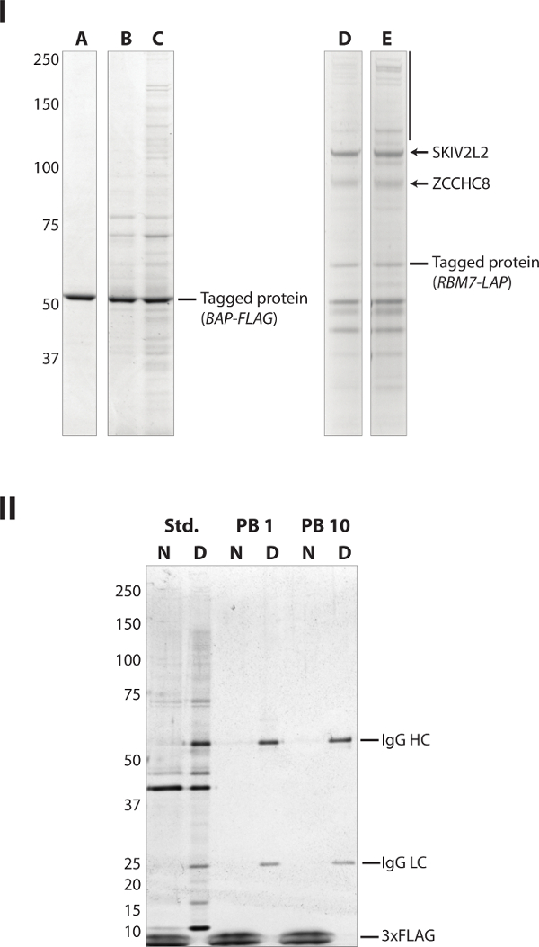
Figure 1: Select Representative Results. (I) This panel has been modified from reference19. Coomassie Blue stained SDS-polyacrylamide gel. (LEFT) Micron-scale magnetic beads show lower off-target binding than agarose-based media. (A) magnetic beads; (B) traditional agarose; (C) iron impregnated (magnetic) agarose; each coupled to anti-FLAG M2 antibodies and used to capture FLAG-tagged bacterial alkaline phosphatase (BAP; labeled) spiked into extracts produced from cryomilled HEK-293 cells. Agarose-based purifications showed higher levels of non-specific adsorption (unlabeled bands). (RIGHT) Extracts produced from cryomilled HeLa cells exhibit lower off-target adsorption upon antibody-coupled magnetic beads than those produced by sonication when used to purify an endogenous protein complex. LAP-tagged44 RBM7 was affinity captured using magnetic beads coupled to polyclonal anti-GFP antibodies to purify the NEXT complex19,37 (D) Extract produced from cryomilled powder; (E) extracts produced by sonication of intact cells. The vertical black bar highlights a region of the gel where high mass non-specific interactors are observed to be enriched in the sample prepared by sonication. The black arrows indicated the expected specific interactors (labeled). Bands observed ~ 50 kDa in lanes D and E are attributable to llama IgG heavy chains. (II) Silver stained SDS-polyacrylamide gel. (Std.) A mock affinity capture carried out on HEK-293T cell extracts in the standard way. After capture, elution of the bound material was achieved via (N) non-denaturing elution with 1 mg/ml 3xFLAG peptide or (D) denaturing elution in SDS-PAGE sample buffer; (PB 1) as Std. but the 3xFLAG peptide was included in the extraction solution at 1 µg/ml to block the α-FLAG paratopes on the affinity medium; (PB 10) as PB 1 but in the presence of 10 µg/ml 3xFLAG peptide. IgG-related bands and 3xFLAG peptide are labeled. Please click here to view a larger version of this figure.
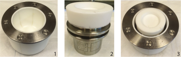
Figure 2: Sleeve-and-puck PTFE insulator. A 250 ml PTFE jar is shown (1). A 50 ml stainless steel milling jar (2) fits inside of the PTFE jar (3) and provides the opportunity to leave the milling jar installed within the mill between cycles. An upper PTFE insulator puck (2) is used between the clamp assembly and the jar lid. To cool the installed jar and insulator assembly (3), LN2 is added directly into the body of the insulator, surrounding the jar. The next cycle of milling can then be initiated. Please click here to view a larger version of this figure.
| Reagent | Suggested Concentration | Notes |
| Sodium chloride | 0.1-0.5 M | High concentrations (>300 mM) tend to improve extraction of total protein and keep background low, but may strip away some otherwise stable interactors. Concentrations below 150 mM are not typically effective at reducing non-specific background. |
| Ammonium acetate | 0.2-2 M | A salt, consisting of two buffers, that yields a neutral pH solution57. Higher concentrations stabilize some protein complexes. Acidic solutions can result from old, improperly stored crystalline stocks on account of ammonia loss. No additional pH buffer or salts are required in extractants containing ammonium acetate. May be combined with NaCl to modulate results obtained. |
| Tween 20 | 0.1% v/v | A non-ionic detergent58; typically combined with NaCl. |
| Triton X-100 | 0.5-1% v/v | A non-ionic detergent58; typically combined with either NaCl or NH4CH3CO2H |
| CHAPS | 5 mM | A zwitterionic detergent58; typically combined with NaCl. |
| Sarkosyl | 1 mM | An anionic detergent58 that reduces background and can strip off stable complex components, potentially revealing binary connectivity; typically combined with NaCl. |
| Tris-Cl | 20 mM | pKa of 8.8 at 4 °C, 8.1 at 25 °C. |
| (pH 8.0) | ||
| HEPES-Na | 20 mM | pKa of 7.8 at 4 °C, 7.5 at 25 °C. NaOH or KOH used for pH equilibration depending on the salt (e.g., NaCl, KCl, or CH3CO2K) used in the extractant. |
| (pH 7.4) |
Table 1: A few suggested reagents useful for purifying protein complexes. This table lists some reagents we have found useful for purifying protein complexes from human cell lines, with suggested working concentrations. A typical extractant formulation contains a pH buffer, a salt, and a detergent1,11,24,45-48. Best practice is to identify the least complex combination of reagents that yields the desired result.
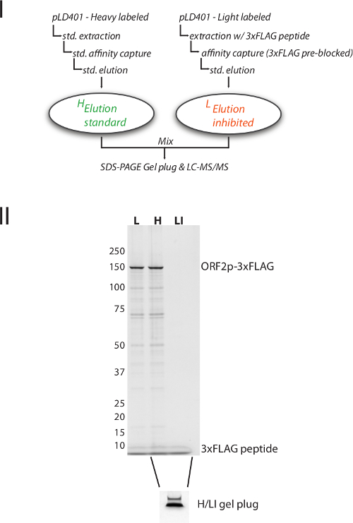
Figure S1: Mix After Purification (MAP-) SILAC analysis of false positives. I. Schematic Diagram: In this experiment 3xFLAG-tagged ORF2p protein complexes (expressed from pLD401) were purified from heavy- and light-labeled HEK-293T cells7, respectively. The light labeled cell extract was spiked with 3xFLAG peptide to prevent affinity capture of the 3xFLAG-tagged ORF2p by competitive inhibition; this is not expected to block the binding of proteins that may occur non-specifically with the magnetic medium or the non-paratopic structures of the antibody. After elution from the magnetic beads, the heavy and light samples were mixed post-purification and analyzed by quantitative MS (MAP-SILAC39) to determine the fraction of proteins associated with either sample; further methodological detail located in Table S1. II. Silver stained gel illustrating that the light- and heavy-labeled materials yield comparable results (compare L with H), and that the purification conducted in the presence of the 3xFLAG spike-in (LI) was competitively inhibited. Below, a Coomassie Blue G-250 stained gel plug consisting of the mixed H and LI fractions, prior to gel-based proteomic analysis, as described in Table S1. Please click here to download a larger version of this figure.
Table S1: MAP-SILAC. This sheet contains the data obtained upon execution of the MAP-SILAC experiment described in Figure S1. Please click here to download this table.
Discussion
These three protocols work in tandem to (1) prepare cells for solid-state breakage by cryomilling, (2) achieve breakage in a planetary ball mill, and (3) produce extracts from cell powder to affinity capture a protein of interest in complex with its physiological interactors. Many different cell lysis techniques exist, including mechanical/physical approaches employing crushing impact, shearing, and/or pressure, as well as chemical/enzymatic approaches, each with different pros and cons49,50. Each investigator is encouraged to explore methods most suitable for their analyses, keeping in mind that any chosen approach for cell breakage and protein extraction is likely to introduce biases51,52 necessitating empirical optimization (discussed below). Mechanical methods may produce high heat and/or shearing forces which can disrupt protein complexes. Cryomilling avoids heating effects by virtue of employing LN2 cooling of samples for the duration of the process. Planetary ball mills are understood to rely upon impact and frictional forces, including shear stress, as components of particle size reduction53,54. At the settings reported we have not observed degeneration of protein complexes. Indeed we have extracted and retrieved apparently intact ~50 MDa nuclear pore complexes11 and enzymatically active retrotransposons exhibiting higher specific activity than preparations employing 'gentle' detergent-based lysis7. Chemical/enzymatic methods of cell lysis suffer from the limitation that the contents of the cell are released into an in vitro milieu that supports the disruption of cell membranes and structural macromolecules but may not be suitable for maintenance of the integrity of the protein complex(es) of interest. Frequently, neither the constituents of the complexes formed with the protein of interest nor the conditions needed to stabilize the target complexes are known in advance. A major benefit of solid-state milling is that breakage and extraction are uncoupled, permitting source material to be prepared free of added liquid, stored (at -80 °C or below), amassed, and conveniently retrieved for on-demand experimentation; e.g., to explore optimized in vitro conditions for affinity capture. Protein interactions are most stable at high concentration55,56, therefore minimizing the volume of extractant can be advantageous for preserving physiological interactions. On the other hand, there are practical considerations — the protein complexes do need to be partitioned out of the cells and into a non-viscous aqueous phase, free of insoluble aggregates, in order to mingle the target complexes with the affinity medium. Moreover, some standardization and control over the in vitro environment (pH, salt concentration, etc.) is needed for systematization and reproducibility. We find that extracts produced in the dilution range of 1:4-1:9 (w:v) satisfy practical and theoretical concerns, yielding quality results. Additionally, the optimal proportion of cell extract to affinity medium needs to be determined. This is done empirically by titration of extracts with varying amounts of affinity medium and can have detectable effects on the signal-to-noise ratio of the experiment, further discussed below. An excellent depletion of the target protein is typically ≥70% of the soluble fraction of the target protein, but >90% is desirable and may be achievable with careful parameterization of extraction conditions. Many such practical considerations are covered in reference1. Paramagnetic beads are manipulated using neodymium magnets in a specialized microcentrifuge tube holder, although homemade alternatives are viable. When placed within the holder, beads collect at the side of the tube under the influence of the magnetic field. Solutions may then be removed without disturbing the beads.
One limitation of the presented cryomilling protocol, developed with the planetary ball mill used here (See Table of Materials), is that a minimum amount of material is required to effectively mill and recover cell powder using this device (>1 g). Such quantities are easily obtained with many microbes, cell lines, and model organisms, and can also often be achieved with tissues excised from laboratory animals. However, certain cell lines may be very difficult to grow in abundance and animal tissues may be scarce. Smaller quantities of material can be comparably milled using other devices utilizing smaller containers, although potentially at the sacrifice of the powder fineness achieved. In addition, the cost of mechanical milling devices may be prohibitively expensive for some laboratories. Cryomilling can be achieved using a number of alternative setups14-19, including, most affordably by hand using a pestle and mortar, although breakage efficiency drops considerably. Affinity capture protocols typically aim for a high efficiency of cell lysis to facilitate maximal protein extraction into solution and hence, maximal potential for capture of the target protein complexes. On the other hand, it has been demonstrated for yeast in vitro splicing extracts that maximal lysis may not equate with maximal in vitro biochemical activity57,58. We have not observed such problems in the systems we have tested thus far, and therefore do not purposefully limit our cell breakage when cryomilling. Nevertheless, this possibility should be borne in mind when optimizing for in vitro enzymatic assays. Although cryomilling is a highly effective method for breaking cells, a limited amount of sonication often benefits the production homogenous whole cell extracts from mammalian tissues because inhomogeneous aggregates can sometimes be observed by visual inspection: typically in extraction conditions using low to moderate salt (100-300 mM) and non-ionic detergent (0.1-1% v/v) concentrations. Because we observed that the presence of these aggregates can reduce the yield and/or quality of the subsequent affinity capture, we routinely implement sonication to disperse them (even when they are not observed with the naked eye). The aggregates are consistent with agglomerated membrane fragments, comparable to those previously reported in extracts from milled yeast cells58. Sonication is also used in some protocols to shear DNA and liberate chromatin fragments into solution, however the degree of sonication applied in this protocol does not appreciably fragment DNA. The limited availability (or the high cost) of an excellent affinity ligand or antibody against the particular protein of interest may be another impediment. A wide array of commercially available affinity reagents may be leveraged when the model organism is amenable to genomic tagging or transfection with ectopic expression vectors, permitting the expression of proteins of interest as affinity-tagged fusions. However, the production of custom antibodies has become increasingly feasible and both polyclonal and monoclonal antibodies can perform excellently in affinity capture applications. These many considerations are also covered in greater detail in reference1.
Examples of how the cell lysis method and choice of affinity medium can affect results are illustrated in Figure 1. Examples of effects exerted by different extractants can be seen in references1,11,24. Because these and other experimental parameters affect the quality of the affinity capture, making it difficult to discriminate FPs, repositories of "bead proteomes" and computational approaches to eliminate non-specific contaminants have been developed to assist in identifying FPs40-43. Nevertheless, such approaches only substitute for optimal sample preparation to a limited degree59. Observing best practices and empirically optimizing the affinity capture experiment will provide the highest quality samples for downstream analyses, further discussed below. An easily implemented practice that can improve sample purity is native elution. Native elution is most frequently used to obtain the affinity isolated protein complex, intact, for further experimentation; but as it frequently enhances sample purity, it may also be used for this reason alone. However, as demonstrated in Figure 1, panel II, the ability of native elution to improve sample purity may depend on an accurate titration of the quantity of affinity medium to the abundance of the target protein in the cell extract — when the medium is in excess, unoccupied paratopes may permit the off-target accumulation of FP proteins observable in the natively eluted fractions. Using the reagents and procedures described here, it has been our experience that the single greatest contributor of FPs to our experiments is the tagged protein itself once removed from the context of the living cell. (Fig. 1.II and Fig. S1). In such cases, parental cell line controls that yield irrelevant proteomes in the absence of the antigen are of no practical value; likewise for tag-only or spike-in controls that are devoid of anything but the target antigen. Therefore, whenever practical we implement I-DIRT7,38, which directly measures the accumulation of post-lysis protein interactions using quantitative MS. As mentioned in Step 3.2.7 of the protocol, procedures for native elution will vary with the details of the affinity system used. Native elution is most commonly achieved by competitive displacement of the tagged protein complex or proteolytic cleavage of the tag, releasing the complex from the affinity medium. Several affinity systems comprised of small epitope tags exist, for which the epitope itself is available as peptide useful for competitive elution of tagged protein complexes60. Likewise, several proteases are available to specifically cleave cognate sites strategically placed in affinity tagged fusion proteins61. Depending on the specifics of the chosen affinity systems, the appropriate elution scheme may be adopted.
Generally, the quality of the results obtained will be significantly impacted by the quality of the sample preparation. It is important to work carefully and precisely through each step of these protocols, and as rapidly as possible while maintaining care and precision. It is advisable to track the partitioning of the protein of interest through each step to understand the efficiency of each manipulation. For example: how abundant is the protein of interest in the cell or tissue in question? Perhaps the protein under study (and its complexes) will be challenging to characterize by mass spectrometry due to low abundance. If the purified complexes are detectable by general protein staining (approximately nanograms range), mass spectrometry is likely to succeed. If the captured protein of interest can only be detected by sensitive enhanced chemiluminescent western blotting (approximately picogram range), mass spectrometry is less likely to be effective. Even if the protein of interest is abundant in the cell, how abundant is it in the cell extract produced? Does the protein partition to the solution or is it in the pellet? If the latter, a new extraction solution can be devised that may improve recovery. Once the cell extract is combined with the affinity medium, how effectively is the protein captured? Does the protein remain bound through subsequent washes? What about other copurifying proteins? By saving an aliquot of each sample at appropriate steps of the protocol these questions can easily be answered, typically by western blotting against the protein of interest or general protein staining, but other assays may also be suitable. Optimizing each step will enhance the yield and purity of captured complexes, although there may be a trade-off to be made in maximizing one attribute or the other.
A typical application of affinity capture for many researchers is to identify candidate in vivo interactors for a small number of proteins of interest; these candidates are commonly subjected to validation by orthogonal approaches in vivo to demonstrate the biological significance of the physical interactors. Affinity capture is also employed by high-throughput studies to generate lists of copurifying proteins observed on a (nearly) proteome-wide basis, facilitating computational inferences concerning putative in vivo complexes. Numerous examples of such studies can be found in the literature. This approach foregoes optimization of the conditions of capture for any given target in favor of exploring many targets; as such, the inferred complexes themselves are rarely retrieved fully intact and highly pure in any given sample. Rather, the partial overlaps in compositions observed between numerous distinct affinity-captured samples are used as a basis of the inferences42. Both approaches contribute valuable data to our understanding of the interactome. Nevertheless, one major benefit of affinity capture is that it does frequently offer the opportunity to obtain the complexes intact and highly purified, provided that efforts are made to optimize the procedure. We believe that the future of the approach lies in increasing the ease and efficiency of optimizing the capture of endogenous protein complexes11, for more accurate assessment of the gamut of physiological interactors and more frequent use in further downstream biochemical, enzymological, and structural studies, e.g., references7,9,23.
Disclosures
The authors declare that they have no competing financial interests.
Acknowledgements
We thank Professor Brian T. Chait for his invaluable advice, support, and access to MS instrumentation, as well as Ms. Kelly R. Molloy for excellent technical support. We thank Ms. Kelly Bare for support with copy editing. This work was supported in part by NIH grants P41GM109824, P41GM103314, and P50GM107632.
Materials
| Name | Company | Catalog Number | Comments |
|---|---|---|---|
| Growth media & cell culturing implements | For producing frozen cells (I) | ||
| 500 cm2 culture dishes | Corning | 431110 | For producing frozen cells (I) |
| Large beaker (1-2 L) | For producing frozen cells (I) | ||
| Rectangular ice pan | Fisher Scientific | 13-900-747 | For producing frozen cells (I), & cryomilling (II) |
| Phosphate buffered saline (PBS) | For producing frozen cells (I) | ||
| Large cell scrapers | Fisher Scientific | 08-100-242 | For producing frozen cells (I) |
| Liquid nitrogen | For producing frozen cells (I), cryomilling (II), & affinity capture (III) | ||
| 50 ml conical tubes | Corning | 352098 | For producing frozen cells (I), & cryomilling (II) |
| 20 ml Luer lock syringe | For producing frozen cells (I) | ||
| Leur lock syringe caps | www.dymax.com | T11182 | For producing frozen cells (I) |
| Refrigerated centrifuge | For producing frozen cells (I) | ||
| 50 ml tube rack, 4 position | For producing frozen cells (I), cryomilling (II), & affinity capture (III) | ||
| Styrofoam box | For producing frozen cells (I), cryomilling (II), & affinity capture (III) | ||
| Planetary ball mill, PM 100 | Retsch | 20.540.0001 | For cryomilling (II) |
| Stainless steel milling jar, 50 ml | Retsch | 01.462.0149 | For cryomilling (II) |
| Stainless steel milling balls, 20 mm Ø | Retsch | 05.368.0062 | For cryomilling (II) |
| Lid gasket: PFA encapsulated spring energized seal, 2.62 mm 44.63 mm | www.marcorubber.com | T1044-2.62x44.63 | For cryomilling (II) |
| mini-LN2 decanter | homemade | see Ref 19 | For cryomilling (II) |
| 250 ml PTFE jar insulator (optional) | homemade | The Rockefeller University | For cryomilling (II); can be produced by The Rockefeller University High Energy Physics Instrument Shop, or design blueprints provided, upon request |
| PTFE puck insulator (optional) | homemade | The Rockefeller University | For cryomilling (II); can be produced by The Rockefeller University High Energy Physics Instrument Shop, or design blueprints provided, upon request. |
| 5 bar/500 kPa pressure relief valve (optional) | http://www.leeimh.com/ | 558 series check valve | For cryomilling (II) |
| 1.5-2 ml microfuge tubes | For affinity capture (III) | ||
| Extractant | For affinity capture (III) | ||
| Complete EDTA free protease inhibitor cocktail tablets | Roche Applied Science | 11873580001 | For affinity capture (III) |
| Vortex mixer | For affinity capture (III) | ||
| Ice bucket | For affinity capture (III) | ||
| Microtip sonicator, Q700 | Qsonica | Q700 | For affinity capture (III) |
| low intensity 1/16” microtip probe | Qsonica | 4717 | For affinity capture (III) |
| Bench-top refrigerated microcentrifuge | For affinity capture (III) | ||
| Dynabeads M-270 Epoxy | Thermo Fisher | 14302D | For affinity capture (III) |
| Antibody or affinity ligand coupled to Dynabeads | homemade | For affinity capture (III); see reference 19 | |
| Sample rotator | For affinity capture (III) | ||
| Neodymium magnet | Life Technologies | 123-21D | For affinity capture (III) |
| SDS-PAGE sample buffer (no reducing agent) | For affinity capture (III) | ||
| DTT | For affinity capture (III) | ||
| SDS-PAGE system | For affinity capture (III) | ||
| SDS-polyacrylamide gel | For affinity capture (III) | ||
| Protein Stain | For affinity capture (III) |
References
- LaCava, J., Molloy, K. R., Taylor, M. S., Domanski, M., Chait, B. T., Rout, M. P. Affinity proteomics to study endogenous protein complexes: Pointers, pitfalls, preferences and perspectives. BioTechniques. 58 (3), 103-119 (2015).
- Devos, D., Russell, R. B. A more complete, complexed and structured interactome. Curr Opin Struct Biol. 17 (3), 370-377 (2007).
- Aitchison, J. D., Rout, M. P. The interactome challenge. J Cell Biol. 211 (4), 729-732 (2015).
- Nie, Y., Viola, C., et al. Getting a grip on complexes. Curr. Genomics. 10 (8), 558-572 (2009).
- Bonetta, L. Protein-protein interactions: Interactome under construction. Nature. 468 (7325), (2010).
- Perkel, J. M. Protein-Protein Interaction Technologies Toward a Human Interactome. Science. 329 (5990), 463-465 (2010).
- Taylor, M. S., LaCava, J., et al. Affinity Proteomics Reveals Human Host Factors Implicated in Discrete Stages of LINE-1 Retrotransposition. Cell. 155 (5), 1034-1048 (2013).
- Liu, J. -J., Bratkowski, M. A., Liu, X., Niu, C. -Y., Ke, A., Wang, H. -W. Visualization of distinct substrate-recruitment pathways in the yeast exosome by EM. Nat Struct Mol Biol. 21 (1), 95-102 (2014).
- Shi, Y., Pellarin, R., et al. A strategy for dissecting the architectures of native macromolecular assemblies. Nat Methods. 12 (12), 1135-1138 (2015).
- Roque, A. C. A., Lowe, C. R. Affinity chromatography: History, perspectives, limitations and prospects. Methods in Molecular Biology. 421 (1), 1-21 (2007).
- Hakhverdyan, Z., Domanski, M., et al. Rapid, optimized interactomic screening. Nat Methods. 12 (6), 553-560 (2015).
- Huttlin, E. L., Ting, L., et al. The BioPlex Network: A Systematic Exploration of the Human Interactome. Cell. 162 (2), 425-440 (2015).
- Hein, M. Y., Hubner, N. C., et al. A Human Interactome in Three Quantitative Dimensions Organized by Stoichiometries and Abundances. Cell. 163 (3), 712-723 (2015).
- Smucker, R. A., Pfister, R. M. Liquid nitrogen cryo-impacting: a new concept for cell disruption. Appl Microbiol. 30 (3), 445-449 (1975).
- Schultz, M. C., Choe, S. Y., Reeder, R. H. Specific initiation by RNA polymerase I in a whole-cell extract from yeast. Proc Natl Acad Sci USA. 88 (3), 1004-1008 (1991).
- Schultz, M., Hockman, D., Harkness, T., Garinther, W., Altheim, B. Chromatin assembly in a yeast whole-cell extract. Proc Natl Acad Sci USA. 94 (17), 9034-9039 (1997).
- Stevens, S. W., Abelson, J. Yeast pre-mRNA splicing: Methods, mechanisms, and machinery. Method Enzymol. 351, 200-220 (2002).
- Oeffinger, M., Wei, K. E., et al. Comprehensive analysis of diverse ribonucleoprotein complexes. Nat Methods. 4 (11), 951-956 (2007).
- Domanski, M., Molloy, K., et al. Improved methodology for the affinity isolation of human protein complexes expressed at near endogenous levels. BioTechniques. 0 (0), 1-6 (2012).
- Sinclair, B. To bead or not to bead: Applications of magnetic bead technology. Scientist. , (1998).
- Cristea, I., Williams, R., Chait, B., Rout, M. Fluorescent proteins as proteomic probes. Mol Cell Proteomics. 4 (12), 1933-1941 (2005).
- Di Virgilio, M., Callen, E., et al. Rif1 prevents resection of DNA breaks and promotes immunoglobulin class switching. Science. 339 (6120), 711-715 (2013).
- Heider, M. R., Gu, M., et al. Subunit connectivity, assembly determinants and architecture of the yeast exocyst complex. Nat Struct Mol Biol. 23 (1), 59-66 (2016).
- LaCava, J., Fernandez-Martinez, J., Hakhverdyan, Z., Rout, M. P. Protein Complex Purification by Affinity Capture. Budding Yeast: A Laboratory Manual. 2016 (21), 383-397 (2016).
- Freshney, R. I. Culture of Animal Cells. , John Wiley & Sons. (2011).
- Uphoff, C. C., Drexler, H. G. Basic Cell Culture Protocols. 946, Humana Press. (2013).
- Taylor, M. S., LaCava, J., et al. Characterization of L1-Ribonucleoprotein Particles. Transposons and Retrotransposons. 1400 (Chapter 20), 311-338 (2016).
- Obado, S. O., Field, M. C., Chait, B. T., Rout, M. P. High-Efficiency Isolation of Nuclear Envelope Protein Complexes from Trypanosomes. The Nuclear Envelope. 1411, 67-80 (2016).
- Ball Mills - Guidelines for sample amount and ball charge. , 1–3at Retsch GmbH. http://www.retsch.com/products/milling/ball-mills/planetary-ball-mill-pm-100/information-downloads/ (2014).
- Cristea, I. M., Chait, B. T. Conjugation of magnetic beads for immunopurification of protein complexes. Cold Spring Harbor Protocols. 2011 (5), (2011).
- Rosenberg, I. M. Electrophoretic Techniques. Protein Analysis and Purification. (4), 63-117 (2005).
- Ornstein, L. Disc Electrophoresis. I. Background and Theory. Ann N Y Acad Sci. 121 (A2), 321-349 (1964).
- Davis, B. J. Disc Electrophoresis. II. Method and Application to Human Serum Proteins. Ann N Y Acad Sci. 121, 404-427 (1964).
- Laemmli, U. K. Cleavage of structural proteins during the assembly of the head of bacteriophage T4. Nature. 227 (5259), 680-685 (1970).
- Shevchenko, A., Tomas, H., Havlis, J., Olsen, J. V., Mann, M. In-gel digestion for mass spectrometric characterization of proteins and proteomes. Nature protocols. 1 (6), 2856-2860 (2006).
- DeGrasse, J. A., Kalkum, M., Krutchinsky, A. N., Padovan, J. C., Zhang, W. MALDI Sample Preparation. , Available from: rockefeller.edu at http://prowl.rockefeller.edu/protocols/in-gel-digestion.html (2006).
- Lubas, M., Christensen, M. S., et al. Interaction profiling identifies the human nuclear exosome targeting complex. Mol Cell. 43 (4), 624-637 (2011).
- Tackett, A. J., DeGrasse, J. A., Sekedat, M. D., Oeffinger, M., Rout, M. P., Chait, B. T. I-DIRT, a general method for distinguishing between specific and nonspecific protein interactions. J Proteome Res. 4 (5), 1752-1756 (2005).
- Wang, X., Huang, L. Identifying dynamic interactors of protein complexes by quantitative mass spectrometry. Mol Cell Proteomics. 7 (1), 46-57 (2008).
- Trinkle-Mulcahy, L., Boulon, S., et al. Identifying specific protein interaction partners using quantitative mass spectrometry and bead proteomes. J Cell Biol. 183 (2), 223-239 (2008).
- Boulon, S., Ahmad, Y., et al. Establishment of a protein frequency library and its application in the reliable identification of specific protein interaction partners. Mol Cell Proteomics. 9 (5), 861-879 (2010).
- Armean, I. M., Lilley, K. S., Trotter, M. W. B. Popular computational methods to assess multiprotein complexes derived from label-free affinity purification and mass spectrometry (AP-MS) experiments. Mol Cell Proteomics. 12 (1), 1-13 (2013).
- Mellacheruvu, D., Wright, Z., et al. The CRAPome: a contaminant repository for affinity purification-mass spectrometry data. Nat Methods. , (2013).
- Cheeseman, I. M., Desai, A. A combined approach for the localization and tandem affinity purification of protein complexes from metazoans. Sci. STKE. 2005 (266), 1(2005).
- Deppert, W. R., Lukaĉin, R. Buffers and Additives. Journal of Chromatography Library. 61 (C), 839(1999).
- Ugwu, S. O., Apte, S. P. The Effect of Buffers on Protein Conformational Stability. Pharmaceutical Technology. 28 (3), 86-108 (2004).
- Linke, D. Detergents: an overview. Methods in Enzymology. 463 (34), 603-617 (2009).
- Zhang, J. Protein-Protein Interactions in Salt Solutions. Protein-Protein Interactions Computational and Experimental Tools. (18), 359-376 (2012).
- Goldberg, S. Mechanical/physical methods of cell disruption and tissue homogenization. Methods in Mole Bio. 424 (Chapter 1), 3-22 (2008).
- Grabski, A. C. Advances in preparation of biological extracts for protein purification. Methods in Enzymology. 463 (C), 285-303 (2009).
- Dhabaria, A., Cifani, P., Reed, C., Steen, H., Kentsis, A. A High-Efficiency Cellular Extraction System for Biological Proteomics. J Proteome Res. 14 (8), 3403-3408 (2015).
- Glatter, T., Ahrné, E., Schmidt, A. Comparison of Different Sample Preparation Protocols Reveals Lysis Buffer-Specific Extraction Biases in Gram-Negative Bacteria and Human Cells. J Proteome Res. 15 (2), 679(2016).
- Zhao, Q. Q., Yamada, S., Jimbo, G. The Mechanism and Grinding Limit of Planetary Ball Milling. KONA. 7, 29-36 (1989).
- Sheng-Yong, L., Qiong-Jing, M., Zheng, P., Xiao-Dong, L., Jian-Hua, Y. Simulation of ball motion and energy transfer in a planetary ball mill. Chinese Phys. B. 21 (7), 078201(2012).
- Fulton, A. B. How crowded is the cytoplasm? Cell. 30 (2), 345(1982).
- Ellis, R. J. Macromolecular crowding: obvious but underappreciated. Trends Biochem Sci. 26 (10), 597-604 (2001).
- Kalkum, M. Using the Retsch MM301 Ball Mill for Cryogenic Disruption of Yeast Cells. 2, City of Hope. (2003).
- Staley, J. Making Whole Cell Extract of Saccharomyces cerevisiae cells using the Retsch MM301 Ball Mill. , Retsch GmbH. (2005).
- Bell, A. W., Nilsson, T., Kearney, R. E., Bergeron, J. J. M. The protein microscope: Incorporating mass spectrometry into cell biology. Nat Methods. 4 (10), 783-784 (2007).
- Zhao, X., Li, G., Liang, S. Several affinity tags commonly used in chromatographic purification. J Anal Methods Chem. 2013 (1), 581093-581098 (2013).
- Waugh, D. S. An overview of enzymatic reagents for the removal of affinity tags. Protein Expr Purif. 80 (2), 283-293 (2011).
Reprints and Permissions
Erratum
Formal Correction: Erratum: Protein Complex Affinity Capture from Cryomilled Mammalian Cells
Posted by JoVE Editors on 1/20/2017. Citeable Link.
A correction was made to: Protein Complex Affinity Capture from Cryomilled Mammalian Cells. The References section has been updated from:
- Cristea, I. M., & Chait, B. T. Conjugation of magnetic beads for immunopurification of protein complexes. Cold Spring Harbor Protocols. 2011 (5) (2011).
- Rosenberg, I. M. Electrophoretic Techniques. Protein Analysis and Purification. (4), 63–117 (2005).
- Ornstein, L. Disc Electrophoresis. I. Background and Theory. Ann N Y Acad Sci. 121 (A2), 321–349 (1964).
- Davis, B. J. Disc Electrophoresis. II. Method and Application to Human Serum Proteins. Ann N Y Acad Sci. 121, 404–427 (1964).
- Laemmli, U. K. Cleavage of structural proteins during the assembly of the head of bacteriophage T4. Nature. 227 (5259), 680–685 (1970).
- Shevchenko, A., Tomas, H., Havlis, J., Olsen, J. V., & Mann, M. In-gel digestion for mass spectrometric characterization of proteins and proteomes. Nature protocols. 1 (6), 2856–2860 (2006).
- DeGrasse, J. A., Kalkum, M., Krutchinsky, A.N., Padovan, J. C., & Zhang, W. MALDI Sample Preparation. rockefeller.edu at <http://prowl.rockefeller.edu/protocols/in-gel-digestion.html> (2006).
- Lubas, M., Christensen, M. S., et al. Interaction profiling identifies the human nuclear exosome targeting complex. Mol Cell. 43 (4), 624–637 (2011).
- Tackett, A. J., DeGrasse, J. A., Sekedat, M. D., Oeffinger, M., Rout, M. P., & Chait, B. T. I-DIRT, a general method for distinguishing between specific and nonspecific protein interactions. J Proteome Res. 4 (5), 1752–1756 (2005).
- Wang, X., & Huang, L. Identifying dynamic interactors of protein complexes by quantitative mass spectrometry. Mol Cell Proteomics. 7 (1), 46–57 (2008).
- Trinkle-Mulcahy, L., Boulon, S., et al. Identifying specific protein interaction partners using quantitative mass spectrometry and bead proteomes. J Cell Biol. 183 (2), 223–239 (2008).
- Boulon, S., Ahmad, Y., et al. Establishment of a protein frequency library and its application in the reliable identification of specific protein interaction partners. Mol Cell Proteomics. 9 (5), 861–879 (2010).
- Armean, I. M., Lilley, K. S., & Trotter, M. W. B. Popular computational methods to assess multiprotein complexes derived from label-free affinity purification and mass spectrometry (AP-MS) experiments. Mol Cell Proteomics. 12 (1), 1–13 (2013).
- Mellacheruvu, D., Wright, Z., et al. The CRAPome: a contaminant repository for affinity purification-mass spectrometry data. Nat Methods. (2013).
- Cheeseman, I. M., & Desai, A. A combined approach for the localization and tandem affinity purification of protein complexes from metazoans. Sci. STKE 2005 (266), pl1 (2005).
- Goldberg, S. Mechanical/physical methods of cell disruption and tissue homogenization. Methods in Molecular Biology. 424 (Chapter 1), 3–22 (2008).
- Grabski, A. C. Advances in preparation of biological extracts for protein purification. Methods in Enzymology. 463 (C), 285–303 (2009).
- Dhabaria, A., Cifani, P., Reed, C., Steen, H., & Kentsis, A. A High-Efficiency Cellular Extraction System for Biological Proteomics. J Proteome Res. 14 (8), 3403–3408 (2015).
- Glatter, T., Ahrné, E., & Schmidt, A. Comparison of Different Sample Preparation Protocols Reveals Lysis Buffer-Specific Extraction Biases in Gram-Negative Bacteria and Human Cells. J Proteome Res. 15 (2), 679 (2016).
- Zhao, Q.-Q., Yamada, S., & Jimbo, G. The Mechanism and Grinding Limit of Planetary Ball Milling. KONA. 7, 29–36 (1989).
- Sheng-Yong, L., Qiong-Jing, M., Zheng, P., Xiao-Dong, L., & Jian-Hua, Y. Simulation of ball motion and energy transfer in a planetary ball mill. Chinese Phys. B 21 (7), 078201 (2012).
- Fulton, A. B. How crowded is the cytoplasm? Cell. 30 (2), 345–347 (1982).
- Ellis, R. J. Macromolecular crowding: obvious but underappreciated. Trends Biochem Sci. 26 (10), 597–604 (2001).
- Kalkum, M. Using the Retsch MM301 Ball Mill for Cryogenic Disruption of Yeast Cells. 2 City of Hope, (2003).
- Staley, J. Making Whole Cell Extract of Saccharomyces cerevisiae cells using the Retsch MM301 Ball Mill. Retsch GmbH: (2005).
- Bell, A. W., Nilsson, T., Kearney, R. E., & Bergeron, J. J. M. The protein microscope: Incorporating mass spectrometry into cell biology. Nat Methods. 4 (10), 783–784 (2007).
- Zhao, X., Li, G., & Liang, S. Several affinity tags commonly used in chromatographic purification. J Anal Methods Chem. 2013 (1), 581093–8 (2013).
- Waugh, D. S. An overview of enzymatic reagents for the removal of affinity tags. 80 (2), 283–293 (2011).
- Williams, R. J., & Lyman, C. M. A neutral buffered standard for hydrogen ion work and accurate titrations which can be prepared in one minute. J Am Chem Soc. 54 (5), 1911–1912 (1932).
- Johnson, M. Detergents: Triton X-100, Tween-20, and More. labome.com. 3 (163) (2013).
- Deppert, W. R., & Lukaĉin, R. Buffers and Additives. Journal of Chromatography Library. 61 (C), 839–862 (1999).
- Ugwu, S. O., & Apte, S. P. The Effect of Buffers on Protein Conformational Stability. Pharmaceutical Technology. 28 (3), 86–108 (2004).
- Linke, D. Detergents: an overview. Methods in Enzymology. 463 (34), 603–617 (2009).
- Zhang, J. Protein-Protein Interactions in Salt Solutions. Protein-Protein Interactions Computational and Experimental Tools. (18), 359–376 (2012).
to:
- Ball Mills - Guidelines for sample amount and ball charge. 1–3at <http://www.retsch.com/products/milling/ball-mills/planetary-ball-mill-pm-100/information-downloads/> Retsch GmbH (2014).
- Cristea, I. M., & Chait, B. T. Conjugation of magnetic beads for immunopurification of protein complexes. Cold Spring Harbor Protocols. 2011 (5) (2011).
- Rosenberg, I. M. Electrophoretic Techniques. Protein Analysis and Purification. (4), 63–117 (2005).
- Ornstein, L. Disc Electrophoresis. I. Background and Theory. Ann N Y Acad Sci. 121 (A2), 321–349 (1964).
- Davis, B. J. Disc Electrophoresis. II. Method and Application to Human Serum Proteins. Ann N Y Acad Sci. 121, 404–427 (1964).
- Laemmli, U. K. Cleavage of structural proteins during the assembly of the head of bacteriophage T4. Nature. 227 (5259), 680–685 (1970).
- Shevchenko, A., Tomas, H., Havlis, J., Olsen, J. V., & Mann, M. In-gel digestion for mass spectrometric characterization of proteins and proteomes. Nature protocols. 1 (6), 2856–2860 (2006).
- DeGrasse, J. A., Kalkum, M., Krutchinsky, A.N., Padovan, J. C., & Zhang, W. MALDI Sample Preparation. rockefeller.edu at <http://prowl.rockefeller.edu/protocols/in-gel-digestion.html> (2006).
- Lubas, M., Christensen, M. S., et al. Interaction profiling identifies the human nuclear exosome targeting complex. Mol Cell. 43 (4), 624–637 (2011).
- Tackett, A. J., DeGrasse, J. A., Sekedat, M. D., Oeffinger, M., Rout, M. P., & Chait, B. T. I-DIRT, a general method for distinguishing between specific and nonspecific protein interactions. J Proteome Res. 4 (5), 1752–1756 (2005).
- Wang, X., & Huang, L. Identifying dynamic interactors of protein complexes by quantitative mass spectrometry. Mol Cell Proteomics. 7 (1), 46–57 (2008).
- Trinkle-Mulcahy, L., Boulon, S., et al. Identifying specific protein interaction partners using quantitative mass spectrometry and bead proteomes. J Cell Biol. 183 (2), 223–239 (2008).
- Boulon, S., Ahmad, Y., et al. Establishment of a protein frequency library and its application in the reliable identification of specific protein interaction partners. Mol Cell Proteomics. 9 (5), 861–879 (2010).
- Armean, I. M., Lilley, K. S., & Trotter, M. W. B. Popular computational methods to assess multiprotein complexes derived from label-free affinity purification and mass spectrometry (AP-MS) experiments. Mol Cell Proteomics. 12 (1), 1–13 (2013).
- Mellacheruvu, D., Wright, Z., et al. The CRAPome: a contaminant repository for affinity purification-mass spectrometry data. Nat Methods. (2013).
- Cheeseman, I. M., & Desai, A. A combined approach for the localization and tandem affinity purification of protein complexes from metazoans. Sci. STKE 2005 (266), pl1 (2005).
- Deppert, W. R., & Lukaĉin, R. Buffers and Additives. Journal of Chromatography Library. 61 (C), 839–862 (1999).
- Ugwu, S. O., & Apte, S. P. The Effect of Buffers on Protein Conformational Stability. Pharmaceutical Technology. 28 (3), 86–108 (2004).
- Linke, D. Detergents: an overview. Methods in Enzymology. 463 (34), 603–617 (2009).
- Zhang, J. Protein-Protein Interactions in Salt Solutions. Protein-Protein Interactions Computational and Experimental Tools. (18), 359–376 (2012).
- Goldberg, S. Mechanical/physical methods of cell disruption and tissue homogenization. Methods in Molecular Biology. 424 (Chapter 1), 3–22 (2008).
- Grabski, A. C. Advances in preparation of biological extracts for protein purification. Methods in Enzymology. 463 (C), 285–303 (2009).
- Dhabaria, A., Cifani, P., Reed, C., Steen, H., & Kentsis, A. A High-Efficiency Cellular Extraction System for Biological Proteomics. J Proteome Res. 14 (8), 3403–3408 (2015).
- Glatter, T., Ahrné, E., & Schmidt, A. Comparison of Different Sample Preparation Protocols Reveals Lysis Buffer-Specific Extraction Biases in Gram-Negative Bacteria and Human Cells. J Proteome Res. 15 (2), 679 (2016).
- Zhao, Q.-Q., Yamada, S., & Jimbo, G. The Mechanism and Grinding Limit of Planetary Ball Milling. KONA. 7, 29–36 (1989).
- Sheng-Yong, L., Qiong-Jing, M., Zheng, P., Xiao-Dong, L., & Jian-Hua, Y. Simulation of ball motion and energy transfer in a planetary ball mill. Chinese Phys. B 21 (7), 078201 (2012).
- Fulton, A. B. How crowded is the cytoplasm? Cell. 30 (2), 345–347 (1982).
- Ellis, R. J. Macromolecular crowding: obvious but underappreciated. Trends Biochem Sci. 26 (10), 597–604 (2001).
- Kalkum, M. Using the Retsch MM301 Ball Mill for Cryogenic Disruption of Yeast Cells. 2 City of Hope, (2003).
- Staley, J. Making Whole Cell Extract of Saccharomyces cerevisiae cells using the Retsch MM301 Ball Mill. Retsch GmbH: (2005).
- Bell, A. W., Nilsson, T., Kearney, R. E., & Bergeron, J. J. M. The protein microscope: Incorporating mass spectrometry into cell biology. Nat Methods. 4 (10), 783–784 (2007).
- Zhao, X., Li, G., & Liang, S. Several affinity tags commonly used in chromatographic purification. J Anal Methods Chem. 2013 (1), 581093–8 (2013).
- Waugh, D. S. An overview of enzymatic reagents for the removal of affinity tags. 80 (2), 283–293 (2011).
