Here, we describe a technique to isolate the side population cells from a zebrafish model of myc-induced T-cell acute lymphoblastic leukemia (T-ALL). This side population assay is highly sensitive and is described for zebrafish T-ALL, but it may be applicable to other malignant and non-malignant zebrafish cell types.
Method Article
Isolation of the Side Population in Myc-induced T-cell Acute Lymphoblastic Leukemia in Zebrafish
In This Article
Summary
Abstract
Heterogeneous cell populations, from either healthy or malignant tissues, may contain a population of cells characterized by a differential ability to efflux the DNA-binding dye Hoechst 33342. This "side population" of cells can be identified using flow cytometric methods after the Hoechst 33342 dye is excited by an ultraviolet (UV) laser. The side population of many cell types contains stem- or progenitor-like cells. However, not all cell types have an identifiable side population. Danio rerio, zebrafish, have a robust in vivo model of T-cell acute lymphoblastic leukemia (T-ALL), but whether these zebrafish T-ALLs have a side population is unknown. The method described here outlines how to isolate the side population cells in zebrafish T-ALL. To begin, the T-ALL in zebrafish is generated via the microinjection of tol2 plasmids into one-cell stage embryos. Once the tumors have grown to a stage at which they expand into more than half of the animal's body, the T-ALL cells can be harvested. The cells are then stained with Hoechst 33342 and examined by flow cytometry for side population cells. This method has broad applications in zebrafish T-ALL research. While there are no known cell surface markers in zebrafish that confirm whether these side population cells are cancer stem cell-like, in vivo functional transplantation assays are possible. Furthermore, single-cell transcriptomics could be applied to identify the genetic features of these side population cells.
Introduction
The side population assay capitalizes on the enhanced ability of certain cells within a tissue to efflux the DNA binding dye Hoechst 33342 due to high levels of the ATP binding cassette (ABC) transporter proteins on the cell membrane. The cells that efflux the Hoechst 33342 dye can be identified using dual wavelength flow cytometric analysis after the dye is excited by a UV laser. This assay was first used to identify murine hematopoietic stem cells (HSCs)1, but it has since been used to identify stem/progenitor cell populations in many tissues and cancers (reviewed in reference 2). However, not all populations of cells have a side population, and not all side populations are enriched for stem/progenitor cells.
The zebrafish is a powerful vertebrate genetic model system for studying human cancer3,4, with a number of advantages over traditional murine models of cancer. Zebrafish embryos are externally fertilized and are optically clear, facilitating transgenesis and the in vivo observation of pathologic processes, including cancer initiation and progression. To date, the side population assay to detect potential stem or progenitor cells has only been applied to the kidney marrow in zebrafish to identify HSCs, and not to any zebrafish cancer models5,6.
The zebrafish model of T-cell acute lymphoblastic leukemia (T-ALL) is morphologically and genetically similar to human T-ALL7,8,11. T-ALL is an aggressive malignancy that, in humans, accounts for 10-15% of pediatric and 25% of adult ALL cases9. While the treatment of T-ALL has improved, relapse is still common and is associated with a poor prognosis. T-ALL tumors are heterogeneous and contain many different tumor cell subpopulations, including leukemia initiating cells (LICs). LICs are defined by their ability to regrow the entire tumor from a single cell, and the frequency of LICs within a tumor cell population can be calculated by transplanting varying cell doses into recipients via a limiting dilution transplantation assay (LDA). While LDA experiments have been performed in zebrafish to calculate the frequency of LICs8,10,11, this determination is made in hindsight and does not allow for the prospective isolation of LICs. Therefore, a method to prospectively isolate a population enriched for cancer stem cell activity is lacking. Identifying and isolating side population cells from zebrafish T-ALLs is the first step towards addressing this deficiency.
The protocol presented here describes how to efficiently generate T-ALL tumors in zebrafish utilizing tol2-mediated transgenesis, harvest the T-ALL tumors cells, and stain them with Hoechst 33342 to identify the side population of cells. Future in vivo experiments with zebrafish T-ALL could include whether the side population cells are enriched for LICs or have other stem- or progenitor-like properties.
Protocol
All procedures with zebrafish have been approved by the Institutional Animal Care and Use Committee (IACUC) at the University of Chicago. The University of Chicago is accredited by the Association for Assessment and Accreditation of Laboratory Animal Care (AAALAC).
1. Generating and Isolating Fluorescently Labeled T-ALL Cells in Zebrafish
- Microinjection into syngeneic zebrafish embryos
- Using commercially available kits, isolate circular rag2:c-myc and rag2:green fluorescent protein (rag2:GFP) plasmid DNA containing inverted terminal repeats recognized by tol2 transposase (see the Table of Materials for source of plasmid DNA)12 and elute the DNA in sterile water.
NOTE: GFP is used in this study, but any fluorescent protein will work. - Prepare pCS2-transposase (tol2) RNA
- Isolate tol2 plasmid DNA using commercially available kits (see the Table of Materials for the source of plasmid DNA)12 and elute the DNA in sterile water.
- Incubate 1 µg of tol2 DNA at 37 °C for 1 h with 5 units of NotI restriction enzyme in the appropriate buffer to linearize the plasmid. Heat-inactivate the NotI at 65 °C for 20 min.
- Transcribe the tol2 RNA with SP6 RNA polymerase from the linearized DNA using a commercially available RNA transcription kit13. Store the RNA in single-use aliquots at -80 °C.
- Prepare the injection mix on ice by adding 250 ng of rag2:c-myc plasmid DNA, 250 ng of rag2:GFP plasmid DNA, 150 ng of tol2 RNA, and 0.5 µL of phenol red. Adjust the volume to 10 µL with nuclease-free water.
- Inject ~1 nL of injection mix into the cell of a one-cell stage syngeneic zebrafish embryo using a microinjector and needle pulled from a glass capillary tube, following previously established methods8,14.
NOTE: Syngeneic zebrafish generated by parthenogenesis15 are used here to eliminate genetic heterogeneity and immune rejection after transplantation. However, wildtype zebrafish or any desired strain can also be used. - Remove dead, underdeveloped, or malformed embryos at 24 h and 48 h post-injection16.
- Place the injected fish on the fish system at 6 days post-injection and raise them according to standard protocol6.
- Using commercially available kits, isolate circular rag2:c-myc and rag2:green fluorescent protein (rag2:GFP) plasmid DNA containing inverted terminal repeats recognized by tol2 transposase (see the Table of Materials for source of plasmid DNA)12 and elute the DNA in sterile water.
- Screening injected zebrafish and monitoring tumor growth
- At 21 days post-injection, place a single injected larva in a droplet of embryo medium on a Petri dish and screen for GFP fluorescence using an epifluorescent microscope (excitation bandpass: 470/40, emission bandpass: 525/50)8.
NOTE: Zebrafish larvae that have integrated both rag2:c-myc and rag2:GFP will have a green fluorescent thymus (positive fish) and may already exhibit the expansion of malignant green fluorescent cells beyond the thymus. All fish with a GFP+ thymus will go on to develop a GFP+ T-ALL. The larvae that do not demonstrate integration of the plasmids will not have any green fluorescent cells (negative fish). - Separate the negative fish (based on a lack of fluorescence) into a different tank and monitor the fish again at 28 and 35 days post-injection. At 28 and 35 days post-injection, anesthetize the fish with 0.02% buffered tricaine (MS-222) in fish system water prior to screening.
- Place each zebrafish larva that is positive for a fluorescently labeled thymus or a fluorescently labeled tumor in an individual tank for monitoring tumor growth three times a week.
- At 21 days post-injection, place a single injected larva in a droplet of embryo medium on a Petri dish and screen for GFP fluorescence using an epifluorescent microscope (excitation bandpass: 470/40, emission bandpass: 525/50)8.
- Collection of fluorescently labeled T-ALL cells from the primary tumor fish
- Prepare at least 20 mL of 10% heat-inactivated fetal bovine serum in 0.9x phosphate buffered saline (FBS/PBS).
- Add 1 mL of FBS/PBS to one vial of heparin sodium salt (300 units).
- Select fish bearing tumors in more than 50% of the body, or those fish having difficulty eating or swimming. Sacrifice the fish by overdosing them with tricaine or by ice water immersion. Confirm death by lack a of gill movement and heartbeat.
- Use a razor blade to remove the head of the fish.
- Wash the cavity of the fish with the heparin solution to remove excess red blood cells.
- Using a pipette, inject 100 µL of heparin solution (prepared in step 1.3.2) into the body cavity of the fish. Remove all liquid that spills out (it will contain blood cells) and pipette it into a 1.5 mL microcentrifuge tube.
- Use a fresh pipette tip to wash the body again. Perform at least three washes, or until the liquid that spills out appears free of blood.
- Discard all liquid from the washes in steps 1.3.5.1 and 1.3.5.2, as it will not be used in further analyses.
- Collect the leukemia cells.
- Inject 100 µL of FBS/PBS into the body cavity of the fish.
- Apply gentle pressure to the outside of the body of the fish with the pipette tip to squeeze/push out the tumor cells.
NOTE: If this step is performed using a dissecting microscope, the tumor cells spilling out of the body cavity will be evident. - Collect the liquid and cells from each wash into a 1.5 mL microcentrifuge tube.
- Repeat steps 1.3.6.1-1.3.6.3 until most of the tumor cells are collected (usually 5-10 times). Collect all tumor cells into the same 1.5-mL microcentrifuge tube. Keep the cells at room temperature.
- Mix the collected tumor cells from step 1.3.6 by gently pipetting them to dissociate the clumps of cells. Filter the cell suspension through a 40-µm mesh filter. Wash the filter 1-2 times with 100 µL of FBS/PBS. Keep the cells at room temperature.
- Mix 2 µL of cell suspension with 18 µL of trypan blue (0.4%, diluted 1:10 and filtered) in a new tube. Load 10 µL on a hemocytometer and count the number of live (non-blue) fluorescent cells to calculate the number of isolated tumor cells.
NOTE: Non-blue GFP-negative cells are likely normal, non-malignant T-cells and should not be counted. At least 2 x 10⁶ cells are required to stain the cells with Hoechst 33342.
2. Staining the Fluorescently Labeled T-ALL Cells with Hoechst 33342 to Identify the Side Population
- Dilute Hoechst 33342 1:10 with nuclease-free H2O to make the 1 mg/mL working concentration. Store the dye at 4 °C and in the dark.
- Prepare a 100-mM solution of verapamil by dissolving 49.1 mg in 1 mL of dimethyl sulfoxide (DMSO).
NOTE: Verapamil is an inhibitor of ABC transporters and is an important control for side population experiments. Adding verapamil to samples will inhibit the ability of the ABC transporters to efflux the Hoechst 33342 dye. Therefore, a true side population will disappear when verapamil is added to the sample. - Set a water bath to 28 °C.
- Prepare samples and controls in the dark by adding cells and solutions to fluorescence-activated cell sorter (FACS) tubes.
- Prepare the samples by adding 106 cells, 15 µL of 1 mg/mL Hoechst 33342, and 2.5 µL of DMSO to a FACS tube. Adjust the volume to 1 mL with FBS/PBS.
- Prepare the controls by adding 106 cells, 15 µL of 1 mg/mL Hoechst 33342, and 2.5 µL of 100 mM verapamil to a FACS tube. Adjust the volume to 1 mL with FBS/PBS.
NOTE: A minimum of 106 cells/sample is recommended, but staining more cells is preferred, especially if the side population cells will be sorted for further analysis. Do not exceed 106 cells/mL, and maintain the Hoechst 33342 concentration at 15 µg/mL. If 10 x 106 cells are stained, the total reaction volume is 10 mL.
- Incubate in a 28 °C water bath in the dark for 120 min.
- After the incubation, place the cells immediately on ice and keep them in the dark.
- Pellet the cells at 4 °C for 6 min at 300 x g. Remove the supernatant. Wash with 1 mL of FBS/PBS.
- Pellet the cells at 4 °C for 6 min at 300 x g. Remove the supernatant. Resuspend the cells in 1 mL of FBS/PBS.
- Keep at 4 °C until cell sorting.
- 15 min prior to cell sorting, add 2 µL of propidium iodide (PI) stock (1 mg/mL) to each 1 mL of FBS/PBS (final concentration: 2 µg/mL). Mix well.
3. Use FACS to Find the Side Population within the Fluorescently Labeled T-ALL Cell Population
- Analyze the stained zebrafish T-ALL cell suspensions on a cell sorter equipped with a UV laser for Hoechst 33342 dye excitation and a 488-nm laser for PI excitation and for the detection of the GFP+ T-ALL cells.
- Determine a gate for the live, GFP+ T-ALL cells.
- Use forward scatter (FSC) and side scatter (SSC) area to gate out debris.
NOTE: FSC is used to distinguish cell size and SSC is used to distinguish the granularity of the cell. - Use FSC and SSC width to discriminate doublets.
NOTE: There are three components identified by the flow cytometer detectors: area, height, and width. Area is typically used to measure the fluorescence of the cell. Width measures the time of flight and is twice as wide if two cells are clumped together. - For live tumor cells, draw a gate around the cells positive for GFP and negative for PI using the appropriate detectors (e.g., FITC for GFP and PerCP Cy5-5 for PI).
- Use forward scatter (FSC) and side scatter (SSC) area to gate out debris.
- In the live cell gate, measure the Hoechst 33342 fluorescence through the Hoechst Blue and Hoechst Red bandpass filters (450/50 and 670/30, respectively).
NOTE: The side population, if present, will be evident after this is set up. Importantly, the side population must disappear in the verapamil control in order for it to be considered a true side population. - If sorting the side population cells for further experiments, use the large nozzle tip (100 µm) and collect the cells into a FACS tube containing 3 mL of 100% FBS. Use a larger nozzle tip (100 µm) instead of the smaller tip (70 µm) to improve cell viability after sorting.
Results
To efficiently generate fluorescent myc-induced T-ALL tumors in zebrafish, circular DNA constructs flanked by tol2 transposase sites can be co-injected with tol2 RNA. Previous studies from the injection of linearized DNA into zebrafish embryos report a 5% transgenesis rate8. With the protocol adjusted to include tol2-mediated transgenesis, transgenesis rates ranging from 10-44% can be observed (Table 1).
In the representative example, a fish with a GFP+ T-ALL was stained with Hoechst 33342 to determine the percentage of cells in the side population. Figure 1 shows the gating scheme used to determine the percentage of side population cells. First, using FSC and SSC, the T-ALL cells are isolated by excluding debris (Figure 1A) and subsequently selecting against doublets (Figure 1B). Next, all live cells are isolated by gating cells that are GFP+ and negative for the dead cell discriminator PI (Figure 1C). Once the single, live T-ALL cells are selected, the side population can be found in the Hoechst 33342 profile. In the main cell population, Hoechst 33342 remains in the cells, and these cells can be separated into groups by DNA content. However, the side population of cells are distinguishable from the main cell population, as the side population cells efflux the dye due to higher numbers of ABC transporters on the cell membrane. To visualize the side population, the Hoechst profile is used to compare two channels, Hoechst Blue and Hoechst Red. The side population is the dim tail of cells extending from the left side of the main cell population towards the Hoechst Blue axis (Figure 1D).
A true side population will be lost when the cells are treated with an inhibitor of ABC transporters, such as verapamil. Figure 2 shows the side populations in several representative T-ALL tumors, as well as when those tumors are treated with verapamil during the staining with Hoechst 33342. To ensure that the population found in the sample is a true side population, those cells must disappear in the presence of the inhibitor, as is shown in Figure 2.
| Stock Number | Number Fish Screened | Number Positive | Rate of Transgenesis |
| 930 | 21 | 3 | 14.3% |
| 934 | 12 | 4 | 33.3% |
| 950 | 12 | 5 | 41.7% |
| 951 | 19 | 3 | 15.8% |
| 960 | 3 | 1 | 33.3% |
| 983 | 9 | 4 | 44.4% |
| 1004 | 30 | 3 | 10.0% |
| 1013 | 4 | 1 | 25.0% |
| 1025 | 35 | 5 | 14.3% |
| 1029 | 10 | 2 | 20.0% |
| 1065 | 5 | 1 | 20.0% |
| 1070 | 6 | 1 | 16.7% |
| 1073 | 19 | 8 | 42.1% |
| 1079 | 8 | 1 | 12.5% |
| 1105 | 38 | 7 | 18.4% |
| 1193 | 30 | 8 | 26.7% |
| 1215 | 9 | 1 | 11.1% |
| 1229 | 10 | 2 | 20.0% |
| 1314 | 6 | 1 | 16.7% |
Table 1: Transgenesis Rates When Using tol2 Transposase for Plasmid Microinjection into Zebrafish Embryos. The calculated rates of transgenesis are shown for nineteen separate injection days. The Number of Fish Screened represents the number of fish that remained alive 21 days post-injection, and the Number Positive represents the number of fish that were positive for fluorescent tumors.
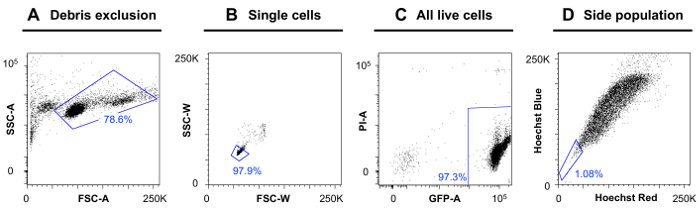
Figure 1: Side Population Gating Scheme. An example of the gating scheme used to identify the side population in a GFP+ T-ALL in zebrafish. (A) First, the debris is excluded using forward scatter (FSC) and side scatter (SSC) area. (B) Next, single cells are isolated by discriminating against doublets using FSC and SSC width parameters. (C) Since this example tumor was GFP+, all live cells must be GFP+ and negative for the dead cell marker Propidium Iodide. (D) The side population can be seen with the Hoechst Red versus Hoechst Blue profile. Please click here to view a larger version of this figure.
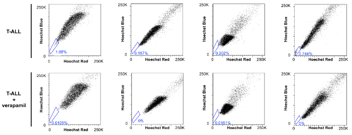
Figure 2: Side Population Disappears When Treated with the ABC Transporter Inhibitor, Verapamil. Gates for the side population are shown. The top panels show four different tumor samples. It is important to note that not all tumors have the same side population percentage, and the side population can look different for each tumor tested. The bottom panels show tumor samples that were treated with verapamil during the staining with Hoechst 33342 to block dye efflux. While the side populations may look different for each tumor, the effect of the verapamil is the same: the side population disappears. Please click here to view a larger version of this figure.
Discussion
The side population assay is highly sensitive; therefore, small changes to the protocol can result in a difficulty in detecting side population cells. First, the temperature during the staining step is specific to each animal/cell system. For mammalian systems, the side population assay is typically performed at 37 °C2. When the zebrafish T-ALL cells were incubated at 37 °C, many of the cells died, which made this incubation temperature unacceptable (data not shown). When Kobayashi and colleagues (2008) dissected zebrafish kidney marrow for the side population assay, the incubations were carried out at 25 °C5. This incubation temperature was not sufficient for zebrafish T-ALL cells, as it resulted in a weak Hoechst 33342 profile (data not shown). In the protocol described here, an incubation temperature of 28 °C was optimal to maintain cell viability and to achieve a strong Hoechst 33342 profile.
A second condition for the side population assay that requires consideration and prior testing is the concentration of Hoechst 33342. If the concentration of Hoechst 33342 is too low, the side population may not be visible, or the side population phenotype (dye efflux capability) may be falsely increased, identifying more side population cells than are actually present2. On the other hand, too high of a concentration of Hoechst 33342 can be toxic to the cells. For zebrafish T-ALL cells, 15 µg/mL Hoechst 33342 incubated for 120 min is the optimal combination. Lower concentrations of Hoechst 33342 were tried for zebrafish T-ALL cells, but the side population was not always evident (data not shown). A total incubation time of 90 min was sufficient to detect a side population, but the maximal number of side population cells was seen after 120 min of incubation time.
As reported previously for mammalian systems, not every tumor will have a detectable side population17,18,19. Using this method, we observed a side population in 14 of 17 T-ALLs tested (data not shown). Having isolated side population cells from T-ALLs, this method is expected to be applicable to other zebrafish tumor models.
Within the heterogeneous zebrafish T-ALL tumor, leukemia initiating cells (LICs) are capable of reinitiating tumor growth and as such are believed responsible for cancer recurrence and metastasis. Currently, there is no method in zebrafish that allows for the prospective isolation of LICs in vivo. Calculating the LIC frequency requires tumor cell transplantation at a limiting dilution8,10. Previously published studies with clonal zebrafish have identified that LICs make up 0.1-1.4% of primary T-ALL cells10. Here, similar variability in the frequency of side population cells (0.2-1.0%) from clonal zebrafish T-ALLs is reported (Figure 2). Further experiments are needed to determine whether the side population is enriched in LICs; however, the similarity between the ranges of LIC frequency and the percentage of side population cells in these T-ALLs supports this hypothesis.
While the side population assay is sensitive and not always easy to perform, the assay allows researchers, for the first time, to isolate a rare population of cells in zebrafish T-ALL that may have stem or progenitor cell properties. There are currently no cell surface markers available in zebrafish to identify stem- or progenitor-like cells; hence, the ability to isolate the side population is very promising. Future applications of this method include limit dilution transplantation experiments of sorted side population T-ALL cells to assess the enrichment of LICs. In addition, this method will allow characteristics that define LICs to be elucidated, including the molecular mechanisms that govern LICs in zebrafish T-ALL. These experiments could provide starting points for the discovery of new therapeutic targets for T-ALL.
Disclosures
The authors have nothing to disclose.
Acknowledgements
This work was supported by the University of Chicago Women's Board, the Wendy Will Case Fund, and the University of Chicago Comprehensive Cancer Center Support Grant (P30 CA014599). We would like to thank the Flow Cytometry Core at the University of Chicago for their help in setting up this experiment, and well as other members of the de Jong lab, particularly Leslie Pedraza and Sean McConnell.
Materials
| Name | Company | Catalog Number | Comments |
|---|---|---|---|
| Syngeneic zebrafish (CG2) | See Mizgireuv et al., 2006 | ||
| rag2:c-myc plasmid | Langenau Laboratory | ||
| rag2:GFP plasmid constructed in de Jong Lab | rag2 promoter from Langenau Laboratory | ||
| pCS2-transposase plasmid | Chien Laboratory | ||
| NotI -HF restriction enzyme | New England Bio-Labs Inc. | R3189S | 500 units in 25 µL |
| mMessage Machine SP6 Transcription Kit | Ambion | Thermo Fisher Scientific - AM1340 | 25 reactions |
| QIAGEN Plasmid Midi Kit | Qiagen, Inc. | 12643 | |
| SteREO Discovery V8 Microscope | Carl Zeiss Microscopy | 495015-0008-000 | |
| Digital microinjector | Tritech Research | MINJ-D | |
| Brass straight-arm needle holder | Tritech Research | MINJ-4 | |
| Magnetic base and stand | Tritech Research | MINJ-HBMB | |
| Micromanipulator | Narishige International | MN153 | |
| Glass capillaries | Sutter Instrument Co. | B100-50-10 | 10 cm without filament |
| Flaming/Brown micropipette puller | Sutter Instrument Co. | P-97 | |
| Microloader pipette tips | Eppendorf North America Inc. | 930001007 | |
| Phenol Red solution | Sigma Life Science | P0290 | 0.5% concentration 100 mL |
| Plastic Transfer pipettes | Fisherbrand | 137119D | graduated 7.5 mL bulb |
| Tricaine methanesulfonate (MS-222) | Western Chemical, Inc. | Fisher Scientific - NC0342409 | 100 g |
| Fetal bovine serum (FBS) | HyClone Laboratories, Inc. | Fisher Scientific - SH3007103 | 500 mL |
| Phosphate-Buffered Saline (PBS) | Gibco by Life Technologies | 1001-023 | pH 7.4 (1x) - 500 mL |
| Heparin sodium salt | Sigma-Aldrich | 2106 | 15 - 300 unit vials |
| 40 µm mesh filter | Falcon Corning Brand | 352340 | Case of 50 |
| Trypan blue | Sigma Life Science | T8154 | 20 mL - Solution (0.4%) |
| Hoechst 33342 | Life Technologies | H3570 | 10 mL |
| Verapamil | Sigma Life Science | V4629 | 1 g |
| Dimethyl sulfoxide (DMSO) | Sigma Life Science | D8418 | 100 mL |
| Propidium Iodide | Life Technologies | P3566 | 10 mL |
| FACSAria Fusion cell sorter | BD Biosciences | ||
| BD FACSDiva 8.0.1 | BD Biosciences | ||
| FlowJo v10.2 | FlowJo, LLC |
References
- Goodell, M., Brose, K., Paradis, G., Conner, A., Mulligan, R. Isolation and functional properties of murine hematopoietic stem cells that are replicating in vivo. J. Exp. Med. 183, 1797-1806 (1996).
- Golebiewska, A., Brons, N. H. C., Bjerkvig, R., Niclou, S. Critical appraisal of the side population assay in stem cell and cancer stem cell research. Cell Stem Cell. 8, 136-147 (2011).
- Dooley, K., Zon, L. I. Zebrafish: a model system for the study of human disease. Curr Opin Genet Dev. 10 (3), 252-256 (2000).
- White, R., Rose, K., Zon, L. Zebrafish cancer: the state of the art and the path forward. Nat Rev Cancer. 13 (9), 624-636 (2013).
- Kobayashi, I., et al. Characterization and localization of side population (SP) cells in zebrafish kidney hematopoietic tissue. Blood. 111 (3), 1131-1137 (2008).
- Tsinkalovsky, O., Vik-Mo, A. O., Ferreira, S., Laerum, O. D., Fjose, A. Zebrafish kidney marrow contains ABCG2-dependent side population cells exhibiting hematopoietic stem cell properties. Differentiation. 75 (3), 175-183 (2007).
- Langenau, D. M., et al. Myc-induced T cell leukemia in transgenic zebrafish. Science. 299, 887-890 (2003).
- Blackburn, J. S., Liu, S., Langenau, D. M. Quantifying the frequency of tumor-propagating cells using limiting dilution cell transplantation in syngeneic zebrafish. J Vis Exp. , e2790(2011).
- Martelli, A. M., et al. Targeting signaling pathways in T-cell acute lymphoblastic leukemia initiating cells. Adv Biol Regul. 56, 6-21 (2014).
- Smith, A. C. H., et al. High-throughput cell transplantation establishes that tumor-initiating cells are abundant in zebrafish T-cell acute lymphoblastic leukemia. Blood. 115 (16), 3296-3303 (2010).
- Blackburn, J. S., et al. Clonal evolution enhances leukemia-propagating cell frequency in T cell acute lymphoblastic leukemia through Akt/mTORC1 pathway activation. Cancer Cell. 25, 366-378 (2014).
- Basic Methods in Cellular and Molecular Biology. Plasmid Purification. JoVE Science Education Database. , JoVE. Cambridge, MA. (2016).
- Kawakami, K. Tol2: a versatile gene transfer vector in vertebrates. Genome Biol. 8, (2007).
- Rosen, J. N., Sweeney, M. F., Mably, J. D. Microinjection of zebrafish embryos to analyze gene function. J Vis Exp. (25), (2009).
- Mizgireuv, I. V., Revskoy, S. Y. Transplantable tumor lines generated in clonal zebrafish. Cancer Research. 66 (6), 3120-3125 (2006).
- Westerfield, M. The Zebrafish Book: A Guide for the Laboratory Use of Zebrafish. , University of Oregan Press. (1993).
- Hirschmann-Jax, C., et al. A distinct "side population" of cells with high drug efflux capacity in human tumor cells. Proc Natl Acad Sci. USA. 101 (39), 14228-14233 (2004).
- Komuro, H., et al. Identification of side population cells (stem-like cell population) in pediatric solid tumor cell lines. J Pediatr Surg. 42 (12), 2040-2045 (2007).
- Moshaver, B., et al. Identification of a small subpopulation of candidate leukemia-initiating cells in the side population of patients with acute myeloid leukemia. Stem Cells. 26 (12), 3059-3067 (2008).
Reprints and Permissions
