In this work, a protocol for signal enhancement of nanoprobe-based biosensing is presented. The protocol is based on the reduction of chloroauric acid onto the surface of existing nanoprobes that consist of gold, silver, silica or iron-oxide nanoparticles.
Method Article
Rapid Nanoprobe Signal Enhancement by In Situ Gold Nanoparticle Synthesis
In This Article
Summary
Abstract
The use of nanoprobes such as gold, silver, silica or iron-oxide nanoparticles as detection reagents in bioanalytical assays can enable high sensitivity and convenient colorimetric readout. However, high densities of nanoparticles are typically needed for detection. The available synthesis-based enhancement protocols are either limited to gold and silver nanoparticles or rely on precise enzymatic control and optimization. Here, we present a protocol to enhance the colorimetric readout of gold, silver, silica, and iron oxide nanoprobes. It was observed that the colorimetric signal can be improved by up to a 10000-fold factor. The basis for such signal enhancement strategies is the chemical reduction of Au3+ to Au0. There are several chemical reactions that enable the reduction of Au3+ to Au0. In the protocol, Good's buffers and H2O2 are used and it is possible to favor the deposition of Au0 onto the surface of existing nanoprobes, in detriment of the formation of new gold nanoparticles. The protocol consists of the incubation of the microarray with a solution consisting of chloroauric acid and H2O2 in 2-(N-morpholino)ethanesulfonic acid pH 6 buffer following the nanoprobe-based detection assay. The enhancement solution can be applied to paper and glass-based sensors. Moreover, it can be used in commercially available immunoassays as demonstrated by the application of the method to a commercial allergen microarray. The signal development requires less than 5 min of incubation with the enhancement solution and the readout can be assessed by naked eye or low-end image acquisition devices such as a table-top scanner or a digital camera.
Introduction
The use of nanomaterials such as gold nanoparticles (AuNPs) or iron oxide nanoparticles (IONPs) has allowed the applications in biosensing with improved sensitivity and versatility.1 The plethora of methods developed for the decoration of the surface of nanoparticles with various ligands, as to take advantage of their high surface to volume ratio, have enabled the design of sensors with improved sensitivity.2 Nevertheless, a biosensing tool is dependent on the number of nanoprobes required to achieve a detectable signal. For example, in the case of 40 nm AuNPs, to acquire a signal through UV-vis spectroscopy, approximately 90 × 106 nanoprobes is required in order to effectively detect the target of interest.3 This nanoprobe density limitation can be circumvented through the use of signal amplification. Such strategies4 can be based on interparticle aggregation or agglomeration, where the density of initial nanoprobes is increased by accumulating a second set of nanoprobes upon the first.5 Increasing the number of nanoparticles at a given location on a sensor surface allows visual or UV-vis signal acquisition. However, the sensitivity of the assay will be inherently linked to the targeting capacity of the second set of nanoparticles towards the initial set of nanoprobes. Other strategies rely on the staining of either gold and silver nanoprobes.6,7 The staining is achieved through the reduction of silver ions onto the surface of the nanoparticles, enabling a visual or UV-vis detection.8 These methods enhance the signal of existing gold or silver nanoprobes by potentiating the surface resonance plasmon signal, not depending on secondary targeting events as in interparticle agglomeration methods. However, silver staining methods have only been reported with use of gold or silver nanoprobes.8,9
In 2005, Zayats et al.10 reported the reduction of Au ions onto the surface of gold nanoprobes to increase the surface plasmon resonance signal. In this enzyme-dependent work, hydrogen peroxide was generated by glucose oxidase catalysis and together with cetyltrimethylammonium chloride allowed the reduction of chloroauric acid.
More recently Wang et al. reported an enhancement method where a gold layer is produced on the surface of existing nanoprobes.11 These enlarged nanoprobes displayed peroxidase-like catalytic activity against the substrate 3′,5,5′-tetramethylbenzidine (TMB) enabling the visualization of a bright blue color by naked eye.
Stevens et al. reported the development of a plasmonic ELISA-based assay.12 After the detection of a prostate-specific antigen (PSA) through a traditional ELISA strategy, a secondary antibody labeled with catalase was incubated with the sensor after which the sensor was immersed in a solution containing hydrogen peroxide and chloroauric acid. The presence of the secondary catalase-modified antibody (positive result) would promote the consumption of hydrogen peroxide, slowing chloroauric acid reduction and yielding quasi-spherical monodispersed AuNPs with a red color. The absence of the catalase-modified antibody (negative result) allowed the hydrogen peroxide concentration to remain intact, thus promoting the rapid chloroauric reduction and yielding AuNPs with ill-defined morphologies responsible for a blue/purple color. The concentration of hydrogen peroxide, determined by the activity of catalase, was shown to be correlated to the concentration of the analyte of interest. The need for a catalase-modified secondary antibody and the control of the conditions of the catalytic activity are two factors that hinder the universality of this method. Moreover, the formation of gold clusters, independent of the existing AuNPs, may introduce background noise issues to the amplification strategy.
The above-mentioned techniques, as well several others13,14,15, have made it possible for nanoprobe-based biosensors to achieve limits of detection on par with traditional techniques.
Here, a novel gold enhancement method is demonstrated, where the signal of an existing nanoprobe is amplified by potentiating or introducing a surface plasmon resonance signal. After the detection is carried out with the use of gold, silver, silica or iron oxide nanoparticles, the sensor is allowed to incubate with a solution of a mixture of hydrogen peroxide and chloroauric acid in 2-(N-Morpholino)ethanesulfonic acid (MES) pH 6 buffer. The concentrations of the components in the enhancement solution were optimized to favor the deposition of Au0 on the surface of existing nanoprobes. For all studied nanoprobe types, i.e. AuNPs, silver nanoparticles (AgNPs), silica nanoparticles (SiNPs) and IONPs, the formation of an ill-defined layer yielded or augmented the scattering of light which resulted in a detectable or increased visible signal. A signal increase with a 100-fold amplification factor was achieved for nanoprobes in paper and glass-based microarrays and the process takes less than 5 min to acquire a signal. The signal acquisition could be done by naked eye, UV-vis spectroscopy or imaging low-end tools such as a digital camera or a table-top scanner. Moreover, it was demonstrated that this enhancement protocol can be readily applied in a commercially available allergy diagnostic assay without requiring specific optimization.
Protocol
Human serum samples were obtained in accordance to the legal and ethical requirements of the country of collection, i.e. with the approval of an ethics committee (or similar) and with written consent from the donor.
1. Enhancement solution preparation:
- Prepare a 10 mM MES pH 6 buffer by suspending MES sodium salt in deionized water. Adjust the pH to 6 using a 4 M NaOH solution.
- Prepare a 5 mM HAuCl4 solution in 10 mM MES pH 6 buffer (solution 1).
- Prepare a 1.027 M H2O2 solution in 10 mM MES pH 6 buffer (solution 2).
NOTE: The volumes of both solution 1 and solution 2 depends on the number and size of microarrays to be enhanced. For example, a microarray of 10 x 10 mm requires 100 μL total volume (solution 1 + solution 2) to ensure that the microarray area is in contact with the enhancement solution. After dilution, the H2O2 should be used within approximately 30-45 days.
2. Nanoparticles preparation:
- Prepare 40 nm AuNPs as described by Bastús et al.16 Briefly, inject 1 mL of aqueous 25 mM HAuCl4 precursor into a boiling solution of 2.2 mM sodium citrate (149 mL). Wait until a red-wine color is observed. Use this solution of nanoparticles as seeds for seed-growth steps.
- At 90 °C, inject 1 mL of 60 mM sodium citrate followed by 1 mL of 25 mM HAuCl4 into the seed containing solution. After 30 min, the reaction is complete. Repeat the seed-growth step five times until 40 nm nanoparticles are obtained, as described by Bastús et al.16
- Measure the UV-vis absorbance of the final solution of nanoparticles for determining the size of the nanoparticles.17 Alternatively, transmission electron micrographs (TEM) can be acquired and used for determining the size of the nanoparticles.
- Prepare 90 nm AgNPs as described by Rivero et al.18 Briefly, add dimethylaminoborane (DMAB) to a vigorously stirred solution of poly(acrylic acid, sodium salt) (PAA) and AgNO3. The final volume of the solution is 50 mL and the final concentration of DMAB, PAA and AgNO3 are 1.6 mM, 10 mM and 3.33 mM, respectively.
NOTE: 100 nm IONPs modified with carboxyl groups and 50 nm SiNPs modified with carboxyl groups were purchased. - Functionalize the AuNPs with antibodies following the protocol described by Puertas et al.19 Briefly, add 1 mg of COOH-PEG-SH (5000 g·mol−1) to a solution of 1.2x10⁻⁹ M AuNPs (10 optical density, OD). Adjust the pH to 12 with a solution of 4 M NaOH.
- Let the solution incubate overnight, at room temperature (~22 °C), under mild stirring conditions.
- Remove the excess of PEG by centrifuging the solution at 13800 x g for 15 min.
- Resuspend the pellet with 500 μL of deionized water.
- Incubate the PEG-modified AuNPs with 5 μmol of N-(3-Dimethylaminopropyl)-N'-ethylcarbodiimide hydrochloride (EDC) and 7.5 μmol of N-hydroxysulfosuccinimide sodium salt (sulfo-NHS) in 10 mM MES pH 6 buffer for 30 min at 37 °C.
- Remove the excess of EDC and sulfo-NHS by centrifuging the solution at 13800 x g for 5 min.
- Resuspend the pellet in 500 µL of 10 mM MES pH 6 buffer and add 100 g·mL-1 of antibody. Let the solution incubate for 2 h at 37 °C.
- Remove the excess of antibody by centrifuging the solution at 13800 x g for 10 min twice and resuspend the pellet in 500 μL of 10 mM sodium phosphate pH 7.5, 0.3 M NaCl buffer. Let the solution incubate for 30 min at 37 °C.
- Remove the unspecific bound antibody by centrifuging the solution at 13800 x g for 10 min twice and resuspend the pellet in 500 μL of 1% bovine serum albumin (BSA) in 10 mM MES pH 6 buffer. Incubate the samples at 4 °C overnight.
- Wash the excess of BSA by centrifuging the solution at 13800 x g for 10 min twice, and resuspending the pellet in 500 μL of 10 mM MES pH 6 buffer.
- Follow the steps 2.5.1 through 2.5.9 for the modification of 5 M AgNPs, 0.5 mg of IONPS and 0.5 mg of SiNPs with antibody, adjusting the quantities of EDC and sulfo-NHS accordingly.
3. Vertical flow paper-based assays:
- Prepare the microarrays by depositing a gradient concentration of protein G on nitrocellulose paper membrane with a robotic printer. After printing, let the microarrays dry overnight at room temperature (~22 °C).
- Carry out the vertical flow assay with the membrane enclosed in a filter holder. Flow an 8 M solution of IgG-modified AuNPs using an ultra-syringe pump at a rate of 1 mL·min−1. Repeat this step with a solution of 17 M IgG-modified AgNPs, 0.1 mg·mL−1 IgG-modified SiNPs and 0.1 mg·mL−1 IgG-modified IONPs.
- Let the microarrays dry at room temperature (~22 °C) for 30 min and scan them in a flatbed scanner at a 4800-dpi resolution. Save the images as 24-bit color TIFF files.
4. Reference commercial glass-based assays:
- Incubate two glass slides for 120 min for detection of allergen components with 4 different human serum samples at room temperature.
- Rinse the microarrays with deionized water.
- Incubate one slide with a fluorescent-conjugated anti-human IgE antibody at room temperature (~22°C) for 30 min. Incubate the second slide with anti-human IgE antibody-modified AuNPs for 30 min at room temperature.
- Rinse the microarrays with deionized water.
- Measure the fluorescence intensity of the microarray that was incubated with the fluorescent anti-human IgE antibody with a laser scanning apparatus. Digitalize the microarray that was incubated with the anti-human IgE antibody-modified AuNPs with a table-top scanner for colorimetric signal acquisition.
- After digitalization, incubate the microarray with the enhancement solution at room temperature (~22°C) for 5 min.
- Rinse the sensor with deionized water and allow it to dry for 10 min at room temperature (~22 °C).
- Digitalize the microarray with a table-top scanner at a 4800-dpi resolution for colorimetric signal acquisition. Save the images as 16-bit grayscale TIFF files.
5. Microarray incubation with enhancement solution:
- Pre-mix the same amount of solution 1 and solution 2 or mix them directly on the surface of the sensor, and ensure that the area of interest on the microarray is covered with the mixture of solution.
- Let the sensor incubate at room temperature with the enhancement solution for up to 5 min. Longer incubation time can yield improved sensitivities as well as higher noise/signal ratio, especially for paper-based microarrays.
- Wash the microarray by submerging it in deionized water once for 5 s. Leave it to dry at room temperature.
NOTE: The time of the drying step depends on the material of the microarray. For a paper-based microarray, the drying step requires approximately 30 min. For a glass-based microarray, the drying step requires approximately 5-10 min.
6. Imaging analysis:
- Analyze the fluorescence intensities with software tool. Consider all results that are equal or greater than 0.3 Isac Standardize Units (ISU), following the directions of use (DfU) of the sensor manufacturer.
- Analyze the colorimetric intensities using a software tool. Invert the image files, measure the intensity of each spot and calculate the mean pixel intensity. Use the values of the triplicate spots to calculate the final signal value as a mean of the three mean spot pixel intensities.
Results
After an assay is carried out, using nanoprobes as detection agents, the sensor detection area may be populated with nanoprobes (Figure 1a). If the number of nanoprobes is below the limit for a visual detection, the detection of the analyte will be, initially, considered negative. Using the protocol presented here (Figure 1b), the signal of the nanoprobes is potentiated by introducing an ill-defined layer of Au (Figure 1c).
A paper-based immunoassay designed for detection of Protein G was carried out as to showcase the effectiveness of the enhancement protocol (Figure 2). AuNPs, AgNPs, SiNPs, and IONPs were modified with IgG antibodies and used and detection agents. Paper microarrays were prepared as to have a gradient of a number of Protein G molecules per spot.
After the assay was carried out, it was observed that IgG modified AuNPs (IgG-AuNPs) were able to detect as low as 2 × 10⁵ molecules per spot (Figure 2a), IgG modified AgNPs (IgG-AgNPs) were able to detect as low as 2 × 10⁴ molecules per spot (Figure 2c), IgG modified IONPs (IgG-IONPs) were able to detect as low as 2 × 10⁷ molecules per spot (Figure 2e) and IgG modified SiNPs (IgG-SiNPs) were not able to provide a visual signal (Figure 2g).
By incubating the microarrays with the enhancement solution, it was possible to increase the signal by 100-fold across all types of nanoparticles used. In the case of SiNPs, the enhancement solution made it possible to observe a signal where no visual signal was observed without the enhancement solution (Figure 2b, 2d,2f and 2h).
The possibility of application of the method to an existing commercial immunoassay was evaluated. The enhancement was carried out on a glass-based allergen component microarray immunoassay. A set of 4 validated and characterized serum samples from allergic patients were evaluated with the microarray (samples were designated as a, b, c and d). The standard fluorometric detection of this assay was compared to staining with anti-IgE labeled AuNPs followed by the enhancement of signal (Figure 3). Prior to enhancement, no spots were detected on the microarrays where the detection was carried out with anti-IgE labeled AuNPs. After enhancement, several spots were visible by naked eye and their intensity was registered by image digitalization (Figure 3). With the fluorescence detection, a variety of spots were detected (Figure 3).
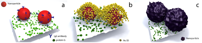
Figure 1. Illustration of the steps a microspot undergoes from analyte detection to enhanced signal. (a) Two antibody-modified nanoprobes after the detection of an analyte immobilized on a microspot, (b) incubation of the microarray with the enhancement protocol and subsequent seed-growth of the antibody-modified nanoprobes and (c) microspot where the size of the nanoprobes increased after incubating 5 min with the enhancement solution. This figure has been dapted from Dias et al.20 Please click here to view a larger version of this figure.
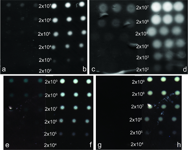
Figure 2. Digitalized image of microarrays for the detection of protein G. On the left of each microarray are the observed microspots prior to enhancement of the detection carried out with (a) IgG-AuNPs, (c) IgG-AgNPs, (e) IgG-IONPs and (g) IgG-SiNPs. On the right of each microarray are the microspots after enhancement of (b) IgG-AuNPs, (d) IgG-AgNPS, (f) IgG-IONPs and (h) IgG-SiNPs. Each concentration of protein G was deposited on the microarray in triplicate. A calculation of the number of protein G was done based on the concentration of the protein printed in each microspot. This figure has been adapted from Dias et al.20 Please click here to view a larger version of this figure.
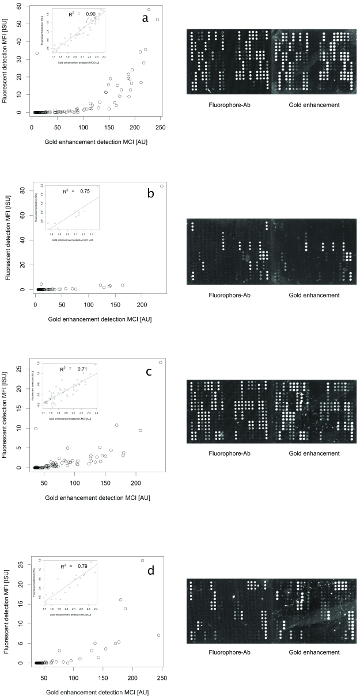
Figure 3. Correlation between fluorescent intensity and colorimetric intensity obtained for the assays carried out with the commercial allergy diagnostic assay. Mean fluorescence intensity (MFI) and mean colorimetric intensity (MCI) measured for the detection of allergens in the samples a, b, c, and d. Insets are the log 10 transformed data in the intervals where a linear correlation between both methods was observed. The intensity of the microspots observed for the detection based on the fluorophore-modified antibody (Fluorophore-Ab) is fluorescence emission. The brightness of the microspots observed for the detection based on the Ab-AuNPs assay was obtained after digitalizing the microarray and inverting the image with imaging software. Microarrays are designed as to have vertical triplicates with positive controls for fluorescence detection on the far-right bottom. The other three corners are used for software evaluation. Images were analyzed in 16-bit grayscale. This figure has been adapted from Dias et al.20 Please click here to view a larger version of this figure.
| R2 prior enhancement | R2 after enhancement | |
| IgG-AuNPs | 8.70E-01 | 7.80E-01 |
| IgG-AgNPs | 9.10E-01 | 9.60E-01 |
| IgG-IONPs | 9.17E-01 | 9.30E-01 |
| IgG-SiNPs | - | 8.50E-01 |
Table 1. R2 values for detection of protein G, prior and after enhancement, using different sets of nanoparticles. The R2 values were obtained after correlating the intensity of the spots with the concentration of analyte measured.
Discussion
Currently, signal enhancement techniques for assays with nanoprobe-based detection are either enzyme-based12, require a second set of nanoparticles to target the detection nanoprobes21 or, in the case of staining techniques, are limited to the use of AuNPs or AgNPs as detection probes.22 Here, a simple, rapid and enzyme-free method is described for signal enhancement of nanoprobe detection-based assays. With this method, it was possible to enhance the colorimetric signal provided by 4 types of nanoprobes: AuNPs, AgNPs, IONPs and SiNPs.
The observed amplification factor for each set of nanoprobes was of 100-fold, except for the SiNPs where quantification of the enhancement factor was not possible due to lack of pre-enhancement signals. This enhancement factor suggests that the efficiency of the protocol is similar across all types of nanoprobes. Moreover, when the enhancement protocol was carried out on a paper support where a dilution series of a stock AuNPs suspension was printed, it was observed a 10000-fold amplification factor. Previous to enhancement, the visible spots with the lowest number of AuNPs were where approximately 10000 nanoparticles were printed, after enhancement the spots harboring less than 10 nanoparticles became visible.20
The conditions established for the enhancement protocol here presented have allowed taking advantage of the reduction of Au3+ to Au0 for signal improving, while maintaining the background noise to a minimum. The enhancement can be performed right after the assay is performed as well as on microarrays that have been stored for up to several months after the assay was performed. The enhancement solution consists of a 1:1 mixture of solution 1 and solution 2. Both solutions can be previously mixed or directly mixed when pipetted onto the microarray. The difference between pre-mixing or direct mixing affects only the shelf-life of the enhancement solution. When pre-mixed the shelf-life is of 5-7 days increasing to 30-45 days if not pre-mixed.
Further analysis of the results has shown that the enhancement method does not interfere with the quantitative analysis of the biosensor. A linear correlation between the intensity observed and the concentration of the analyte was maintained (Table 1). The data obtained for 4 pre-characterized clinical samples using the glass-based allergen component microarray immunoassay, with standard fluorescence detection and colorimetric detection, showed a good concordance between the two detection methods. Comparing the mean fluorescence intensity (MFI) and the mean colorimetric intensity (MCI), an average R2 = 0.79 +/- 0.08 was obtained when the data is 10-logged on both axes. This experiment showed that the enhancement method can be applied to a commercial kit if it uses the nanoprobes for detection or can be adapted for nanoprobe-based detection.
The enhancement method efficacy relies on two critical aspects. It is important to assure that the mixture of solution 1 and solution 2 is homogenous. Without a homogenous mixture, the sensor will be in contact with fractions of the enhancement solution where solution 1 or 2 is predominant relative to the other. That will reduce the efficiency of the reduction of Au3+ to Au0, thus damaging the efficiency of the enhancement. It is also important to have the entire area of interest of the microarray in contact with the enhancement solution during the incubation period.
To achieve ultrasensitive enhancement of a signal, a longer incubation time (approximately 5 min) with the enhancement solution will be required. To avoid the development of background noise that can interfere with the signal acquisition, the microarray surface should be previously blocked (e.g. BSA, PEG) to prevent rapid unspecific deposition of Au0 and consequent formation of a gold layer.
A limitation to the enhancement method is the intrinsic efficacy of the assay. The assay requires having negative and positive controls as to ensure that the signal enhancement observed can be attributed to true-positive results. If there is a non-specific interaction of the nanoprobes with the target, signals acquired after enhancement will not be valid.
Other enhancement techniques are based on either nanoparticle aggregation or staining of the nanoparticles with a material that allows improved signal acquisition. By promoting the gathering of the number of nanoparticles at the detection site, the signal acquisition will be possible either visually or UV-Vis measurements.5 These signal enhancement techniques rely on a two-step detection system, the initial detection of the analyte of interest and a second step where the reporter is required to bind to the initial detection construct. Such strategies lack universality due to the requirement of specific enhancement protocol optimization for each type of assay.
The staining enhancement techniques, such as silver staining, much like the protocol here presented, allow the direct enhancement of the signal detection constructs. However, unlike the protocol that is shown here, silver staining has only been shown to be applied to either AuNPs or AgNPs.6,7,8,9
The applicability of this method could likely span any assay that uses AuNPs, AgNPs, IONPs or SiNPs as detection agents. Further work is being carried out to study the application of the method in sensors consisting of other materials.
Disclosures
The authors have nothing to disclose.
Acknowledgements
Funding from the European Research Council under the European Union's Seventh Framework Programme (FP/2007-2013)/ERC Grant Agreement No. 615458, the Swedish Research Council and Pronova excellence center for protein technology (VINNOVA - Swedish Governmental Agency for Innovation Systems) is gratefully acknowledged.
Materials
| Name | Company | Catalog Number | Comments |
|---|---|---|---|
| Chloroauric acid | Sigma-Aldrich | 254169 ALDRICH | |
| Sodium citrate | Sigma-Aldrich | S4641 SIGMA-ALDRICH | |
| Dimethylaminoborane | Sigma-Aldrich | 476668 ALDRICH | |
| Poly(acrylic acid, sodium salt) | Sigma-Aldrich | 416037 ALDRICH | |
| Silver nitrate | Sigma-Aldrich | 204390 ALDRICH | |
| 100 nm magnetic nanoparticles | Ademtech | 021111 | |
| 50 nm silica nanoparticles | Corposcular | 140510-10 | |
| COOH-PEG-SH | RAPP Polymere | 135000-4-32 | |
| Sodium hydroxide | Sigma-Aldrich | 1.06462 EMD MILLIPORE | |
| N-(3-Dimethylaminopropyl)-N’-ethylcarbodiimide hydrochloride | Sigma-Aldrich | 03449 SIGMA | |
| N-Hydroxysulfosuccinimide sodium salt | Sigma-Aldrich | 56485 ALDRICH | |
| Hydrogen peroxide solution | Sigma-Aldrich | 95313 SIGMA-ALDRICH | |
| 2-(N-Morpholino)ethanesulfonic acid hydrate | Sigma-Aldrich | M8250 SIGMA | |
| IgG antibody | Abcam | ||
| Sodium phosphate | Sigma-Aldrich | 342483 ALDRICH | |
| Sodium chloride | Sigma-Aldrich | S7653 SIGMA-ALDRICH | |
| Bovine serum albumin | VWR | 422351S | |
| Protein G | Thermo Scientific | 21193 | |
| Nitrocellulose paper membrane | Whatman | 10402096 | |
| Nanoplotter 2.1 robotic printer | GeSIM | ||
| Filter holder | Merck | XX3001200 | |
| PhD2000 Ultrasyring pump | Harvard | ||
| Flatbed scanner CannonScan 9000F Mark II | Cannon | ||
| Anti-human IgE antibody-modified AuNPs | Thermofisher | ||
| Laser scanning LuxScan 10/K | Capital Bio | ||
| Table-top scanner Image Scanner Epson Expression 1600 pro | Epson | ||
| Glass slide ImmunoCAP ISAC sIgE 112 | Thermofisher | 81-1011-01 | |
| Fluorescent-conjugated anti-human IgE antibody | Thermofisher | 81-1011-01 | |
| Phadia Microarray Image Analyzer MIA 1.2.4 | Thermofisher |
References
- Holzinger, M., Le Goff, A., Cosnier, S. Nanomaterials for biosensing applications: a review. Front. Chem. 2, 1(2014).
- Conde, J., Dias, J. T., Grazú, V., Moros, M., Baptista, P. V., de la Fuente, J. M. Revisiting 30 years of biofunctionalization and surface chemistry of inorganic nanoparticles for nanomedicine. Front. Chem. 2, 48(2014).
- Sato, K., Onoguchi, M., Sato, Y., Hosokawa, K., Maeda, M. Non-cross-linking gold nanoparticle aggregation for sensitive detection of single-nucleotide polymorphisms: Optimization of the particle diameter. Anal. Biochem. 350 (1), 162-164 (2006).
- Cao, X., Ye, Y., Liu, S. Gold nanoparticle-based signal amplification for biosensing. Anal. Biochem. 417 (1), 1-16 (2011).
- Fraire, J. C., Motrich, R. D., Coronado, E. A. Design of a novel plasmonic nanoconjugated analytical tool for ultrasensitive antigen quantification. Nanoscale. 8 (39), 17169-17180 (2016).
- Taton, T. A. Scanometric DNA Array Detection with Nanoparticle Probes. Science. 289 (5485), 1757-1760 (2000).
- Cao, C., Gontard, L. C., Thuy Tram, L. L., Wolff, A., Bang, D. D. Dual Enlargement of Gold Nanoparticles: From Mechanism to Scanometric Detection of Pathogenic Bacteria. Small. 7 (12), 1701-1708 (2011).
- Lu, Y., Shi, W., Qin, J., Lin, B. Low cost, portable detection of gold nanoparticle-labeled microfluidic immunoassay with camera cell phone. Electrophoresis. 30 (4), 579-582 (2009).
- Zhang, M., Wittstock, G., Shao, Y., Girault, H. H. Scanning electrochemical microscopy as a readout tool for protein electrophoresis. Anal. Chem. 79 (13), 4833-4839 (2007).
- Zayats, M., Baron, R., Popov, I., Willner, I. Biocatalytic Growth of Au Nanoparticles: From Mechanistic Aspects to Biosensors Design. Nano Lett. 5 (1), 21-25 (2005).
- Wang, S., Chen, Z., Choo, J., Chen, L. Naked-eye sensitive ELISA-like assay based on gold-enhanced peroxidase-like immunogold activity. Anal. Bioanal. Chem. 408 (4), 1015-1022 (2016).
- de la Rica, R., Stevens, M. M. Plasmonic ELISA for the ultrasensitive detection of disease biomarkers with the naked eye. Nat. Nanotechnol. 7 (12), 821-824 (2012).
- Liu, D., Yang, J., et al. Glucose Oxidase-Catalyzed Growth of Gold Nanoparticles Enables Quantitative Detection of Attomolar Cancer Biomarkers. Anal. Chem. 86 (12), 5800-5806 (2014).
- Qu, W., Liu, Y., Liu, D., Wang, Z., Jiang, X. Copper-mediated amplification allows readout of immunoassays by the naked eye. Angew. Chem. Int. Edit. 50 (15), 3442-3445 (2011).
- Wang, X., Niessner, R., Knopp, D. Controlled growth of immunogold for amplified optical detection of aflatoxin B1. Analyst. 140 (5), 1453-1458 (2015).
- Bastús, N. G. N., Comenge, J. J., Puntes, V. V. Kinetically controlled seeded growth synthesis of citrate-stabilized gold nanoparticles of up to 200 nm: size focusing versus Ostwald ripening. Langmuir. 27 (17), 11098-11105 (2011).
- Haiss, W., Thanh, N. T. K., Aveyard, J., Fernig, D. G. Determination of Size and Concentration of Gold Nanoparticles from UV-Vis Spectra. Anal. Chem. 79 (11), 4215-4221 (2007).
- Rivero, P. J., Goicoechea, J., Urrutia, A., Arregui, F. J. Effect of both protective and reducing agents in the synthesis of multicolor silver nanoparticles. Nanoscale Res. Lett. 8 (1), 101(2013).
- Puertas, S., Batalla, P., et al. Taking Advantage of Unspecific Interactions to Produce Highly Active Magnetic Nanoparticle-Antibody Conjugates. ACS Nano. 5 (6), 1-8 (2011).
- Dias, J. T., Svedberg, G., Nystrand, M., Andersson-Svahn, H., Gantelius, J. Rapid signal enhancement method for nanoprobe-based biosensing. Sci. Rep. 7 (1), 6837(2017).
- Shen, G., Zhang, S., Hu, X. Signal enhancement in a lateral flow immunoassay based on dual gold nanoparticle conjugates. Clin. Biochem. 46 (16-17), 1734-1738 (2013).
- Gupta, S., Huda, S., Kilpatrick, P. K., Velev, O. D. Characterization and optimization of gold nanoparticle-based silver-enhanced immunoassays. Anal. Chem. 79 (10), 3810-3820 (2007).
Reprints and Permissions
