We present a protocol for efficient, bulk, and rapid chemical reversion of conventional lineage-primed human pluripotent stem cells (hPSC) into an epigenomically-stable naïve preimplantation epiblast-like pluripotent state. This method results in decreased lineage-primed gene expression and marked improvement in directed multilineage differentiation across a broad repertoire of conventional hPSC lines.
Method Article
Chemical Reversion of Conventional Human Pluripotent Stem Cells to a Naïve-like State with Improved Multilineage Differentiation Potency
* These authors contributed equally
In This Article
Summary
Abstract
Naïve human pluripotent stem cells (N-hPSC) with improved functionality may have a wide impact in regenerative medicine. The goal of this protocol is to efficiently revert lineage-primed, conventional human pluripotent stem cells (hPSC) maintained on either feeder-free or feeder-dependent conditions to a naïve-like pluripotency with improved functionality. This chemical naïve reversion method employs the classical leukemia inhibitory factor (LIF), GSK3β, and MEK/ERK inhibition cocktail (LIF-2i), supplemented with only a tankyrase inhibitor XAV939 (LIF-3i). LIF-3i reverts conventional hPSC to a stable pluripotent state adopting biochemical, transcriptional, and epigenetic features of the human pre-implantation epiblast. This LIF-3i method requires minimal cell culture manipulation and is highly reproducible in a broad repertoire of human embryonic stem cell (hESC) and transgene-free human induced pluripotent stem cell (hiPSC) lines. The LIF-3i method does not require a re-priming step prior to the differentiation; N-hPSC can be differentiated directly with extremely high efficiencies and maintain karyotypic and epigenomic stabilities (including at imprinted loci). To increase the universality of the method, conventional hPSC are first cultured in the LIF-3i cocktail supplemented with two additional small molecules that potentiate protein kinase A (forskolin) and sonic hedgehog (sHH) (purmorphamine) signaling (LIF-5i). This brief LIF-5i adaptation step significantly enhances the initial clonal expansion of conventional hPSC and permits them to be subsequently naïve-reverted with LIF-3i alone in bulk quantities, thus obviating the need for picking/subcloning rare N-hPSC colonies later. LIF-5i-stabilized hPSCs are subsequently maintained in LIF-3i alone without the need of anti-apoptotic molecules. Most importantly, LIF-3i reversion markedly improves the functional pluripotency of a broad repertoire of conventional hPSC by decreasing their lineage-primed gene expression and erasing the interline variability of directed differentiation commonly observed amongst independent hPSC lines. Representative characterizations of LIF-3i-reverted N-hPSC are provided, and experimental strategies for functional comparisons of isogenic hPSC in lineage-primed vs. naïve-like states are outlined.
Introduction
The 2i (MEK/ERK and GSK3β inhibitor) culture system was originally developed to refine heterogeneous serum-based mouse embryonic stem cells (mESC) cultures to a uniform ground state of pluripotency akin to the mouse preimplantation epiblast1. However, 2i does not support the stable maintenance of human pluripotent stem cell (hPSC) lines2. The various complex small molecule, growth factor-supplemented, and transgenic approaches have recently been reported to capture putatively similar human naïve-like pluripotent molecular states2. However, many of the "naïve-like" states created with these methods also exhibited karyotypic instability, epigenomic defects (e.g., global loss of parental genomic imprinting), or impaired differentiation potential.
In contrast, the cocktail of triple chemical inhibition of GSK3β, ERK and tankyrase signaling and leukemia inhibitory factor (LIF-3i) was sufficient for the stable naïve-like reversion of a broad repertoire of conventional hPSC lines3. LIF-3i-reverted naïve hPSC (N-hPSC) maintained normal karyotypes and increased their expressions of naïve-specific human preimplantation epiblast genes (e.g.,NANOG, KLF2, NR5A2, DNMT3L, HERVH, Stella (DPPA3), KLF17, TFCP2L1). LIF-3i reversion also conferred hPSC with an array of molecular and biochemical characteristics unique to mESC-like naïve pluripotency that included increased phosphorylated STAT3 signaling, decreased ERK phosphorylation, global 5-methylcytosine CpG hypomethylation, genome-wide CpG demethylation at embryonic stem cell (ESC)-specific gene promoters, and dominant distal OCT4 enhancer usage. Moreover, in comparison to other naïve reversion methods that resulted in aberrantly hypomethylated imprinted genomic loci, LIF-3i-reverted N-hPSC were devoid of systematic loss of imprinted CpG patterns or loss of DNA methyltransferase expression (e.g., DNMT1, DNMT3A, DNMT3B)3.
A direct LIF-3i culture of a broad array of conventional human embryonic stem cells (hESC) and human induced pluripotent stem cells (hiPSC) grown on either feeders or E8 feeder-free conditions achieved rapid and bulk reversion to a naïve epiblast-like state. However, direct LIF-3i naïve reversion may be inefficient in some unstable conventional hPSC lines due to the inherent genomic and lineage-primed variabilities arising from the genetically diverse donor backgrounds.
Thus, to broaden the utility of the LIF-3i method, a stepwise optimization was developed and is presented herein, that allows universal naïve reversion with almost any conventional hESC or transgene-free hiPSC line cultured on feeders. This universalized naïve reversion method employs a transient initial culture step in conventional hPSC that supplements the LIF-3i cocktail with two additional small molecules (LIF-5i) that potentiate protein kinase A (forskolin) and sonic hedgehog (sHH) (purmorphamine) signaling. One initial passage of conventional hPSC in LIF-5i adapts them to subsequent stable LIF-3i reversion in bulk quantities. Initial LIF-5i adaptation significantly augments the initial single cell clonal proliferation of conventional hPSC grown on E8 or feeders (prior to their subsequent stable, continuous passage in LIF-3i alone). Conventional hPSC lines adapted first to one passage in LIF-5i tolerate subsequent bulk clonal passaging of naïve-reverted cells in LIF-3i conditions, which obviates the need for picking and subcloning of the rare stable colonies, or the routine use of anti-apoptotic molecules or Rho-associated protein kinase (ROCK) inhibitors.
The LIF-3i method has been successfully employed to stably expand and maintain a broad repertoire of >30 independent, genetically-diverse conventional hPSC lines for >10–30 passages using either non-enzymatic or enzymatic dissociation methods, and without evidence of induction of chromosomal or epigenomic abnormalities, including abnormalities at imprinted gene loci. Additionally, sequential LIF-5i/LIF-3i culture is the only naïve reversion method that has thus far been reported that improves the functional pluripotency of a broad repertoire of conventional hPSC lines by decreasing their lineage-primed gene expression and dramatically improving their multipotent differentiation potency. The LIF-3i naïve reversion method erases the inherent interline variability of differentiation of lineage-primed, conventional hPSC lines, and will have a great utility of application in regenerative medicine and cellular therapies.
Protocol
All animal procedures were performed in accordance with animal care guidelines and protocols approved by the Johns Hopkins School of Medicine Institute of Animal Care and Use Committee (IACUC).
1. Preparation of Mouse Embryonic Fibroblasts (MEF) for Feeder-dependent Conventional (hESC medium/MEF) or Naïve-reverted (LIF-3i medium/MEF) hPSC Culture
- Purchase or prepare in-house low-passage supplies of MEF feeders from CF1 or CF1 x DR4 hybrid E13.5 mouse embryos following published protocols4.
- Cryopreserve low passage (p1-p2) MEF cultures pre- (for long term storage) or post- (for up to 6 months) irradiation and store in liquid nitrogen as previously described4.
- Irradiate (5,000 rad) bulk expanded MEF under p5 using a γ- or X-ray- irradiator and prepare aliquots at 1.5 x 10⁶ cells per vial for short-term storage (less than 6 months) at -80 °C in an ultra-low temperature freezer.
- Plate between 1.2 x 10⁶ (freshly irradiated non-cryopreserved) and 1.5 x 10⁶ (irradiated cryopreserved) MEF per one 6-well gelatinized plate for preparing feeder cultures, as described below.
- Prepare gelatin-coated 6-well sterile tissue culture-treated plates by adding 1.5 mL of sterile 0.1% gelatin solution to each well in a biological safety cabinet.
- Incubate gelatin-coated plates at 37 °C for at least 1 h or overnight in a laboratory CO2 incubator.
- On the next day, thaw low passage (e.g., P2 to P4) DMSO-cryopreserved and irradiated (5,000 rad) MEF according to the steps indicated below.
NOTE: Non-irradiated MEF should also be thawed, expanded, and irradiated immediately prior to usage. - Place a cryopreserved MEF aliquot in a 37 °C water bath. Upon thawing, sterilize the tube with ethanol and immediately dilute the DMSO cryoprotectant at least 10-fold with MEF medium (Table 1) within a biosafety cabinet (e.g. transfer 1 mL DMSO-MEF aliquot into 9 mL MEF medium in a sterile 15 mL conical).
- Centrifuge the diluted MEF cells at 200 x g for 5 min in sterile 15 mL conical tubes.
- In a biosafety cabinet, aspirate and discard the cell-free supernatant, and resuspend the cell pellet in 1–2 mL fresh MEF medium.
- Gently discard the gelatin solution and add 2 mL of MEF re-suspended in the MEF medium to each well of a gelatinized 6-well plate, as indicated above. Count MEF cells and plate 1.2 x 10⁶ (freshly irradiated non-cryopreserved) or 1.5 x 10⁶ (irradiated cryopreserved) MEF per one 6-well gelatinized plate.
NOTE: All cell culture plates that are transferred from the CO2 incubator to the biosafety cabinet can be gently wiped with ethanol-sprayed paper but should not be directly sprayed on with 70% ethanol solution to avoid ethanol dissemination in the wells. - Incubate MEF plates at 37 °C overnight in a laboratory CO2 incubator (5% CO2, humid atmosphere) to allow attachment, prior to use.
2. Bulk Stabilization of Conventional hPSC Cultures for Subsequent Naïve Reversion with a Brief LIF-5i Adaptation Step
- Validate all conventional hPSC lines for possessing a normal karyotype by G-banding, prior to beginning LIF-5i/LIF-3i reversion.
NOTE: LIF-3i reversion of high-passage conventional hPSC lines (e.g., P>40–50) should be avoided, since these cultures may already harbor genomic aberrations that may negatively impact stable, efficient, and bulk LIF-3i reversion of primed, convetional hPSC lines. - Maintain and expand conventional hPSC cultures with validated normal karyotypes in either a MEF-based culture system (as outlined in Section 1), or a feeder-free culture system (according to the investigator's preference).
NOTE: Both feeder-dependent hPSC/MEF cocultures (e.g., hESC medium (Table 1) supplemented with 4–10 ng/mL bFGF) or feeder-free (e.g., E8 5 or mTSER 6 media (according to manufacturer's instructions on vitronectin-coated plates) methods are compatible with bulk naïve reversion using the LIF-5i/LIF-3i/MEF system (Figure 1). Non-enzymatic methods are preferred for the passaging of conventional hPSC prior to preparing them for reversion. LIF-5i and LIF-3i media formulations do not contain antibiotics or antifungal agents. Standard operation rules for the biosafety cabinet sterility and maintenance are expected to be rigorously observed to avoid any bacterial or fungal contamination. - For the MEF- based culture system, prepare MEF feeders in the gelatinized 6-well plate as described in Section 1 at least one day before passaging of LIF-5i-adapted conventional hPSC cultures.
- After conventional hPSC cultures have reached ~50% confluency (i.e., 3–5 days after initial plating), replace the standard hESC culture medium with LIF-5i medium (2 mL per well; Table 1).
NOTE: Perform these steps in a biosafety cabinet. - Culture and maintain conventional hPSC/MEF for up to 2 days in LIF-5i in the CO2 incubator (5% CO2, humid atmosphere). Change the LIF-5i medium daily to adapt them for their subsequent passage and stable reversion in LIF-3i.
- Alternatively, for the feeder-free conventional cultures (e.g., in E8), culture hPSC in LIF-5i only overnight before passaging the next morning.
NOTE: LIF-5i and LIF-3i cultures can both be maintained with either atmospheric (21% O2) or physiologic (5% O2) oxygen levels in the CO2 incubator (5% CO2, humid atmosphere). - Prior to passaging, place the culture plates in a biosafety cabinet, wash LIF-5i-adapted conventional hPSC once with PBS, and add 1 mL of cell dissociation reagent to each well. Incubate for 5 min at 37 °C in a CO2 incubator, and gently triturate with a pipette into a single cell suspension back in the biosafety cabinet.
NOTE: Non-enzymatic dissociation buffers may alternatively be used for preparing single cells for passaging. - Collect the cell suspension in hESC medium (at least 2-fold dilution) in a sterile 15 mL conical tubes, and gently triturate cells by pipetting to obtain a single cell suspension.
- Centrifuge at 300 x g for 5 min, aspirate/discard the supernatant, and resuspend the cell pellet in 1–2 mL of the LIF-5i medium in the biosafety cabinet. Count the cells using a hemocytometer or an automatic cell counter.
- Wash the pre-plated MEF plate twice with PBS (2 mL per well in 6-well plates) and distribute 1–2 x 10⁶ cells (in 2 mL LIF-5i medium) onto 1 well of the PBS-washed MEF plate.
- Adjust and optimize initial plating densities for each individual hPSC line to be naïve-reverted, as replating efficiencies can be highly variable. Place the plate in a CO2 incubator (5% CO2, humid atmosphere).
NOTE: The routine use of anti-apoptotic reagents is not recommended for most hPSC lines with this method. The LIF-5i system already significantly enhances initial bulk clonal re-plating efficiencies of conventional hPSC lines. However, a one-time use of ROCK inhibitor (5–10 μM Y-27632) may further improve the initial LIF-5i clonal re-plating efficiency of conventional hPSC cultures in the first passage for some unstable lineage-primed hPSC lines with the propensity for spontaneous differentiation. - If starting from feeder-free cultures (e.g., E8), directly transfer one well of the dissociated conventional hPSC adapted in LIF-5i into one irradiated MEF-plated well (i.e., 1:1 passage).
- The next day, gently swirl the plate to lift all non-attached cells, aspirate the medium (PBS wash is optional) and replace with 2 mL of LIF-5i medium. Perform this step in a biosafety cabinet daily for 3–5 days or until cells are 60–70% confluent (Figure 1). Place the plate in a CO2 incubator (5% CO2, humid atmosphere).
3. Long-term Maintenance and Expansion of N-hPSC in LIF-3i Medium
- Following initial LIF-5i adaptation, passage subsequent stable LIF-3i cultures every 3–4 days in a biosafety cabinet, or when cultures become 60–70% confluent (Figure 1).
NOTE: LIF-3i cultures require rigorous maintenance and allowing N-hPSC cultures to reach high confluency/cell density from prolonged culture (e.g., >4 days) decreases subsequent clonal re-plating efficiency, and promotes spontaneous differentiation. - In a biosafety cabinet, discard the culture medium and wash each well of LIF-5i/LIF-3i cultures by gently adding 2 mL of PBS. Discard PBS and add 1 mL of cell detachment solution. Incubate for 5 min at 37 °C in a CO2 incubator (5% CO2, humid atmosphere).
- Collect the cell suspension, add hESC medium (without inhibitors or growth factors (Table 1); at least 2-fold dilution) to recover all hPSC and gently triturate cells by pipetting to obtain a single cell suspension. Transfer the suspension to a sterile 15 mL conical tube.
- Centrifuge at 300 g for 5 min and aspirate/discard the supernatant. Re-suspend the cell pellet in LIF-3i medium. Count the cells using a hemocytometer or an automatic cell counter.
- Plate ~2 x 10⁵ cells per well onto the irradiated MEF in the gelatinized 6-well plate for routine passaging of LIF-3i cultures. For the initial LIF-5i-adapted cultures, plate an initially higher density (4 x 10⁵ cells/well) prior to the first passage into LIF-3i/MEF.
- Re-plate and distribute LIF-5i-adapted hPSC onto fresh PBS-washed irradiated MEF feeder plates (prepared the previous day as above) in the LIF-3i medium. Replace the LIF-3i medium daily.
- Passage N-hPSC for least 4–7 continuous bulk passages in the LIF-3i medium prior to use of N-hPSC in functional studies or cryopreservation. Record the number of passages of N-hPSC in either conventional or LIF-3i media.
NOTE: LIF-3i reversion of high-passage (e.g. >p40) lineage-primed, conventional hPSC lines is not recommended. An effort should be made to revert conventional hPSC lines at the lowest possible passage that they are available. Additionally, the use of LIF-3i-reverted hPSC that have undergone greater than 10 LIF-3i passages is not recommended for functional studies, since such N-hPSC cultures may harbor karyotypically-abnormal clones due to prolonged clonal cell culture selection. Fresh LIF-5i/LIF-3i reversions of low-passage conventional hPSC lines should be conducted for functional studies, if stocks of N-hPSC with <10 passages in LIF-3i are not available.
4. Cryopreservation and Thawing of LIF-3i-reverted N-hPSC
- Expand reverted N-hPSC for at least 5-7 passages in LIF-3i, as indicated above, prior to use in functional studies or long-term cryopreservation. Record the number of passages in conventional conditions and in LIF-3i conditions on each cryopreserved vial.
NOTE: Excess LIF-3i-reverted N-hPSC not used in functional assays can be cryopreserved at each passage, but freezing of lower post-reversion passages (e.g., <p3) may result in poor, or highly variable post-thaw recovery efficiencies. - In a biosafety cabinet, aspirate the culture medium, wash cells in PBS (2 mL per well), aspirate PBS and dissociate hPSC colonies into single cells using cell detachment solution (1 mL per well). Place the plate for 5 min at 37 °C in a CO2 incubator (5% CO2, humid atmosphere). Dilute the cell detachment solution with the LIF-3i medium (2-fold), collect hPSC in a sterile 15 mL conical, centrifuge cells at 200 x g for 5 min and resuspend the cell pellet in LIF-3i medium (1–2 mL per well-equivalent). Count the number of cells using a hemocytometer or an automatic cell counter.
- Centrifuge N-hPSC in the LIF-3i medium (200 x g for 5 min) and resuspend cells in a biosafety cabinet in the freezing solution (Table 2), at a density of at least 1 x 10⁶ cells/mL.
- Transfer cells into the long-term storage cryogenic tubes and place into a slow-freezing container. Allow the samples to freeze overnight in a -80 °C freezer.
- The next day, transfer the cryovials into a liquid nitrogen freezer for long-term storage.
- For thawing, place the frozen vial into a 37 °C water bath for ~2 min. Sterilize the vial (i.e., ethanol spray), transfer hPSC in a sterile 15 mL conical and slowly dilute the cells 10-fold in hESC medium (Table 1) supplemented with 5 μM of Rho-associated protein kinase (ROCK) inhibitor Y-27632 within a sterile biological safety hood cabinet.
- Centrifuge at 200 x g for 5 min. In a biosafety cabinet, discard cell-free supernatant and resuspend the cell pellet in LIF-3i medium (1–2 mL) supplemented with 5 μM ROCK inhibitor Y-27632.
NOTE: Exclusion of Y-27632 will result in poor post-thawing recovery efficiencies (Figure 2). - Transfer the thawed cells resuspended in LIF-3i/ROCK Inhibitor onto the PBS-washed MEF-plated wells. Cryopreserve LIF-3i cultures at a density of 1 x 10⁶ cells per vial. Thaw each of these vials onto feeders in one well of a gelatinized 6-well plate.
- The next day, start regular LIF-3i medium expansion without Rho kinase inhibitor.
5. Feeder Removal for the Collection of N-hPSC Samples
- To prepare samples from LIF-3i/MEF (or hESC/MEF conventional) cultures (i.e., for gene expression (e.g., quantitative RT-PCR, microarrays) or protein (e.g., Western blot) analyses, deplete LIF-3i cultures from MEF feeders using Magnetic Activated Cell Sorting (MACS) with anti-TRA-1-81 antibody coated microbeads according to the manufacturer's protocol.
- Alternatively, utilize the following simple pre-plating method to deplete cultures of feeders, as described below.
- In a sterile biological safety hood cabinet, discard the cell-free supernatant, wash the pellet with 2 mL sterile PBS per well. Detach adherent LIF-3i/MEF cultures using cell detachment solution (1 mL/well). Incubate for 5 min in a CO2 incubator (5% CO2, humid atmosphere). Collect hPSC in a sterile 15 mL conical. Wash 2-fold in hESC medium, transfer cells into the sterile 15 mL conical and centrifuge at 200 x g for 5 min.
- In a biosafety cabinet, re-suspend each well of LIF-3i/MEF dissociated cells into 2 mL of the LIF-3i medium, and directly transfer onto a new well of a 6-well plate that has been freshly coated with 0.1% gelatin.
- Incubate LIF-3i/MEF hPSC cells for 1 h at 37 °C in a CO2 incubator (5% CO2, humid atmosphere). Collect non-adherent cells with a pipette in a new conical tube. Gently add 2 mL of hESC medium to each well and swirl to collect the remaining non-adherent cells.
NOTE: The majority of the irradiated MEF will attach to the gelatinized plate in 1 h, leaving the majority of the hPSC in suspension. - Combine and centrifuge cells at 300 x g for 5 min. Wash in PBS. Snap-freeze the cell pellet in liquid nitrogen after centrifugation, or alternatively re-suspend pellets in a lysing buffer that is compatible with the downstream protein or nucleic acid analysis (Figure 2B).
- Perform characterizations of LIF-3i hPSC lines with a matching conventional primed isogenic hPSC control.
NOTE: Because of intrinsic variability between and within primed cultures, relevant controls are prepared at a matching timepoint in culture or prior to naïve reversion. Detailed protocols and materials for downstream immunofluorescent bioimaging, flow cytometry, Western blotting, gene expression (RT-PCR assays and microarrays), methylation studies (dot blot, CpG methylation microarray), OCT4 proximal and distal enhancer predominance reporter assays and signaling inhibitor assays are provided elsewhere3.
6. Post Naïve reversion: Validation of Genomic Integrity and Retention of Parental Imprints of LIF-3i-reverted N-hPSC Prior to Use in Functional Assays
- Screen the starting primed, conventional hPSC cultures for possession of a normal karyotype (e.g., with Giemsa-band staining analysis using published methods 7) before initiating LIF-5i/LIF-3i reversion.
NOTE: This is to eliminate conventional hPSC populations that may harbor abnormal genomic alterations which may drive artefactual selective survival advantage in clonal LIF-3i conditions. - For optimal results, freshly revert conventional hPSC cultures to a naïve-like state with LIF-3i several weeks prior to their use in functional studies or directed differentiation.
NOTE: Routine prolonged 'maintenance' culture in LIF-3i conditions for more than 10 passages following naïve reversion is not recommended. Routine expansion and maintenance of hESC and hiPSC lines should be performed using conventional culture systems (e.g., in E8, or MEF/hESC medium with bFGF). - Assess post-reverted N-hPSC lines for the retention of normal karyotypes 5–7 passages after LIF-3i reversion (e.g., with Giemsa-band staining analysis7, or any other method of choice).
- Assess all reverted N-hPSC lines for retention of normal parental genomic imprints by a DNA methylation analysis of choice (e.g., protocols for CpG DNA microarray analysis of parental imprints in LIF-3i-reverted N-hPSC are provided elsewhere3) after 5–10 passages of LIF-3i reversion.
7. Post Naïve Reversion: Experimental Design Guidelines for Quantitative Directed Differentiation Assays Using LIF-3i-reverted N-hPSC
- Directly utilize LIF-3i N-hPSC into established directed differentiation protocols without additional cell culture manipulations.
NOTE: Re-priming (i.e., converting N-hPSC back to conventional primed conditions prior to their use in directed differentiation assays) is not necessary with the LIF-3i method and is not recommended. - To control for the impacts of the assay and interline variability in the functional testing of individual hPSC lines, cross-validate the lineage-specific differentiation potencies by employing independent differentiation protocols with at least three hPSC lines derived from independent genetic backgrounds (i.e., multiple donor-derived hiPSC and hESC).
- For functional comparisons, set up sibling cultures, at equivalent passage number, and from the same (isogenic) hPSC line in parallel to the conventional lineage-primed and LIF-3i-reverted hPSC cultures. Maintain primed/naïve sibling isogenic hPSC cultures in their respective media (e.g., E8 vs. LIF-3i), and simultaneously differentiate using identical differentiation protocols and materials, to eliminate any experimental bias (Figure 4).
- For isogenic primed vs. naïve hPSC comparisons, adjust and optimize initial plating densities for each individual differentiation assay.
NOTE: Detailed protocols for neural progenitor, definitive endoderm and hemato-endothelial directed differentiation of LIF-3i-reverted N-hPSC are provided elsewhere 3. LIF-3i-reverted N-hPSC have more robust proliferative and differentiation capacity in the directed differentiation assays than conventional hPSC. N-hPSC typically requires a lower initial plating concentration than the conventional hPSC, and unlike their conventional primed hPSC counterparts, do not require the use of anti-apoptotic reagents to enhance their clonal survival following enzymatic digestion in differentiation assays.
Results
This protocol optimizes efficient naïve-like reversion with LIF-3i in both feeder-dependent and feeder-independent lineage-primed conventional hPSC cultures (Figure 1). The detailed protocol, described herein, outlines sequential adaptation to LIF-3i starting from either feeder-dependent or feeder-free conventional hPSC conditions (e.g. E8 medium).
Representative results for the LIF-3i reversion of several conventional hESC and transgene-free hiPSC lines are presented in Figures 1–3. These typical results can be validated with the commercially available hESC line H9, or with a commercially available transgene-free, cord blood-derived episomal hiPSC line (6.2) derived in the Zambidis laboratory8,9. Introduction of an initial LIF-5i adaptation step permits highly efficient subsequent, bulk clonal propagation of conventional hPSC cultures in LIF-3i, and does not require anti-apoptotic agents or ROCK inhibitors10 (Figure 1A, B). Multiple plates of naïve-like hPSC samples can be rapidly collected for downstream analyses or multilineage directed differentiation only after 5–7 passages in LIF-3i. Alternatively, LIF-3i cultures can be cryopreserved for future applications. Post-thawing cell recovery can be improved (Figure 2) via inclusion of a ROCK inhibitor in cryopreservation and post-thaw media11.
The determinants of molecular and functional pluripotency, both in vitro and in the embryo were recently reviewed2. These factors include the genetic background, culture-associated acquisition of mutations for key developmental genes, and differences in hESC and hiPSC derivation and culture methodologies. Provided below is a summary of standard assays that can be employed for characterization and validation of the phenotypic, molecular, and functional pluripotencies of LIF-3i-reverted hPSC.
Colony morphology
The transition between primed, conventional and LIF-3i-reverted culture systems is accompanied by distinct physical changes in hPSC colony morphology (Figure 1B). Conventional hPSC cells proliferate as flat, wide monolayer colonies that expand rapidly from small cell clumps (on MEF or feeder-free conditions), but poorly as single cells. Exposure of conventional hPSC lines to LIF-3i promotes the growth and expansion and of smaller, tightly-packed, dome-shaped colonies that arise clonally from single cells. These morphological changes are completely reversible, and LIF-3i-reverted dome-shaped colonies can spontaneously transition back to a conventional monolayer morphology if LIF-3i is withdrawn and cells are re-cultured in standard conventional hESC medium supplemented with bFGF. Additionally, expansion of LIF-3i-reverted cells at high confluent densities (or prolonged culture without frequent passaging) results in the spontaneous reacquisition of the flat, conventional morphology with reduced clonal efficiency; emphasizing the need for diligent maintenance and care of LIF-3i-reverted hPSC (e.g., <40–60% confluence).
Live immunofluorescence staining and flow cytometric analysis of surface pluripotency markers
Evaluation of pluripotency markers during the transition from conventional to a naïve-like state following continuous LIF-3i culture can be monitored non-invasively using live antibody staining without detecting any negative effects on LIF-3i/MEF expansion (e.g., live-staining fluorochrome-conjugated antibodies against TRA-1-81, TRA-1-60, and SSEA4).
Retention of pluripotency during LIF-3i reversion can also be routinely monitored by flow cytometric analysis of pluripotency-associated surface marker expression of TRA-1 and SSEA antigens during single cell passaging (Figure 1C) or immunofluorescence of intact, fixed colonies in situ (Figure 3). Although these markers do not discriminate between conventional and LIF-3i states, their levels inversely correlate with the frequency of spontaneous differentiation that may occur in hPSC when transitioning from conventional hPSC to LIF-3i conditions. Additional surface antigens that may more specifically mark human naïve-like states in vitro2,12,13 can also be employed to detect effective LIF-3i hPSC reversion.
Validation of molecular pluripotency of N-hPSC
Because the genetic background of hPSC lines has been characterized as a strong contributor to interline variability, it is important to rigorously assess isogenic cultures at matching culture timepoints when comparing hPSC culture systems (Figure 4). Since some of these systems have already been shown to generate hPSC populations with aberrant genomic and epigenetic configurations2, any naïve reversion method should be assayed with a number of N-hPSC of independent genetic backgrounds, in a manner that is sufficient to validate biological reproducibility and exclude non-developmentally relevant "pseudo-pluripotent" states (i.e., with apparent hallmarks of molecular pluripotency but lacking functional differentiation abilities). Zimmerlin et al. further extended the validation of the LIF-3i culture system to include assaying the molecular and functional pluripotencies of reverted N-hiPSC derived from various reprogramming methods, which is another known putative contributor of functional variability between pluripotent states2.
Accordingly, most studies of human naïve culture systems have focused on assaying molecular pluripotency of N-hPSC at 1) the epigenetic level (e.g., histone marks by ChIP sequencing or ChIP-PCR, global DNA methylation by immunoblots or whole genomic bisulfite sequencing, allele-specific CpG methylation microarrays, OCT4 enhancer predominant usage by reporter systems, global activity at regulatory elements by DNAse I hypersensitivity, and repeat element profiling by RNA-sequencing), 2) transcriptomic level (RNA-sequencing, expression microarrays, and quantitative RT-PCR), protein expression analysis (e.g., FACS, immunofluorescent microscopy, and Western blotting) and 3) via metabolic studies (e.g., glycolysis, oxidative phosphorylation and nicotinamide metabolism). Representative examples of immunofluorescence stains and Western blot detection of expression for key markers of molecular pluripotency are shown (Figure 3B, C). For example, shown in Figure 3B are activated phosphorylated (phospho) and total isoforms of STAT3 and ERK1/2, which are key molecular hallmarks of mouse ESC-like naïve pluripotency. These were detected using anti-STAT3 and anti-ERK1/2 primary antibodies. Additionally, functional pluripotency in teratoma differentiation assays is demonstrated in conventional vs. LIF-3i-reverted hPSC teratoma tissues dissected 10 weeks following injection and fixed in 4% formaldehyde and paraffin embedded (Figure 3D).
Preparation of LIF-3i/MEF or hESC/MEF/bFGF samples for downstream molecular analysis should include MEF depletion. Two approaches are described above for MEF depletion that have been successfully employed in our laboratory. MEF pre-plating is a simple, reliable, and cost-efficient method to eliminate feeders from N-hPSC samples for molecular studies and is a preferred alternative to FACS or MACS separation (i.e., anti-TRA-1-81 or anti-TRA-1-60 antibody-based separation). Additionally, spontaneously-differentiating TRA-1-negative adherent cells in LIF-5i/LIF-3i cultures can be rapidly eliminated from LIF-5i/LIF-3i N-hPSC cultures prior to subsequent LIF-3i passage onto fresh MEF by pre-plating the enzymatically-digested single cells for 1 h on plates pre-coated with 0.1% gelatin (Figure 2).
Evaluation of functional pluripotency of N-hPSC
The most rigorous assay of functional pluripotency of PSC is the blastocyst injection chimera assay, which is limited in the testing of N-hPSC lines for ethical reasons. Alternatively, several groups that have reported the generation of N-hPSC with various other methods have attempted to generate interspecies chimeras. However, these attempts have yielded extremely low or unsuccessful contribution of differentiated N-hPSC lineages to murine or porcine embryos, in comparison to the chimera-generating capacity of standard mouse ESC2.
Additional functional studies have investigated directed in vitro differentiation of putative N-hPSC derived via various methods, but have revealed biased, defective, or diminished multilineage differentiation capacity, with concomitant harboring of epigenetic abnormalities2,13. Similar epigenomic aberrations, especially at imprinted loci, have been detected in mouse ESC following prolonged exposure to the LIF-2i cocktail14. Interestingly, some reversion culture systems have reported global improvements in specific attributes of functional pluripotency of PSC such as the enhanced capacity for in vivo trophectoderm contribution2,15.
Using a broad collection of independently-derived LIF-3i-reverted hPSC, Zimmerlin et al. employed multilineage differentiation assays to show that the LIF-3i system dramatically improves the functional pluripotency of conventional hPSC lines3. This allows systematic analysis of conventional vs LIF-3i hPSC lines in isogenic pairs to eliminate interline-dependent variations (Figure 4). LIF-3i-reverted hPSC lines do not require a re-priming step prior to EB differentiation. However, LIF-3i hPSC proliferate at significantly higher clonal rates than isogenic cells expanded in E8, and thus, initial lower plating densities require adjustment to allow each culture to reach confluence at a similar timepoint.
Investigators should routinely utilize multiple assays to demonstrate the improved functionality of LIF-3i-reverted hPSC that includes not only in vivo teratoma assays but also in vitro directed differentiation assays to neural, definitive endoderm and hematovascular lineages3 using multiple assays (e.g., 2D APEL3,16 and 3D embryoid body17,18 systems). To control for assay-dependent reproducibility, at least two different differentiation methods should be performed in replicate for each isogenic pair of primed/LIF-3i hPSC cultures (e.g., Figure 4, APEL, embryoid body differentiation protocols). The experimental design should include a robust number (e.g., >3–5) primed/LIF-3i isogenic pairs of hPSC lines from multiple, independent donor genetic backgrounds (Figure 4).
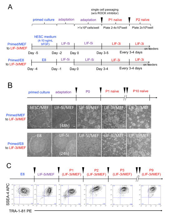
Figure 1: A stepwise transition of conventional, lineage-primed hPSC cultures to naïve-like conditions with the LIF-3i method. (A) Schema of protocols for stepwise LIF-3i reversion. (Top schematic) A general method for the transition of primed, conventional hPSC to LIF-3i cultures. (Bottom schematics) Two summarized strategies for LIF-3i naïve reversion of conventional (primed) hPSC cultured on either feeder (i.e., Primed/MEF to LIF-3i/MEF), or feeder-free conditions (i.e, Primed/E8 to LIF-3i/MEF). (B) Human PSC morphologies. Shown are representative photomicrographs of hPSC during LIF-3i reversion using a commercially available human episomal iPSC line (6.2). Shown are the transitions observed between initial conventional flat monolayer colonies, and the subsequent dome-shaped clonogenic colonies that arise following passage in intermediate LIF-5i and stable LIF-3i culture conditions. Scale bars = 200 µm. (C) Representative flow cytometric analyses of pluripotency surface markers. Shown are TRA-1-81 and SSEA-4 surface expressions in conventional hPSC line 6.2 (p40) expanded in E8, the initial passage in LIF-5i/MEF, and following 1 to 9 passages (P1-P9) in LIF-3i conditions. Please click here to view a larger version of this figure.
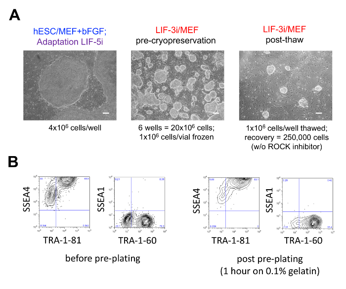
Figure 2: Cryopreservation of N-hPSC and sample preparation by MEF pre-plating. (A) Example of a freeze/thaw cycle of LIF-3i/MEF cultures. The conventional cord blood-derived, non-integrated, transgene-free human iPSC line E5C3 was derived and expanded on MEF feeders in hESC medium supplemented with 4ng/mL bFGF for 18 passages. Conventional, lineage-primed E5C3 were adapted in LIF-5i (left) and transitioned into LIF-3i medium for 3 passages (center). The E5C3 LIF-3i cells shown in the center panel were cryopreserved using DMSO-based cryoprotectant medium (Table 2; 1 x 10⁶ cells per vial) and stored in liquid nitrogen). One vial was thawed a month later and E5C3 cells were transferred in a feeder-coated well of a 6-well plate (right). Cell recovery can be enhanced by supplementing the LIF-3i medium with 5 µM Y-27632 for only one day post-thaw. Scale bars = 200 µm. (B) Elimination of MEF (and also adherent TRA-negative differentiated cells) in LIF-3i/MEF hPSC cultures by the pre-plating method. Shown are flow cytometric analyses before and after pre-plating of PE-conjugated anti-TRA-1-81 and TRA-1-60 antibodies, APC-conjugated SSEA-4 antibodies and SSEA-1/CD15 antibodies demonstrating depletion of TRA-1 antigen negative and MEF cells using the one-hour pre-plating method. Please click here to view a larger version of this figure.
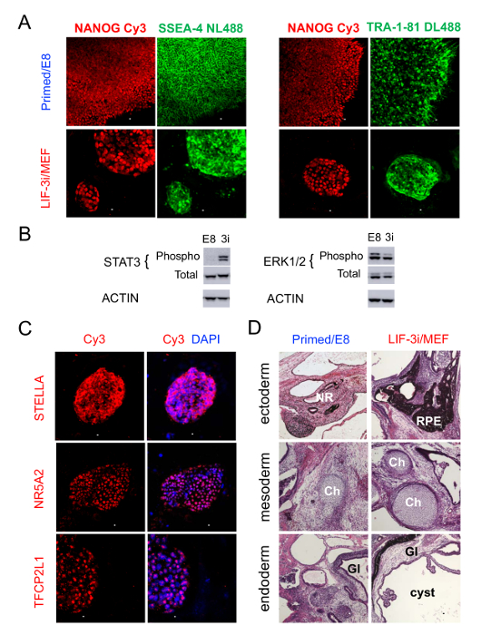
Figure 3: Characterization of pluripotency in LIF-3i/MEF N-hPSC cultures. (A) Surface and nuclear pluripotency markers. Expression of pluripotency factor NANOG in the same (isogenic) TRA-1-81+SSEA4+ conventional, primed cord blood-derived hiPSC line E5C3 (p39) expanded in either primed (E8), or naive LIF-3i (+p8 in LIF-3i/MEF) conditions. Immunofluorescent stains of representative hPSC cultures on chamber slides revealed the uniform, nuclear expression of the core pluripotency factor NANOG in SSEA-4+TRA-1-81+ hPSC cultured in primed, conventional (E8 medium) or LIF-3i/MEF conditions. Scale bar = 100 µm. (B) Western blot analysis. STAT3 (left), ERK (center), and control beta-ACTIN protein expression for a representative hPSC line (hESC line H9) in conventional (E8 medium) or LIF-3i/MEF conditions (3i). (C) Expression of naïve pluripotency-associated transcription factors. Shown are STELLA/DPPA3, NR5A2, and TFCP2L1 by immunofluorescence in a representative LIF-3i/MEF culture (cord blood-derived hiPSC line E5C3 at p33 (hESC/MEF) +p8 (LIF-3i/MEF). Scale bar = 100 µm. (D) Teratoma assays of conventional and LIF-3i-reverted hPSC. Validation of functional pluripotency in an isogenic representative hiPSC line (cord blood-derived E32C6) cultured in either E8 (p9) or LIF-3i/MEF (+p12) by teratoma assay. 10 x 106 cells of isogenic parallel-cultured conventional, primed vs LIF-3i-reverted hPSC were injected subcutaneously into the limbs of immunodeficient NSG mice. Hematoxylin and eosin stains of teratoma microsections revealed robust differentiation into all three germ layers with well-structured ectoderm (neural rosette: NR, retinal pigmented epitheliam: RPE), mesoderm (chondroblasts: Ch), and endoderm (glandular tissue: GI) lineages. Scale bars = 100 µm. Please click here to view a larger version of this figure.
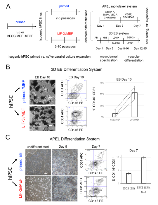
Figure 4: Comparison of functional pluripotency between isogenic primed and naïve states. (A) Schematic of strategy for assessing functional pluripotency from distinct pluripotent states in isogenic conventional vs. LIF-3i cultured hPSC in independent differentiation protocols. Shown are two hemato-vascular progenitor differentiation systems (APEL monolayer and 3D embryoid body (EB) systems) that were previously employed to assess differentiation potency of conventional vs. LIF-3i-reverted in the same (isogenic) hPSC line cultured in parallel post LIF-3i reversion with same passage numbers. LIF-3i-reverted hPSC lines do not require a re-priming step prior to EB differentiation and are subjected to the differentiation protocol directly. (B) EB vascular progenitor (VP) differentiation system. The EB 3D differentiation system employed for this study was previously described17,18. Shown are the representative results at day 10 of EB differentiation (left panels) for isogenic cultures of the same cord blood (CB)-derived E5C3 hPSC line9, cultured in either conventional hESC/MEF (Primed/ MEF) conditions or LIF-3i/MEF naïve conditions. Flow cytometry analysis of these EB cells show dramatic increases of CD31+CD146+ VP populations following LIF-3i reversion of the E5C3 line prior to differentiation. The histogram displays the mean ±SD of CD31+CD146+ VP cell percentages recovered at day 10 in this EB system using three isogenic pairs of independent hPSC lines (i.e., two CB-hiPSC and one adult fibroblast-derived hiPSC, dotted lines connect isogenic pairs). Results demonstrate significant improvement of VP differentiation efficiencies in the EB system across genetic backgrounds of multiple hPSC lines. (C) APEL monolayer vascular progenitor differentiation system. Conventional (primed E8) and LIF-3i-reverted hPSC can be directly differentiated using the same culture conditions, growth factors, cytokines and small molecules of the stepwise APEL endothelial differentiation protocol 3,16. Shown are independent APEL differentiation experiments using the E5C3 cord blood-derived hiPSC line, and the percentage of CD31+CD146+ vascular progenitor populations at day 7 of APEL differentiation. Please click here to view a larger version of this figure.
| Mouse Embryonic Fibroblast (MEF) Medium (500mL) | human embryonic stem cell (hESC) Medium (500mL) | LIF-5i (500mL) | LIF-3i (500mL) | ||
| media and supplements | DMEM high glucose | 439.5mL | - | - | - |
| DMEM/F12 | - | 391.5mL | 391.5mL | 391.5mL | |
| fetal bovine serum | 50mL | - | - | - | |
| KnockOut serum replacement | - | 100mL | 100mL | 100mL | |
| L-Glutamine (200mM) | 2.5mL | 2.5mL | 2.5mL | 2.5mL | |
| MEM Non-essential amino acid (MEM NEAA), 10mM | 5mL | 5mL | 5mL | 5mL | |
| Penicillin/streptomycin (100X) | 2.5mL | - | - | - | |
| β-mercaptoethanol (55mM) | 0.5mL (0.055mM) | 910µL (0.1mM) | 910µL (0.1mM) | 910µL (0.1mM) | |
| cytokines and small molecules | recombinant basic FGF (100µg/mL) | - | 50µL (10ng/mL) | 50µL (10ng/mL) | - |
| recombinant human LIF (100µg/mL) | - | - | 100µL (20ng/mL) | 100µL (20ng/mL) | |
| CHIR99021 (100mM) | - | - | 15µL (3µM) | 15µL (3µM) | |
| PD0325901 (100mM) | - | - | 5µL (1µM) | 5µL (1µM) | |
| XAV939 (100mM) | - | - | 20µL (4µM) | 20µL (4µM) | |
| Forskolin (100mM) | - | - | 50µL (10µM) | - | |
| Purmorphamine (10mM) | - | - | 100µL (2µM) | - |
Table 1:
| LIF-3i crypreservation medium (10mL) | |
| hESC medium (Table 1) | 40% |
| KnockOut serum replacement | 50% |
| dimethyl sulfoxide (DMSO) | 10% |
| Y-27632 | 5 µM |
Table 2:
Discussion
The LIF-3i system applies a modified version of the classic murine 2i naïve reversion cocktail1 to human pluripotent stem cells. The self-renewal of hPSC (which cannot expand in 2i alone) is stabilized in LIF-2i by supplementing this cocktail with the tankyrase inhibitor XAV939. LIF-3i culture allows bulk and efficient reversion of the conventional hPSC to a pluripotent state resembling the human preimplantation epiblast3. Although the mechanisms of action of XAV939 in hPSC are likely complex and synergistic with 2i, they likely include at a minimum, an important stabilization and augmentation of hPSC self-renewal via WNT signaling pathways3.
Conventional hPSC cultures normally adopt a spectrum of pluripotent states with highly variable lineage-primed gene expressions and post-implantation epiblast epigenetic marks that may result in inconsistent or diminished functional pluripotency2. This inherent lineage priming of conventional hPSC cultures may also interfere with the successful LIF-3i reversion of some hPSC lines2. However, the inclusion of the initial LIF-5i adaptation step (Table 1) in the LIF-3i method universally broadens the efficiency of LIF-3i reversion among a multitude of hPSC lines and promotes bulk naïve reversion of conventional hPSC lines in a manner that circumvents tedious picking and subcloning of rare naïve-reverted colonies, or the need for use of routine anti-apoptotic molecules to stabilize their viability.
The LIF-5i/LIF-3i naïve reversion method is reproducible in a broad variety of hESC and hiPSC lines. It requires minimal training with basic cell culture skill. Zimmerlin et al. successfully employed this sequential strategy to revert >30 independent hESC and transgene-free hiPSC lines from a broad array of donors 3. A single passage in LIF-5i (Figure 1) is sufficient for most hPSC lines to undergo efficient naïve reversion in bulk hPSC cultures, and further advance their subsequent stable maintenance and expansion in LIF-3i alone.
Furthermore, the LIF-3i/MEF system supports robust bulk clonal expansion efficiencies throughout all the steps between lineage-primed conventional hPSC culture all the way to completed naïve-like hPSC reversion (i.e., adaptation, transition and expansion for 7–10 passages in LIF-3i/MEF alone). Although the current formulation of this culture system requires feeder support, a simple and affordable method to deplete feeders by the pre-plating technique for analysis of LIF-3i cultures is presented (Figure 2).
Multiple other culture systems have also been reported to promote conventional hPSC to similar naïve-like pluripotent states2. Although these hPSC culture systems have also relied on the utilization of the classical mouse naïve 2i conditions, in most cases these single-cell passaging methods also required additional chemical modulation for stabilizing an inherently unstable/metastable human naïve state. Importantly, most of these other methods demonstrated impaired functional pluripotency following differentiation and/or acquired abnormal epigenomic imprints or karyotypes2. Although the emergence of abnormal karyotypes within conventional primed hPSC cultures is already well documented19, prolonged, enzymatic single-cell passaging methods that are routinely employed in most naïve reversion methods. This has also been shown to potentiate the generation of abnormal chromosomal configurations20,21; more sensitive techniques (e.g., copy number variations, single nucleotide polymorphism) may reveal additional alterations22,23.
In contrast, a wide repertoire of LIF-3i-reverted hPSC lines were confirmed to possess normal karyotypes at low-medium passages (e.g., p5-p15), and also at high passage numbers (e.g., >p30) following LIF-3i culture3. Additionally, epigenomic imprints in LIF-3i-reverted hPSC were found reproducibly normal and intact at 5–10 passages post LIF-3i reversion2. Using the sensitive allele-specific commercial methylation array platform, it was previously demonstrated that CpG methylation marks at imprinted loci of a wide repertoire of LIF-3i-reverted hPSC lines (following 4–7 passages in LIF-3i) were found to be grossly normal in structure1. Since abnormal genomic imprints and karyotypes may ultimately impair the functional capacity of hPSC, prerequisite guidelines were outlined in this protocol that encourages researchers to validate hPSC cultures before and after naïve reversion, using this method as well as others.
The LIF-3i method improved functional pluripotency across germ layers in a large repertoire of hESC and non-transgenic hiPSC lines (Figure 3). Unlike other naïve reversion protocols, the LIF-3i method does not require a re-priming step for subsequent differentiation of N-hPSC (i.e., converting N-hPSC back to conventional primed conditions prior to their use in directed differentiation assays). LIF-3i-reverted N-hPSC display significantly more efficient differentiation capacities than their isogenic conventional hPSC counterparts in both teratoma assays (Figure 3D), and directed differentiation protocols of lineages of all three germ layers2. Due to assay-dependent and interline variations in functional testing of conventional hPSC, lineage-specific differentiation should be evaluated using independent directed differentiation protocols and hPSC derived from multiple genetic backgrounds. Using careful experimental design, a broad array of hPSC lines can be expected to significantly improve their multilineage differentiation efficiencies compared to their isogenic conventional counterparts following 4–10 passages in LIF-3i conditions.
In summary, this method rapidly and clonally expands the numbers of human PSC, improves their downstream differentiation efficiency, increases the lineage-committed progenitor cell numbers following differentiation, and decreases interline variability among conventional, lineage-primed hPSC lines. These N-hPSC with improved functionality may further have a wide impact for their potential to contribute functional tissues to a developing embryo. For example, stable N-hESC may be employed for developing transplantable human organs and adult stem cells in developing animal chimeras, or for generating humanized gene-targeted animal models of disease. The further optimization of this tankyrase inhibitor-utilizing LIF-3i method in defined feeder-free GMP-compliant culture conditions will facilitate efficient clinically useful generation of a broad array of functional and engraftable cell types for therapeutic use.
Disclosures
Under a licensing agreement between Life Technologies and the Johns Hopkins University (JHU), Dr. Zambidis is entitled to a share of royalty received by the University for licensing of stem cells. The terms of this arrangement are managed by JHU in accordance with its Conflict of Interest policies. This does not alter authors' adherence to journal policies on sharing data and materials.
Acknowledgements
This work was supported by grants from NIH/NEI (R01EY023962), NIH/NICHD (R01HD082098), RPB Stein Innovation Award, The Maryland Stem Cell Research Fund (2018-MSCRFV-4048, 2014-MSCRFE-0742), Novo Nordisk Science Forum Award, and The Moseley Foundation.
Materials
| Name | Company | Catalog Number | Comments |
|---|---|---|---|
| anti- SSEA-1/CD15 antibody, APC conjugated | BD Biosciences | 561716 | use 5µL per assay (FACS) |
| anti- TFCP2L1 antibody | Sigma Aldrich | HPA029708 | use at a 1:100 dilution (immunostainings) |
| anti-beta-Actin antibody | Abcam | ab6276 | use at 1:5000 (Western blot) |
| anti-CD146 antibody, PE conjugated | BD Biosciences | 550315 | use 5µL per assay (FACS) |
| anti-CD31 antibody, APC conjugated | eBioscience | 17-0319-42 | use 2µL per assay (FACS) |
| anti-CD31 microbead kit | Miltenyi Biotec | 130-091-935 | |
| anti-NANOG antibody | Abcam | ab109250 | use at a 1:100 dilution (immunostainings) |
| anti-NR5A2 antibody | Sigma Aldrich | HPA005455 | use at a 1:100 dilution (immunostainings) |
| anti-p44/42 MAPK (Erk1/2) antibody | Cell Signaling | 4695 | use at 1:1000 (Western blot), for detection of total protein |
| anti-phospho-p44/42 MAPK (Erk1/2) (Thr202/Tyr204 antibody | Cell Signaling | 4370 | use at 1:1000 (Western blot) |
| anti-phospho-STAT3 (Tyr705) antibody | Cell Signaling | 9145 | use at 1:1000 (Western blot) |
| anti-rabbit immunoglobulin antibody, biotinylated | Agilent | E0432 | use at a 1:500-1:1000 dilution (immunostainings) |
| anti-SSEA-4 antibody, APC conjugated | R&D System | FAB1435A | use 5µL per assay (FACS) |
| anti-SSEA-4 GloLIVE antibody, NL493 conjugated | R&D System | NLLC1435G | use at 1:50 dilution (live and fixed immunostainings) |
| anti-STAT3 antibody | Cell Signaling | 9139 | use at 1:1000 (Western blot), for detection of total protein |
| anti-STELLA/DPPA3 antibody | Millipore | MAB4388 | use at a 1:50 dilution (immunostainings) |
| anti-TRA-1-60 GloLIVE antibody, NL557 conjugated | R&D System | NLLC4770R | use at a 1:50 dilution (live and fixed immunostainings) |
| anti-TRA-1-60 StainAlive Antibody, DyLight 488 conjugated | Stemgent | 09-0068 | use at a 1:100 dilution (live and fixed immunostainings) |
| anti-TRA-1-81 StainAlive Antibody, DyLight 488 conjugated | Stemgent | 09-0069 | use at a 1:100 dilution (live and fixed immunostainings) |
| anti-TRA1-60 antibody, PE conjugated | BD Biosciences | 560193 | use 10µL per assay (FACS) |
| anti-TRA1-81 antibody, PE conjugated | BD Biosciences | 560161 | use 10µL per assay (FACS) |
| APEL2-Li | StemCell Technologies | 5271 | |
| Bovine Serum Albumin | Sigma Aldrich | A3311 | |
| CellAdhere dilution buffer | StemCell Technologies | 7183 | dilutent for Vitronectin XF™ matrix |
| CF1 mouse | Charles river | 023 | |
| CHIR99021 | R&D System | L5283 | reconstitute at 100mM in DMSO |
| confocal microscope system | Zeiss | LSM 510 | |
| cord blood CD34+ derived iPSC line | Thermo Fisher Scientific | A18945 | also referred as 6.2 line |
| Corning Costar tissue culture-treated 6-well plates | Corning | 3506 | |
| Countess cell counting chamber slide | Thermo Fisher Scientific | C10228 | |
| Countess automated cell counter | Thermo Fisher Scientific | AMQAX1000 | |
| DMEM (Dulbecco's Modified Eagle Medium) | Thermo Fisher Scientific | 11995-065 | |
| DMEM-F12 | Thermo Fisher Scientific | 11330-032 | |
| DMSO (dimethyl sulfoxide) | Sigma Aldrich | D2650 | |
| DR4 mouse | The Jackson Laboratory | 3208 | |
| Essential 8 (E8) medium | StemCell Technologies | 5940 | |
| Fetal bovin serum (FBS) | Thermo Fisher Scientific | SH30071.03 | |
| Forskolin | Stemgent | 04-0025 | reconstitute at 100mM in DMSO |
| Gelatin (porcine) | Sigma Aldrich | G1890-100G | resuspend in water and sterilize with an autoclave |
| KnockOut Serum Replacement | Thermo Fisher Scientific | 10828-028 | |
| L-Glutamine (100X) | Thermo Fisher Scientific | 25030-081 | |
| MEM Non-essential amino acid (MEM NEAA) (100X) | Thermo Fisher Scientific | 11140-050 | |
| mTeSR1 medium | StemCell Technologies | 85850 | |
| Nalgene cryogenic vials | Thermo Fisher Scientific | 5000-0020 | |
| Nunc Lab-Tek II Chamber Slide System | Fisher Scientific | 154534 | |
| Paraformaldehyde (PFA) solution , 4% in PBS | USB Corporation | 19943 | |
| PD0325901 | Sigma Aldrich | PZ0162 | reconstitute at 100mM in DMSO |
| Penicillin/streptomycin (10,000 U/mL) | Thermo Fisher Scientific | 15140-122 | |
| Phosphate buffered saline (PBS) | Biological Industries | 02-023-1A | |
| Purmorphamine | Stemgent | 04-0009 | reconstitute at 10mM in DMSO |
| recombinant human Activin A | Peprotech | AF-120-14E | |
| recombinant human Bone morphogenetic protein (BMP)-4 | Peprotech | 120-05ET | resupend at 100ug/mL in 0.1% bovine serum albumin in PBS |
| recombinant human FGF-basic (bFGF) | Peprotech | 100-18B | resupend at 100ug/mL in 0.1% bovine serum albumin in PBS |
| recombinant human LIF | Peprotech | 300-05 | resupend at 100ug/mL in 0.1% bovine serum albumin in PBS |
| SB431542 | Stemgent | 04-0010-05 | reconstitute at 100mM in DMSO |
| Stemolecule Y27632 in Solution | Stemgent | 04-0012-02 | ROCK inhibitor in solution (10mM) |
| StemPro Accutase Cell Dissociation Reagent | Thermo Fisher Scientific | A11105-01 | |
| Streptavidin-Cy3 conjugate | Sigma Aldrich | S6402 | use at 1:500-1:1000 dilution (immunostainings) |
| Thermo Scientific Mr. Frosty Freezing Container | Thermo Fisher Scientific | 5100-0001 | |
| Vascular endothelial growth factor (VEGF)-165 | Peprotech | 100-21 | resupend at 100ug/mL in 0.1% bovine serum albumin in PBS |
| Vitronectin XF matrix | StemCell Technologies | 7180 | dilute at 40µL/mL in CellAdhere™ dilution buffer |
| XAV939 | Sigma Aldrich | X3004 | reconstitute at 100mM in DMSO |
| β-mercaptoethanol | Thermo Fisher Scientific | 21985-023 | light sensitive |
References
- Ying, Q. L., et al. The ground state of embryonic stem cell self-renewal. Nature. 453 (7194), 519-523 (2008).
- Zimmerlin, L., Park, T. S., Zambidis, E. T. Capturing Human Naive Pluripotency in the Embryo and in the Dish. Stem Cells Dev. 26 (16), 1141-1161 (2017).
- Zimmerlin, L., et al. Tankyrase inhibition promotes a stable human naive pluripotent state with improved functionality. Development. 143 (23), 4368-4380 (2016).
- Jozefczuk, J., Drews, K., Adjaye, J. Preparation of mouse embryonic fibroblast cells suitable for culturing human embryonic and induced pluripotent stem cells. J Vis Exp. (64), e3854(2012).
- Chen, G., et al. Chemically defined conditions for human iPSC derivation and culture. Nat Methods. 8 (5), 424-429 (2011).
- Ludwig, T. E., et al. Derivation of human embryonic stem cells in defined conditions. Nat Biotechnol. 24 (2), 185-187 (2006).
- Howe, B., Umrigar, A., Tsien, F. Chromosome preparation from cultured cells. J Vis Exp. (83), e50203(2014).
- Burridge, P. W., et al. A universal system for highly efficient cardiac differentiation of human induced pluripotent stem cells that eliminates interline variability. PLoS One. 6 (4), 18293(2011).
- Park, T. S., et al. Growth factor-activated stem cell circuits and stromal signals cooperatively accelerate non-integrated iPSC reprogramming of human myeloid progenitors. PLoS One. 7 (8), 42838(2012).
- Watanabe, K., et al. A ROCK inhibitor permits survival of dissociated human embryonic stem cells. Nat Biotechnol. 25 (6), 681-686 (2007).
- Li, X., Krawetz, R., Liu, S., Meng, G., Rancourt, D. E. ROCK inhibitor improves survival of cryopreserved serum/feeder-free single human embryonic stem cells. Hum Reprod. 24 (3), 580-589 (2009).
- Collier, A. J., et al. Comprehensive Cell Surface Protein Profiling Identifies Specific Markers of Human Naive and Primed Pluripotent States. Cell Stem Cell. 20 (6), 874-890 (2017).
- Liu, X., et al. Comprehensive characterization of distinct states of human naive pluripotency generated by reprogramming. Nat Methods. 14 (11), 1055-1062 (2017).
- Choi, J., et al. Prolonged Mek1/2 suppression impairs the developmental potential of embryonic stem cells. Nature. 548 (7666), 219-223 (2017).
- Yang, Y., et al. Derivation of Pluripotent Stem Cells with In Vivo Embryonic and Extraembryonic Potency. Cell. 169 (2), 243-257 (2017).
- Orlova, V. V., et al. Generation, expansion and functional analysis of endothelial cells and pericytes derived from human pluripotent stem cells. Nat Protoc. 9 (6), 1514-1531 (2014).
- Park, T. S., Zimmerlin, L., Zambidis, E. T. Efficient and simultaneous generation of hematopoietic and vascular progenitors from human induced pluripotent stem cells. Cytometry A. 83 (1), 114-126 (2013).
- Park, T. S., et al. Vascular progenitors from cord blood-derived induced pluripotent stem cells possess augmented capacity for regenerating ischemic retinal vasculature. Circulation. 129 (3), 359-372 (2014).
- Taapken, S. M., et al. Karotypic abnormalities in human induced pluripotent stem cells and embryonic stem cells. Nat Biotechnol. 29 (4), 313-314 (2011).
- Mitalipova, M. M., et al. Preserving the genetic integrity of human embryonic stem cells. Nat Biotechnol. 23 (1), 19-20 (2005).
- Beers, J., et al. Passaging and colony expansion of human pluripotent stem cells by enzyme-free dissociation in chemically defined culture conditions. Nat Protoc. 7 (11), 2029-2040 (2012).
- Laurent, L. C., et al. Dynamic changes in the copy number of pluripotency and cell proliferation genes in human ESCs and iPSCs during reprogramming and time in culture. Cell Stem Cell. 8 (1), 106-118 (2011).
- Hussein, S. M., et al. Copy number variation and selection during reprogramming to pluripotency. Nature. 471 (7336), 58-62 (2011).
Reprints and Permissions
