Here, we describe an optimized, Langendorff-based procedure for the isolation of single-cell atrial cardiomyocytes from a rat model of metabolic syndrome-related heart failure with preserved ejection fraction. A manual regulation of intraluminal pressure of cardiac cavities is implemented to yield functionally intact myocytes suitable for excitation-contraction-coupling studies.
Method Article
Isolation of Atrial Cardiomyocytes from a Rat Model of Metabolic Syndrome-related Heart Failure with Preserved Ejection Fraction
Opens in a new tab
In This Article
Summary
Loading...
$$\rightleftharpoonup{xx}$$
$$\longleftharp{xx}$$,
$$\longrightharp{xx}$$,
Abstract
Loading...
$$\rightleftharpoonup{xx}$$
$$\longleftharp{xx}$$,
$$\longrightharp{xx}$$,
In this article, we describe an optimized, Langendorff-based procedure for the isolation of single-cell atrial cardiomyocytes (ACMs) from a rat model of metabolic syndrome (MetS)-related heart failure with preserved ejection fraction (HFpEF). The prevalence of MetS-related HFpEF is rising, and atrial cardiomyopathies associated with atrial remodeling and atrial fibrillation are clinically highly relevant as atrial remodeling is an independent predictor of mortality. Studies with isolated single-cell cardiomyocytes are frequently used to corroborate and complement in vivo findings. Circulatory vessel rarefication and interstitial tissue fibrosis pose a potentially limiting factor for the successful single-cell isolation of ACMs from animal models of this disease.
We have addressed this issue by employing a device capable of manually regulating the intraluminal pressure of cardiac cavities during the isolation procedure, substantially increasing the yield of morphologically and functionally intact ACMs. The acquired cells can be used in a variety of different experiments, such as cell culture and functional Calcium imaging (i.e., excitation-contraction-coupling).
We provide the researcher with a step-by-step protocol, a list of optimized solutions, thorough instructions to prepare the necessary equipment, and a comprehensive troubleshooting guide. While the initial implementation of the procedure might be rather difficult, a successful adaptation will allow the reader to perform state-of-the-art ACM isolations in a rat model of MetS-related HFpEF for a broad spectrum of experiments.
Introduction
Loading...
$$\rightleftharpoonup{xx}$$
$$\longleftharp{xx}$$,
$$\longrightharp{xx}$$,
MetS describes a cluster of risk factors for diabetes mellitus type-2 and cardiovascular disease and includes an increased arterial blood pressure, dyslipidemia (raised triglycerides and lowered high-density lipoprotein cholesterol), increased fasting glucose, and central obesity1. The worldwide prevalence of MetS is estimated to be 25–30% and constantly rising2. HFpEF is a heterogenous clinical syndrome often associated with MetS. The cardiac remodeling during HFpEF and its preceding phases (i.e., hypertensive heart disease) is also accompanied by a remodeling of the atria3. Reduced contractile function and structural changes of the left atrium have been associated with increased mortality, atrial fibrillation, and new-onset heart failure4. Atrial remodeling is characterized by changes in the ion channel function, Ca2+ homeostasis, atrial structure, fibroblast activation, and tissue fibrosis5. Left atrial remodeling in MetS-related HFpEF and its underlying pathological mechanisms are still poorly understood and require a further in-depth investigation. Animal models have proven to be a valuable tool and lead to many advances in the field of atrial cardiomyopathies6,7,8,9.
Studies with isolated single-cell cardiomyocytes are frequently used to corroborate and complement in vivo findings. An isolation, and the potential subsequent cell culture, allow for the investigation of signaling pathways, ionic channel currents, and excitation-contraction-coupling. Under physiologic conditions, cardiomyocytes do not proliferate. The fusion between the transcriptional regulatory sequences of an atrial natriuretic factor and a simian virus 40 large T antigen in transgenic mice led to the creation of the first immortalized ACMs, named AT-110. The further development of AT-1 cells gave rise to HL-1 cells, which cannot only be serially passaged but also contract spontaneously11. They do, however, show structural and functional differences compared to freshly isolated cells, such as a less organized ultrastructure, a high occurrence of developing myofibrils11, and a hyperpolarization-activated inward current12. The isolation of ventricular cardiomyocytes (VCM) in rats and mice from a variety of models is well established13,14,15,16,17,18,19. Generally, the excised heart is mounted to a Langendorff apparatus and retrogradely perfused with a Ca2+-free buffer containing digestive enzymes, such as collagenases and proteases. Calcium is then reintroduced in a stepwise manner to the physiological conditions. However, even though protocols dedicated to the isolation of ACMs are available20,21, due to increased fibrosis and pressure-related differences, their usefulness in disease models with atrial remodeling is limited.
In this article, we have implemented a protocol for the isolation of atrial single-cell cardiomyocytes from animals that show atrial remodeling (i.e., in particular for the ZFS1 rat model for MetS-related HFpEF)22. Existing isolation protocols were optimized and complemented by a simple, custom-made device to control and modify the intraluminal pressure of the cardiac cavities, leading to higher yields of morphologically and functionally intact cardiomyocytes. The following protocol provides the researcher with a step-by-step guide, a detailed description of the custom-made equipment, a list of solutions, as well as a comprehensive troubleshooting guide.
Access restricted. Please log in or start a trial to view this content.
Protocol
Loading...
$$\rightleftharpoonup{xx}$$
$$\longleftharp{xx}$$,
$$\longrightharp{xx}$$,
All experiments were approved by the local Ethics Committee (TVA T0060/15 and T0003-15) and performed in agreement with the Guidelines for the Care and Use of Laboratory Animals (National Institute of Health, U.S.A.).
NOTE: A simplified flowchart of the procedure is shown in Figure 1.
1. Prearrangements
- Prepare the buffers according to Table 1.
| Solution | PB | CB | DB | SB | S1 | S2 | S3 | NT | |
| Reagent (mM) | |||||||||
| NaCl | 135 | 135 | 135 | 135 | 135 | 135 | 135 | 135 | |
| KCl | 4.7 | 4.7 | 4.7 | 4.7 | 4.7 | 4.7 | 4.7 | 4 | |
| KH2PO4 | 0.6 | 0.6 | 0.6 | 0.6 | 0.6 | 0.6 | 0.6 | ||
| Na2HPO4 | 0.6 | 0.6 | 0.6 | 0.6 | 0.6 | 0.6 | 0.6 | ||
| MgSO4 | 1.2 | 1.2 | 1.2 | 1.2 | 1.2 | 1.2 | 1.2 | ||
| MgCl | 1 | ||||||||
| HEPES | 10 | 10 | 10 | 10 | 10 | 10 | 10 | 10 | |
| Taurine | 30 | 30 | 30 | 30 | 30 | 30 | 30 | ||
| Glucose | 10 | 10 | 10 | 10 | 10 | 10 | 10 | ||
| BDM | 10 | 10 | 10 | 10 | 10 | 10 | 10 | ||
| CaCl2 | 1 | 0.01 | 0.125 | 0.25 | 0.5 | 1 | |||
| BSA | 150 | 70 | 70 | 70 | |||||
| Purified enzyme blend (medium Thermolysin) | 0.195 Wünsch units/mL | ||||||||
| pH adjusted to | 7.4 | 7.4 | 7.4 | 7.4 | 7.4 | 7.4 | 7.4 | 7.4 | |
| pH adjusted at | 37 °C | 4 °C | 37 °C | 37 °C | 37 °C | 37 °C | 37 °C | 37 °C | |
| pH adjusted with | NaOH | NaOH | NaOH | NaOH | NaOH | NaOH | NaOH | NaOH |
Table 1: List of Buffers. PB: perfusion buffer, which can be stored for 3 days at 4 °C (300 mL per animal). CB: cannulation buffer, which can be stored for 3 days at 4 °C (200 mL per animal). DB: digestion buffer, which has to be used within the day (40 mL per animal). SB: stopping buffer, which has to be used within the day (2 mL per animal). S1: Step 1 buffer, which has to be used within the day (2 mL per animal). S2: Step 2 buffer, which has to be used within the day (2 mL per animal). S3: Step 3 buffer, which has to be used within the day (2 mL per animal). NT: normal Tyrode, which has to be used within the day (50 mL per animal).
- Prepare the Langendorff apparatus (Figure 2A).
- Flush the system with 100 mL of 70% ethanol, followed by 2 flushes of 100 mL distilled water. Fill the system with 200 mL of perfusion buffer (PB).
- Set the flow rate of the peristaltic pump to 3 mL/min.
- Calibrate the temperature of the PB leaving the Langendorff apparatus to 39 °C.
- Place the custom-made cannula [16 G, 25 mm long, sharp tip removed (Figure 3A)] on top of the Langendorff apparatus and start the flow. Turn on the heating module and adjust the value of the heating module to reach the desired temperature of the PB at the tip of the cannula. Once the temperature calibration is completed, move the custom-made cannula to the syringe used for the cannulation (Figure 2B).
- Prepare the pressure control device (Figure 3C) next to the Langendorff system. Mount the butterfly needle onto the tripod clamp and prepare 3 blocking knots.
NOTE: Collagenase enzyme activity varies with temperature. A temperature monitoring of the left atrium, as well as a dynamic adjustment thereof, is required later in the procedure.
- Set up the equipment for the organ excision and the cannulation according to Figure 2B. Fill up the beaker, syringe, and Petri dish with an ice-cold cannulation buffer (CB) and prepare the cannulation knots.
- Heparinize the rat with 500 I.U. of heparin per 100 g of the rat's body weight as follows.
- Put 2 mL of 100% isoflurane in an anesthesia induction chamber suitable for rodents. Transfer the 21 week-old ZFS-1 obese rat from the cage to the induction chamber. Allow the rat to enter deep anesthesia, indicated by a deceleration of the breathing to half of its initial frequency.
- Remove the rat from the induction chamber. Inject 500 I.U. of heparin per 100 g of the rat's body weight into the peritoneum using a heparin syringe.
- Return the rat into its cage and allow for it to wake up.
NOTE: It is not necessary to maintain sterility during step 1.4. Wait 20 min before proceeding to the next step. An incomplete anticoagulation can cause blood clotting, leading to micro-infarctions, which can substantially impact the quality and yield of isolated cardiomyocytes.
2. Heart Preparation
- Put 2 mL of 100% isoflurane in an anesthesia induction chamber suitable for rodents. Transfer the 21 week-old ZFS-1 obese rat from the cage to the induction chamber. Allow the rat to enter deep anesthesia, indicated by a deceleration of the breathing to half of its initial frequency.
- Euthanize the animal by decapitation using a guillotine suitable for rodents. Fixate the limbs of the rat on a polystyrene foam surface. Lift and remove the skin covering the xiphoid process with surgical scissors. Open the peritoneum below the rib cages on both sides and expose the diaphragm.
- Open the diaphragm by making an incision along the anterior arc using fine scissors. Cut through the ribs on both sides along the linea mediaclavicularis to the clavicular bone, using surgical scissors, to expose the mediastinum in situ.
- Remove the lungs at the distal ends of the hila with fine scissors. Cut out the thymus to expose the aortic arch.
- Pinch the base of the heart using forceps and gently pull down towards the tail of the animal. Cut across the aorta while maintaining a pull on the heart, leaving a 5 mm long segment of the aorta attached to the heart. Quickly transfer the heart into the 50 mL ice-cold CB in the 50 ml beaker (Figure 2B).
NOTE: During steps 2.5–3.3, the heart is effectively in ischemic conditions. Avoid exceeding a total of 3 min for these steps in order not to damage the cardiomyocytes.
3. Cannulation
- Wait approximately 10 s for the heart to cool down and seize contractions. Transfer the heart into a Petri dish containing 50 mL of fresh, ice-cold CB. Carefully remove the fatty tissue surrounding the aorta using forceps and scissors.
- Insert the custom-made cannula (the same as used in step 1.2.3), which is attached to the 10 mL syringe filled with ice-cold CB, 3 mm into the aorta. Fixate the aorta onto the cannula by tying one of the two cannulation knots (Figure 3B) in the indentation proximal to the tip of the cannula.
NOTE: Be careful not to damage the aortic valve by a penetration with the cannula. - Gently flush the aorta with 5 mL of ice-cold CB using the syringe attached to the cannula until no more blood is visible in the coronary arteries. Gently massage the left atrium using forceps and inject the remaining 5 mL of CB, allowing for any excess blood to be removed from the cavity into the Petri dish.
- Tie the second cannulation knot (suture: USP 3/0, silk) in the indentation distal to the tip of the cannula. Unmount the cannula with the attached heart from the syringe. Mount the cannula with the attached heart to the Langendorff using an appropriate adapter.
NOTE: This protocol employs elevated pressure in the ventricular cavity during the perfusion at the Langendorff apparatus. The additional cannulation knot is required to maintain this pressure by avoiding any anterograde buffer leakage through the aorta.
4. Pressure Manipulation and Digestion
- Start the peristaltic pump of the Langendorff apparatus to initiate the perfusion of the cardiac tissue with PB. Tie a double overhand knot (suture: USP 3/0, silk) around the base of the heart, excluding the aorta. Repeat this step until an inflation of the right and left atrium, as well as the coronary sinus, is noticed.
- Puncture the atrium with the butterfly needle of the pressure control device (Figure 3D) and allow the atrium to deflate. Manipulate the intraluminal pressure of the atrium by adjusting the elevation of the butterfly hose. Keep the atrium slightly inflated throughout the rest of the procedure; monitor and adjust accordingly.
NOTE: This step needs to be performed swiftly, as a prolonged inflation of the left atrium will result in cardiomyocyte death. - Measure the approximate temperature of the left atrium by positioning a temperature probe between the left atrium and the left ventricle. Adjust the temperature of the Langendorff heating module accordingly, targeting an approximate temperature of 37 °C of the left atrium.
- Perfuse the heart with PB for a total of 3 min.
- Switch the perfusion to a digestion buffer (DB) for approximately 14–18 min.
NOTE: The digestion is complete when the left atrial structure collapses and the tissue acquires a milky texture. - Pinch the atrium using forceps and enact a slight pull. Remove the left atrium using fine scissors and transfer the atrium into a large weighing boat containing 2 mL of stopping buffer (SB), fully submerging the tissue.
- Dispose of the remaining cardiac tissue following the guidelines of the laboratory where the procedure is performed.
5. Cell Processing and Calcium Re-adaptation
- Mince the atrial tissue into small pieces of roughly 2 mm x 2 mm using fine scissors.
- Disperse the tissue by a gentle suction and ejection of the tissue chunks using a transfer pipette. Continue this procedure for approximately 5 min until a macroscopic dissociation of the tissue can be observed.
NOTE: Avoid any air bubbles during this step, as exposing the cells to air will result in cardiomyocyte death. - Transfer the cells to a 15 mL conical tube. Allow the tissue chunks to settle for 30 s. Transfer the supernatant into another 15 mL conical tube. Allow the cells to settle for 15 min.
- Remove and discard the supernatant. Add 2 mL of Step 1 buffer (see Table 1). Allow the cardiomyocytes to settle for 10 min.
NOTE: The discarded supernatant also includes fibroblasts and endothelial cells. Please refer to other protocols if it is desired to use these cells for experiments14,23. The pellet will contain mostly atrial cardiomyocytes, which can be confirmed by light microscopy. The microscopic characteristics of atrial cardiomyocytes are discussed in step 6.1. - Remove and discard the supernatant. Add 2 mL of Step 2 buffer. Allow the cardiomyocytes to settle for 10 min.
- Remove and discard the supernatant. Add 2 mL of Step 3 buffer. Allow the cardiomyocytes to settle for 10 min.
- Remove and discard the supernatant. Add 250 µL of normal Tyrode (NT) containing 1 mM Ca2+.
- Transfer 50 µL of the normal Tyrode containing atrial cardiomyocytes onto a glass-bottom dish, which has been coated with 25% laminin (and allowed to dry beforehand). Allow the cardiomyocytes to settle for 10 min.
- Fill a glass-bottom dish with 500 µL of NT containing 1 mM CaCl2.
6. Functional Evaluation of Excitation-contraction-coupling
- Evaluate the cell morphology and viability under a light microscope using a 20X magnification (Figure 4A and 4B). Randomly select approximately 100 cells and classify them as either viable or unviable in order to estimate the viability of the cell isolation procedure.
NOTE: Viable cells are characterized by symmetric sarcomere structure, the absence of membrane blebs, and a rod shape. - Load the cells with the Ca2+-sensitive fluorescent dye within 20 min of completing step 5.9.
- Add 10 µM Fluo4-AM to 500 µL of NT. Remove the supernatant from the glass-bottom dish (from step 5.9). Add the NT containing Fluo4-AM to the glass-bottom dish. Incubate the mixture for 20 min at room temperature. Remove and discard the supernatant. Wash the sample 2x using 500 µL of NT.
- Visualize the Ca2+-excitation with a confocal microscope as follows.
- Transfer the glass-bottom dish to a confocal microscope and visualize the Ca2+-excitation with a confocal microscope (laser intensity at 5.8%, excitation at 488 nm, emission at 515 nm) using a 40X magnification.
- Perfuse the cells with NT heated to 37 °C using an appropriate superfusion device. Alternatively, keep the cells warm using a microscope-mounted heat incubator.
- Stimulate the cardiomyocytes in an electrical field with commercially available, microscope-mounted stimulator electrodes at a frequency of 1 Hz and an electrical current of 24 A. Wait for 1 min to allow the cells to reach a steady-state of Ca2+ handling.
- Place the scan line parallel to the transversal axis, half-way between the nucleus and the edge of a randomly selected, macroscopically contracting cell. Acquire line scan images by repetitive scanning.
- Use freely available imaging software to estimate the signal intensity immediately before an electric stimulation (F0) across the entire cell. Plot the signal intensity across the entire cell over the course of 1 stimulation cycle (F). Divide (F) by (F0) to obtain the respective Ca2+ transient.
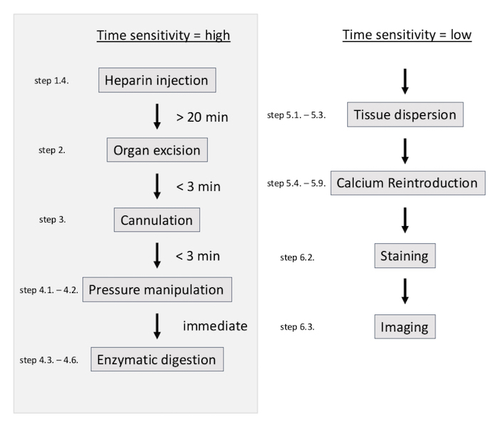
Figure 1: Simplified flowchart of the isolation procedure. The procedure is highly time-sensitive until the enzymatic digestion of the myocytes is completed. Please click here to view a larger version of this figure.
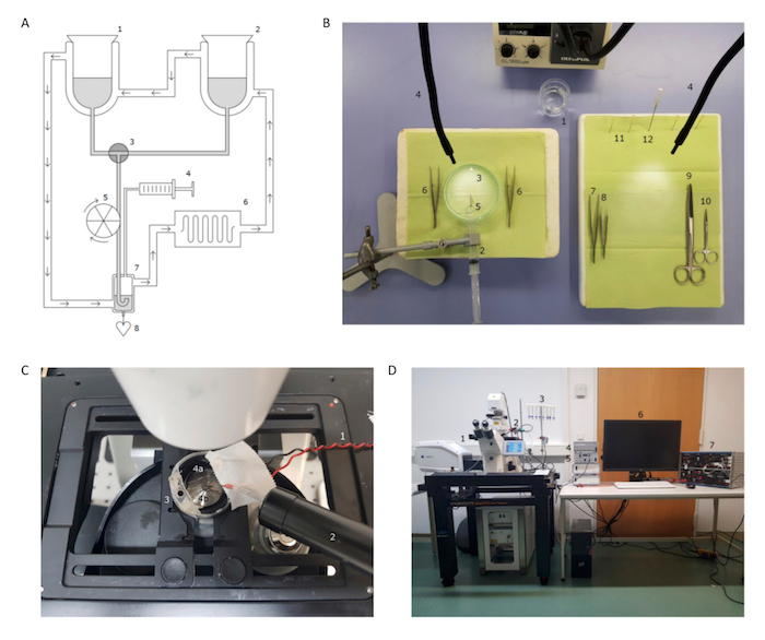
Figure 2: Preparation of equipment prior to the isolation. (A) This panel shows a self-made Langendorff apparatus: (1) a jacketed reaction vessel with PB; (2) a jacketed reaction vessel with CB; (3) a 3-way stopcock; (4) a syringe; (5) a peristaltic pump; (6) a heating immersion; (7) a jacketed bubble trap; and (8) the cannula and heart. (B) This panel shows the set-up for the cannulation and organ excision for an optimized work flow: (1) a 100 mL beaker with 50 mL of ice-cold CB; (2) a 10 mL syringe with ice-cold CB; (3) a Petri dish with ice-cold CB; (4) a light source; (5) the custom-made cannula with a cannulation knot (see also Figure 3A and 3B); (6) fine, curved forceps; (7) tissue forceps; (8) fine forceps, angled 45°; (9) abdominal surgical scissors; (10) fine surgical scissors; (11) 4 x 30 G needles; and (12) a 15 G needle. (C) This panel shows the microscope-mounted equipment for the confocal imaging: (1) electric stimulator electrodes; (2) a superfusion pen; (3) a glass-bottom dish with the ACMs; and (4) immersed platinum electric stimulator electrodes. (D) This panel shows the microscope set-up for the confocal imaging: (1) the confocal microscope; (2) a superfusion pen; (3) a superfusion buffer reservoir; (4) a superfusion flow regulator; (5) a superfusion heating module; (6) a computer workstation; and (7) an electric stimulator. Please click here to view a larger version of this figure.

Figure 3: Custom-made equipment. (A) This panel shows the manipulated 15 G cannula with a Luer lock. The arrows indicate two indentations for the cannulation knots. (B) These are the cannulation knots, two double overhand knots placed on top of each other for rapid tightening. The arrows indicate where and in which order the knot needs to be tightened. (C) This panel shows the assembled pressure control device. A 21 G butterfly needle is hooked into a tripod clamp. The hose is kept at the same height as the needle. The screw top is opened. The elevation of the butterfly hose can be altered as indicated by the arrows in order to change the intraluminal pressure of the left atrium. (D) The left atrium is punctured with the pressure control device. This picture shows an ideally inflated left atrium. The ellipse marks the placement of the overhand knot. (E) The left atrium is punctured with the pressure control device. This picture shows an over-inflated left atrium, which will result in a lower yield of viable ACMs. The ellipse marks the placement of the overhand knot. Please click here to view a larger version of this figure.
Access restricted. Please log in or start a trial to view this content.
Results
Loading...
$$\rightleftharpoonup{xx}$$
$$\longleftharp{xx}$$,
$$\longrightharp{xx}$$,
At 21 weeks of age, 60–90% of viable ACMs (estimated as described in step 6.1), after the calcium re-adaptation (step 5.4–5.7), can be isolated from ZSF-1 obese rats by this method (Figure 4A). In rats, ACMs are characterized by a different and more heterogenous phenotype compared to VCMs24,25. Figure 4B shows an individual ACM with preserved membranes and sarcomere struc...
Access restricted. Please log in or start a trial to view this content.
Discussion
Loading...
$$\rightleftharpoonup{xx}$$
$$\longleftharp{xx}$$,
$$\longrightharp{xx}$$,
Here, we first described a protocol for the isolation of single-cell ACMs from a rat model of MetS-related HFpEF that shows marked atrial remodeling22. The procedure is uniquely challenging as excessive fatty tissue can make the surgical preparation, as well as the cannulation of the aorta, increasingly difficult. The troubleshooting guide provided in Table 2 addresses the most common issues of the isolation procedure.
Access restricted. Please log in or start a trial to view this content.
Disclosures
Loading...
$$\rightleftharpoonup{xx}$$
$$\longleftharp{xx}$$,
$$\longrightharp{xx}$$,
The authors have nothing to disclose.
Acknowledgements
Loading...
$$\rightleftharpoonup{xx}$$
$$\longleftharp{xx}$$,
$$\longrightharp{xx}$$,
This research was supported by the DZHK (German Centre for Cardiovascular Research, D.B.), the EKFS (Else-Kröner-Fresenius Stiftung, F.H.), and by the BMBF (German Ministry of Education and Research), as well as the BIH-Charité clinical scientist program funded by the Charité - Universitätsmedizin Berlin and the Berlin Institute of Health (F.H.).
Access restricted. Please log in or start a trial to view this content.
Materials
| Name | Company | Catalog Number | Comments |
|---|---|---|---|
| ZSF-1 Obese rat | Charles River Laboratories, Inc. | 21 weeks old | |
| Fine Iris Scissors | Fine Science Tools GmbH | 14094-11 | |
| Surgical Scissors | Fine Science Tools GmbH | 14001-18 | |
| Micro Dressing Forceps (curved, serrated) | Aesculap, Inc. | BD312R | |
| Tissue Forceps (straight, 1 x 2 teeth) | Aesculap, Inc. | BD537R | |
| Tying Forceps (angled) | Aesculap, Inc. | MA624R | |
| Rodent and Small Animal Guillotine | Kent Scientific Corp. | DCAP | |
| Low Cost Induction Chamber 3.0 L | Kent Scientific Corp. | SOMNO-0730 | |
| Butterfly Winged Infusion Set 21 G | Hospira, Inc. | 181106101 | |
| Abbocath 16 G | Hospira, Inc. | 0G7149702 | |
| Microlance Hypodermic Needle | Becton Dickinson GmbH | 301300 | modify needle to make cannula |
| Braun Original Perfusor Syringe 50 ml | B. Braun Melsungen AG | 8728810F | |
| Braun Inject Solo Syringe 10 ml | B. Braun Melsungen AG | 2057926 | |
| Beaker 50ml | Duran Group (DWK Life Sciences GmbH) | 21 106 17 | |
| Duroplan petri dish (100 x 20 mm) | Duran Group (DWK Life Sciences GmbH) | 21 755 48 | |
| Seraflex Suture USP 3/0 | SERAG-WIESSNER GmbH & Co. KG | IC208000 | |
| VWR disposable Square Weighin Boats 100ml | VWR, Inc. | 10803-148 | |
| Styrofoam surface | |||
| Sodium chloride | Sigma-Aldrich, Inc. | 71380 | |
| Potassium chloride | Sigma-Aldrich, Inc. | P4504 | |
| Potassium phosphate monobasic | Sigma-Aldrich, Inc. | P5379 | |
| Sodium phosphate dibasic | Sigma-Aldrich, Inc. | S0876 | |
| Magensium sulfate heptahydrate | Sigma-Aldrich, Inc. | 230391 | |
| Magensium chloride | Sigma-Aldrich, Inc. | M8266 | |
| HEPES | Sigma-Aldrich, Inc. | H3375 | |
| Taurine | Sigma-Aldrich, Inc. | T0625 | |
| Glucose | Sigma-Aldrich, Inc. | G7528 | |
| 2,3-Butanedione monoxime | Sigma-Aldrich, Inc. | B0753 | |
| Calcium chloride solution (1 M) | Sigma-Aldrich, Inc. | 21115 | |
| Bovine Serum Albumin | Sigma-Aldrich, Inc. | A9647 | |
| Liberase | Roche (Sigma-Aldrich, Inc.) | LIBTM-RO | |
| Heparin | Rotexmedica GmbH | 3862357 | |
| Forene (Isoflurane) | Abbvie Deutschland GmbH & Co. KG | 10182054 | |
| Laminin from Engelbreth-Holm-Swarm murine sarcoma basement membrane | Sigma-Aldrich, Inc. | L2020 | |
| WillCo glass-bottom dish 500µl 0.005mm | WillCo Wells B.V. | HBST-3522 | |
| Fluo4 AM | Invitrogen (Thermo Fisher Scientific, Inc.) | F14201 | 5µM for 20min at RT |
| Di-8-ANNEPS | Invitrogen (Thermo Fisher Scientific, Inc.) | D3167 | 10µM for 45 min at 37° C |
| Mitotracker RED FM | Invitrogen (Thermo Fisher Scientific, Inc.) | M22425 | 20nM for 30 min at 37° C |
| Jacketed reaction vessel 500 ml | Gebr. Rettberg GmbH | 107024414 | |
| Jacketed reaction vessel 1000 ml | Gebr. Rettberg GmbH | 107025414 | |
| Jacketed bubble trap | Gebr. Rettberg GmbH | 134720001 | |
| ED heating immersion circulator | Julabo GmbH | 9116000 | |
| Reglo Digital MS-2/6 peristaltic pump | Ismatec (Cole-Parmer Gmbh) | ISM 831 | |
| Voltcraft Thermometer 302 K/J | Conrad Electronic SE | 030300546 | |
| Tubing | |||
| LSM 700 microscope | Carl Zeiss, Inc. | ||
| ZEN 2.3 imaging software | Carl Zeiss, Inc. | 410135-1011-240 | |
| Single channel heater controller TC-324B | Warner Instruments, LLC | 64-2400 | |
| 8 channel perfusion system | Warner Instruments, LLC | 64-0185 | |
| 8 channel Multi-Line In-Line Solution Heaters | Warner Instruments, LLC | 64-0105 |
References
Loading...
$$\rightleftharpoonup{xx}$$
$$\longleftharp{xx}$$,
$$\longrightharp{xx}$$,
- Alberti, K. G., et al. Harmonizing the metabolic syndrome: a joint interim statement of the International Diabetes Federation Task Force on Epidemiology and Prevention; National Heart, Lung, and Blood Institute; American Heart Association; World Heart Federation; International Atherosclerosis Society; and International Association for the Study of Obesity. Circulation. 120 (16), 1640-1645 (2009).
- International Diabetes Federation Task Force on Epidemiology and Prevention. IDF Consensus Worldwide Definition of the Metabolic Syndrome. , Available from: https://www.idf.org/e-library/consensus-statements/60-idfconsensus-worldwide-definitionof-the-metabolic-syndrome.html (2006).
- Melenovsky, V., et al. Left atrial remodeling and function in advanced heart failure with preserved or reduced ejection fraction. Circulation: Heart Failure. 8 (2), 295-303 (2015).
- Goette, A., et al. EHRA/HRS/APHRS/SOLAECE expert consensus on atrial cardiomyopathies: definition, characterization, and clinical implication. EP Europace. 18 (10), 1455-1490 (2016).
- Schotten, U., Verheule, S., Kirchhof, P., Goette, A. Pathophysiological mechanisms of atrial fibrillation: a translational appraisal. Physiological Reviews. 91 (1), 265-325 (2011).
- Hohendanner, F., DeSantiago, J., Heinzel, F. R., Blatter, L. A. Dyssynchronous calcium removal in heart failure-induced atrial remodeling. American Journal of Physiology-Heart and Circulatory Physiology. 311 (6), H1352-H1359 (2016).
- Hohendanner, F., et al. Inositol-1,4,5-trisphosphate induced Ca2+ release and excitation-contraction coupling in atrial myocytes from normal and failing hearts. The Journal of Physiology. 593 (6), 1459-1477 (2015).
- Tada, Y., et al. Role of mineralocorticoid receptor on experimental cerebral aneurysms in rats. Hypertension. 54 (3), 552-557 (2009).
- Iwasaki, Y. K., et al. Atrial fibrillation promotion with long-term repetitive obstructive sleep apnea in a rat model. Journal of the American College of Cardiology. 64 (19), 2013-2023 (2014).
- Field, L. J. Atrial natriuretic factor-SV40 T antigen transgenes produce tumors and cardiac arrhythmias in mice. Science. 239 (4843), 1029-1033 (1988).
- Claycomb, W. C., et al. HL-1 cells: a cardiac muscle cell line that contracts and retains phenotypic characteristics of the adult cardiomyocyte. Proceedings of the National Academy of Sciences of the United States of America. 95 (6), 2979-2984 (1998).
- Sartiani, L., Bochet, P., Cerbai, E., Mugelli, A., Fischmeister, R. Functional expression of the hyperpolarization-activated, non-selective cation current I(f) in immortalized HL-1 cardiomyocytes. The Journal of Physiology. 545 (Pt 1), 81-92 (2002).
- Louch, W. E., Sheehan, K. A., Wolska, B. M. Methods in cardiomyocyte isolation, culture, and gene transfer. Journal of Molecular and Cellular Cardiology. 51 (3), 288-298 (2011).
- Gunduz, D., Hamm, C. W., Aslam, M. Simultaneous Isolation of High Quality Cardiomyocytes, Endothelial Cells, and Fibroblasts from an Adult Rat Heart. Journal of Visualized Experiments. (123), e55601(2017).
- Li, D., Wu, J., Bai, Y., Zhao, X., Liu, L. Isolation and culture of adult mouse cardiomyocytes for cell signaling and in vitro cardiac hypertrophy. Journal of Visualized Experiments. (87), e51357(2014).
- Graham, E. L., et al. Isolation, culture, and functional characterization of adult mouse cardiomyoctyes. Journal of Visualized Experiments. (79), e50289(2013).
- Roth, G. M., Bader, D. M., Pfaltzgraff, E. R. Isolation and physiological analysis of mouse cardiomyocytes. Journal of Visualized Experiments. (91), e51109(2014).
- Thum, T., Borlak, J. Isolation and cultivation of Ca2+ tolerant cardiomyocytes from the adult rat: improvements and applications. Xenobiotica. 30 (11), 1063-1077 (2000).
- Egorova, M. V., Afanas'ev, S. A., Popov, S. V. A simple method for isolation of cardiomyocytes from adult rat heart. Bulletin of Experimental Biology and Medicine. 140 (3), 370-373 (2005).
- Kohncke, C., et al. Isolation and Kv channel recordings in murine atrial and ventricular cardiomyocytes. Journal of Visualized Experiments. (73), e50145(2013).
- Wagner, E., Brandenburg, S., Kohl, T., Lehnart, S. E. Analysis of tubular membrane networks in cardiac myocytes from atria and ventricles. Journal of Visualized Experiments. (92), e51823(2014).
- Hohendanner, F., et al. Cellular mechanisms of metabolic syndrome-related atrial decompensation in a rat model of HFpEF. Journal of Molecular and Cellular Cardiology. 115, 10-19 (2017).
- Seluanov, A., Vaidya, A., Gorbunova, V. Establishing primary adult fibroblast cultures from rodents. Journal of Visualized Experiments. (44), e2033(2010).
- Bootman, M. D., Higazi, D. R., Coombes, S., Roderick, H. L. Calcium signalling during excitation-contraction coupling in mammalian atrial myocytes. Journal of Cell Science. 119 (Pt 19), 3915-3925 (2006).
- Smyrnias, I., et al. Comparison of the T-tubule system in adult rat ventricular and atrial myocytes, and its role in excitation-contraction coupling and inotropic stimulation. Cell Calcium. 47 (3), 210-223 (2010).
- Pritchett, A. M., et al. Diastolic dysfunction and left atrial volume: a population-based study. Journal of the American College of Cardiology. 45 (1), 87-92 (2005).
- Linz, D., et al. Cathepsin A mediates susceptibility to atrial tachyarrhythmia and impairment of atrial emptying function in Zucker diabetic fatty rats. Cardiovascular Research. 110 (3), 371-380 (2016).
- Ackers-Johnson, M., et al. A Simplified, Langendorff-Free Method for Concomitant Isolation of Viable Cardiac Myocytes and Nonmyocytes From the Adult Mouse Heart. Circulation Research. 119 (8), 909-920 (2016).
- Ramanathan, T., Skinner, H. Coronary blood flow. Continuing Education in Anaesthesia Critical Care & Pain. 5 (2), 61-64 (2005).
- Bond, M. D., Van Wart, H. E. Characterization of the individual collagenases from Clostridium histolyticum. Biochemistry. 23 (13), 3085-3091 (1984).
- Deel, E. D., et al. In vitro model to study the effects of matrix stiffening on Ca(2+) handling and myofilament function in isolated adult rat cardiomyocytes. The Journal of Physiology. 595 (14), 4597-4610 (2017).
- Wuensch, E., Heidrich, H. G. [On the Quantitative Determination of Collagenase]. Hoppe-Seyler's Zeitschrift für physiologische Chemie. 333, 149-151 (1963).
- Conceicao, G., Heinonen, I., Lourenco, A. P., Duncker, D. J., Falcao-Pires, I. Animal models of heart failure with preserved ejection fraction. Netherlands Heart Journal. 24 (4), 275-286 (2016).
- Horgan, S., Watson, C., Glezeva, N., Baugh, J. Murine models of diastolic dysfunction and heart failure with preserved ejection fraction. Journal of Cardiac Failure. 20 (12), 984-995 (2014).
Access restricted. Please log in or start a trial to view this content.
Reprints and Permissions
Request permission to reuse the text or figures of this JoVE article
Request Permission