A protocol for the preparation of poly(pentafluorophenyl acrylate) (poly(PFPA)) grafted silica beads is presented. The poly(PFPA) functionalized surface is then immobilized with antibodies and used successfully for the protein separation through immunoprecipitation.
A subscription to JoVE is required to view this content. Sign in or start your free trial.
Method Article
Preparation of Poly(pentafluorophenyl acrylate) Functionalized SiO2 Beads for Protein Purification
In This Article
Erratum Notice
Important: There has been an erratum issued for this article. View Erratum Notice
Summary
Abstract
We demonstrate a simple method to prepare poly(pentafluorophenyl acrylate) (poly(PFPA)) grafted silica beads for antibody immobilization and subsequent immunoprecipitation (IP) application. The poly(PFPA) grafted surface is prepared via a simple two-step process. In the first step, 3-aminopropyltrimethoxysilane (APTMS) is deposited as a linker molecule onto the silica surface. In the second step, poly(PFPA) homopolymer, synthesized via the reversible addition and fragmentation chain transfer (RAFT) polymerization, is grafted to the linker molecule through the exchange reaction between the pentafluorophenyl (PFP) units on the polymer and the amine groups on APTMS. The deposition of APTMS and poly(PFPA) on the silica particles are confirmed by X-ray photoelectron spectroscopy (XPS), as well as monitored by the particle size change measured via dynamic light scattering (DLS). To improve the surface hydrophilicity of the beads, partial substitution of poly(PFPA) with amine-functionalized poly(ethylene glycol) (amino-PEG) is also performed. The PEG-substituted poly(PFPA) grafted silica beads are then immobilized with antibodies for IP application. For demonstration, an antibody against protein kinase RNA-activated (PKR) is employed, and IP efficiency is determined by Western blotting. The analysis results show that the antibody immobilized beads can indeed be used to enrich PKR while non-specific protein interactions are minimal.
Introduction
Reactive polymer brushes have received much interest in recent years. They can be used to immobilize functional molecules on organic or inorganic materials to create activated surfaces with applications in areas such as detection and separation1,2,3,4,5. Among the reactive polymers reported, those containing pentafluorophenyl ester units are particularly useful due to their high reactivity with amines and resistance toward hydrolysis6. One such polymer is poly(PFPA), and it can be readily functionalized post-polymerization with molecules containing primary or secondary amines7,8,9,10. In one example, poly(PFPA) brushes were reacted with amino-spiropyrans to create light-responsive surfaces7.
The preparation of poly(PFPA) and its applications have been described in a number of previous publications6,7,8,9,10,11,12,13,14,15,16,17. In particular, Theato and co-workers reported the synthesis of poly(PFPA) brushes via both "grafting to" and "grafting from" methods7,8,10,11,12. In the "grafting to" approach, a poly(methylsilsesquioxane)-poly(pentafluorophenyl acrylate) (poly(MSSQ-PFPA)) hybrid polymer was synthesized8,10,11,12. The poly(MSSQ) component was able to form strong adhesion with a number of different organic and inorganic surfaces, thus allowing the poly(PFPA) component to form a brush layer on the coated material surface. In the "grafting from" approach, surface initiated reversible addition and fragmentation chain transfer (SI-RAFT) polymerization was employed to prepare poly(PFPA) brushes7. In this case, a surface immobilized chain transfer agent (SI-CTA) was first covalently attached to the substrate via silica-silane reaction. The immobilized SI-CTA then participated in the SI-RAFT polymerization of PFPA monomers, generating densely packed poly(PFPA) brushes with stable covalent linkage to the substrate.
By utilizing the poly(PFPA) brushes synthesized via SI-RAFT polymerization, we recently demonstrated the immobilization of antibodies on poly(PFPA) grafted silica particles and their subsequent application in protein purification18. The use of poly(PFPA) brushes for antibody immobilization was found to resolve a number of issues associated with current protein separation through IP. Conventional IP relies on the use of Protein A/G as a linker for antibody immobilization19,20,21. Since the use of Protein A/G allows the antibodies to be attached with a specific orientation, high target antigen recovery efficiency is achieved. However, the use of Protein A/G suffers from non-specific protein interaction as well as the loss of antibodies during protein recovery, both of which contribute to a high level of background noise. To resolve these shortcomings, direct crosslinking of the antibodies to a solid support has been explored22,23,24. The efficiency of such techniques is typically low due to the random orientation of the crosslinked antibodies. For the poly(PFPA) grafted substrate, the immobilization of antibodies is permanent, achieved through exchange reaction between PFP units and amine functionalities on antibodies. Although the antibody orientation is still random, the system benefits from having many reactive PFP sites, controllable by the degree of polymerization. Furthermore, we showed that by partial substitution of PFP units with amino-PEG, surface hydrophilicity can be tuned, further improving the protein recovery efficiency of the system18. Overall, the poly(PFPA) grafted silica particles were shown to be an effective alternative to traditional IP with reasonable efficiency as well as much cleaner background.
In this contribution, we report an alternative method to prepare poly(PFPA) grafted surface for antibody immobilization and IP application. In a simple two-step process, as illustrated in Figure 1, an APTMS linker molecule is first deposited onto the silica surface, then the poly(PFPA) polymer is covalently attached to the linker molecule through the reaction between the PFP units on the polymer and the amine functions on APTMS. This preparation method allows for the permanent crosslinking of poly(PFPA) to a substrate surface, but avoids the many complications associated with SI-CTA synthesis and SI-RAFT polymerization of poly(PFPA) brushes. Partial substitution of the PFP units with amino-PEG can still be performed, allowing fine-tuning of the polymer brush surface properties. We show the poly(PFPA) grafted silica beads thus prepared can be immobilized with antibodies and used for protein enrichment via IP. The detailed bead preparation procedure, antibody immobilization, and IP testing are documented in this article, for readers interested in seeking an alternative to conventional Protein A/G based IP.
Access restricted. Please log in or start a trial to view this content.
Protocol
1. Preparation of Poly(PFPA) Homopolymer
- Recrystallization of AIBN
- Combine 5 g of 2,2’-azobis(2-methylpropionitrile) (AIBN) with 25 mL of methanol in a 250 mL beaker. Immerse the beaker in a 60 °C oil bath, then vigorously stir the mixture with a stir bar until AIBN is fully dissolved.
- Filter the warm solution through filter paper (5-8 μm particle retention) and store the filtrate at 4 °C to allow the crystals to form slowly.
- Collect the recrystallized AIBN by filtration. Combine the collected product with 25 mL of fresh methanol and repeat the recrystallization process.
- Dry the 2x recrystallized AIBN in a vacuum oven at room temperature (RT) overnight. Store the product in the dark at < -10 °C.
- Synthesis of benzyl dithiobenzoate25
- Prepare a 500 mL three-neck round-bottom flask equipped with a magnetic stir bar, a refluxing condenser, a dropping funnel, and a rubber septum. Connect the flask to the nitrogen gas line through the refluxing condenser and flush out the inside air with nitrogen. Insert a thermometer through the septum. Add 41 mL (0.041 mol) of 1 M solution of phenylmagnesium bromide in tetrahydrofuran (THF) via a syringe through the same septum.
- Warm the phenylmagnesium bromide solution to 40 °C in an oil bath. Then add 3.1 g (0.041 mol) of carbon disulfide through the dropping funnel slowly, maintaining the solution temperature at 40 °C.
- Add 7.1 g (0.042 mol) of benzyl bromide to the resultant mixture through the dropping funnel over 15 min. Increase the reaction temperature to 50 °C. Continue stirring at this temperature for 45 min.
- Transfer the reaction mixture into a separatory funnel and dilute with 15 mL of ice cold water. Extract the product by adding 15 mL of diethyl ether and remove the lower water layer. Repeat the extraction with diethyl ether two more times.
- Wash the combined organic phases with copious amount of water, then brine (solution of 50% (w/v) NaCl in water) and dry the product over anhydrous magnesium sulfate.
- Remove the solvent in vacuum at 35 °C using a rotary evaporator.
- Purify the product by column chromatography using 400 mL of silica gel (pore size 60 Å, 63-200 mesh particle size) and petroleum ether as the eluent, yielding 5 g of benzyl dithiobenzoate (BDB) as red oil. Confirm the product purity by 1H NMR (400 MHz, CDCl3): δ 8.02-7.99 (2H, m), 7.55-7.50 (1H, m), 7.41-7.29 (7H, m), 4.60 (2H, s).
- Synthesis of poly(PFPA) via RAFT polymerization9,26
- Commercially available PFPA monomer contains small amount of inhibitors. Before polymerization, remove the inhibitors by passing the monomer through a disposable syringe packed with basic alumina.
- Add 0.4 mg (0.0024 mmol) of recrystallized AIBN, 4.3 mg (0.018 mmol) of BDB, 1012 mg (4.25 mmol) of inhibitor-free PFPA, and 0.7 mL of anhydrous anisole to a 20 mL Schlenk flask.
- Connect the flask to Schlenk line and degas the mixture with at least three freeze-pump-thaw cycles. Briefly, freeze the reaction mixture in a liquid nitrogen bath. Apply vacuum to remove the gas in headspace. Seal the flask then remove away from liquid nitrogen to allow the content to thaw at RT.
- Place the flask in a 70 °C oil bath, and react for 4 h under N2 purge.
- To terminate the reaction, remove the flask from the oil bath and expose the reaction content to air.
- Precipitate the polymer in cold methanol, then dry the recovered polymer in a vacuum oven at 40 °C overnight.
- To measure the polymer molecular weight, use gel permeation chromatography (GPC). Use THF as the mobile phase at 35 °C with a 1 mL/min flow rate and construct the calibration curve using monodisperse polystyrene standards. To acquire GPC measurement, dissolve the polymer in THF (1-2 mg/mL) and filter through 0.2 μm disposable polytetrafluoroethylene (PTFE) filter. Inject 100 μL of the sample into the GPC instrument. Convert the measured sample retention time to molecular weight using the polystyrene calibration curve.
2. Preparation of Poly(PFPA) Functionalized SiO2 Beads
- Treatment of SiO2 beads with APTMS
- SiO2 particles are available in the form of a 5% (w/v) aqueous suspension. Combine 0.8 mL of SiO2 suspension with 40 mg of APTMS and 8 mL of methanol in a 20 mL scintillation vial equipped with a stir bar.
- Allow the reaction to proceed at RT for 5 h with vigorous stirring.
- Transfer the solution to a conical tube. To isolate the APTMS functionalized SiO2 beads, centrifuge the solution at 10,000 x g for 5 min, then remove the supernatant. Wash the beads by re-dispersing them in 3 mL of fresh methanol. Shake the tube by hand for mixing, but if necessary, improve the dispersion by sonication in a water bath for a few seconds. Centrifuge the beads at 10,000 x g for 5 min. Remove the supernatant and repeat the wash step one more time.
- Combine the methanol washed SiO2 beads with 3 mL of dimethyl sulfoxide (DMSO). Shake the mixture by hand, or if necessary sonicate for a few seconds, until the beads are fully dispersed in DMSO. Centrifuge the beads at 10,000 x g for 5 min, then remove the supernatant. Repeat the step to ensure complete solvent exchange from methanol to DMSO.
NOTE: The final suspension contains the APTMS functionalized SiO2 beads dispersed in 4 mL of DMSO. - To check the particle size distribution, perform DLS analysis. Take one drop of the suspension prepared in Step 2.1.4 and place into a disposable UV cuvette. Dilute the sample by filling the cuvette with fresh DMSO until it is 2/3 full. Insert the sample into the cell holder to begin data acquisition. For particle size measurement, use the following setup parameters: Temperature: 25 °C; Equilibration Time: 120 s; Measurement Duration: Automatic.
- To check the surface composition, perform XPS analysis. Dry a small sample from the suspension prepared in Step 2.1.4 in vacuum oven at 40 °C overnight. Take the dried polymer and pack evenly onto a 0.5 cm x 0.5 cm sample holder. Load the sample into the high vacuum chamber (10-8 torr) and begin data acquisition. For the particular XPS instrument used, generate the photoelectrons using a monochromatic Al Kα X-ray operated at 15 kV and 6.7 mA, and collect using hybrid mode magnification with the analyzer at a 50 eV pass energy for high resolution spectra, and a 100 eV pass energy for elemental surveys.
- Grafting poly(PFPA) to APTMS functionalized SiO2 beads
- Prepare the poly(PFPA) solution by dissolving 20 mg of poly(PFPA) in 2 mL of DMSO in a 20 mL scintillation vial.
NOTE: In this study, a relatively low molecular weight poly(PFPA) (20 kg/mol) is used. Thus, despite the high polymer concentration (10 mg/mL), no evidence of polymer crosslinking is observed. If a higher molecular weight polymer is used, then polymer solution concentration may need to be adjusted to avoid possible crosslinking. - Add 1 mL of APTMS functionalized SiO2 beads suspended in DMSO (from Step 2.1.4) to the poly(PFPA) solution. React at RT for 1 h with vigorous stirring.
- Isolate the poly(PFPA) grafted SiO2 beads by centrifugation at 10,000 x g for 5 min, followed by the removal of the supernatant. Wash the beads by adding 3 mL of DMSO and mix by either shaking with hand or few seconds of sonication. Centrifuge the beads at 10,000 x g for 5 min, then remove the supernatant. Repeat washing of the poly(PFPA) grafted SiO2 beads with DMSO twice.
- Wash the beads two times more with triple distilled water (TDW). In this step, combine the beads with 3 mL of TDW, then mix by shaking with hand or few seconds of sonication. Centrifuge the beads at 10,000 x g for 5 min, then remove the supernatant.
- To check the particle size distribution, perform DLS following the procedure described in Step 2.1.5. To check the surface chemistry, perform XPS following the procedure described in Step 2.1.6.
- Prepare the poly(PFPA) solution by dissolving 20 mg of poly(PFPA) in 2 mL of DMSO in a 20 mL scintillation vial.
3. Preparation of SiO2 Beads Grafted with PEG-Substituted Poly(PFPA)
- To prepare the poly(PFPA) solution, dissolve 20 mg of poly(PFPA) in 2 mL of DMSO in a 20 mL scintillation vial.
- To prepare PEG solution, dissolve amine-functionalized PEG in 1 mL of DMSO. The exact amount of PEG used is determined by the desired degree of PFP substitution, determined by the equation shown below:
Amount of amino-PEG (g/g-poly(PFPA)) = (N_poly(PFPA) x % PEG-Sub) x (MW_PEG / MW_poly(PFPA))
where N_poly(PFPA) = poly(PFPA) degree of polymerization
% PEG-Sub = percent PEG substitution
MW_PEG = molecular weight of amino-PEG
MW_ poly(PFPA) = molecular weight of poly(PFPA) - Transfer the PEG solution to the poly(PFPA) solution. React at RT for 1 h with vigorous stirring.
- To prepare APTMS functionalized SiO2 beads suspended in DMSO, follow the same steps shown in Step 2.1. Transfer 1 mL of the bead suspension into the PEG-substituted poly(PFPA) solution prepared in Step 3.3. Allow the grafting between poly(PFPA) and APTMS functionalized SiO2 beads to proceed at RT for 1 h with vigorous stirring.
- Isolate the beads by centrifugation at 10,000 x g for 5 min, followed by the removal of the supernatant. Wash the beads by adding 3 mL of DMSO and mix by either shaking with hand or few seconds of sonication. Centrifuge the beads at 10,000 x g for 5 min, then remove the supernatant. Repeat the DMSO wash twice.
- Wash the beads two times more with TDW. In this step, combine the beads with 3 mL TDW, then mix by shaking with hand or few seconds of sonication. Centrifuge the beads at 10,000 x g for 5 min, then remove the supernatant.
- Dry the beads at 40 °C in a vacuum oven overnight.
4. Antibody Immobilization on Poly(PFPA) Grafted SiO2 Beads
NOTE: The same procedure is used regardless of percent PEG substitution on poly(PFPA). Prepare phosphate buffered saline (PBS) by dissolving PBS tablet in TDW. Prepare 0.1% (v/v) phosphate buffered saline with Tween-20 (PBST) by adding 1/1000 of Tween-20 to PBS.
- Add 5 mg of poly(PFPA) grafted SiO2 beads to a 1.5 mL microcentrifuge tube.
- Wash the beads by adding 800 µL of PBS and mix well by vortexing. Centrifuge the beads at 10,000 x g at RT for 1 min. Remove the supernatant and repeat the wash step three times.
- Add 350 µL of fresh PBS, 50 µL 0.1% (v/v) PBST, and 6.67 µg of the antibody. Incubate ~20 h on a rotator at 4 °C.
- Wash the beads to remove unbound antibodies. Centrifuge the beads at 400 x g and 4 °C for 1 min. Remove the supernatant and add 400 µL of lysis buffer carefully. Gently re-suspend the beads by pipetting up and down for five times.
NOTE: Lysis buffer used to wash the beads should be the same one used during cell lysis and IP, except that the addition of dithiothreitol and protease inhibitor are optional, (see Step 5). - Repeat this wash step three times. After the final wash, remove the supernatant as much as possible.
5. Cell Lysis and Immunoprecipitation
- Preparation of lysis buffer and wash buffer
- Prepare the lysis buffer (50 mM Tris-HCl (pH 8.0), 100 mM KCl, 0.5% (v/v) NP-40, 10% (v/v) glycerol, 1 mM dithiothreitol (DTT), and protease inhibitor cocktail).
- Prepare the wash buffer (50 mM Tris-HCl (pH 8.0), 100 mM KCl, 0.1 % (v/v) NP-40, and 10% (v/v) glycerol).
- Store the buffer solutions at 4 °C.
- Preparation of the cells
- Seed the cells (HeLa cells) one or two days before IP experiment, and grow the cells at 37 °C and 5% CO2.
- Collect about 1.4 x 10⁷ cells with a cell scraper and transfer into a 15 mL conical tube. Centrifuge the cells at 380 x g at RT for 3 min. Remove the supernatant and re-suspend with 1 mL of cold PBS and transfer into a 1.5 mL microcentrifuge tube.
- Centrifuge the cells at 10,000 x g at 4 °C for 30 s. Remove the supernatant cleanly. Cell pellets can be stored at -80 °C after removing the supernatant.
- Preparation of cell lysates
- Re-suspend the cell pellet with 400 µL of the lysis buffer. Sonicate the cells using an ultrasonicator.
- After the sonication, vortex briefly and centrifuge the lysate at 20,000 x g at 4 °C for 10 min.
- Transfer the supernatant to a new 1.5 mL centrifuge tube.
- Immunoprecipitation
- Transfer 300 µL of cell lysate to previously prepared antibody incubated poly(PFPA) grafted SiO2 beads. Retain 30 µL of the cell lysate as the input sample in a new microcentrifuge tube. Store the input sample at 4 °C.
NOTE: The total amount of protein in cell lysate should be approximately 4 mg. - Incubate the lysate/beads mixture for 3 h on a rotator at 4 °C.
- Centrifuge the mixture at 400 x g at 4 °C for 1 min. Remove the supernatant and add 400 µL of wash buffer carefully. Gently re-suspend the beads by pipetting up and down about five times.
- Repeat this wash step three times. After the final wash, remove the supernatant as much as possible.
- Prepare 2x sodium dodecyl sulfate (SDS) loading dye (25% (v/v) glycerol, 0.1% (w/v) bromo phenol blue (BPB), 60 mM Tris-HCl (pH 6.8), 2% (w/v) SDS, and 2.75 mM 2-mercaptoethanol). Store 2x SDS loading dye at -20 °C. Add 30 µL of 2x SDS loading dye to the beads and the stored input sample, and heat them for 10 min at 95 °C.
- After heating, analyze the sample using Western blotting27, or store the sample at -20 °C.
- Transfer 300 µL of cell lysate to previously prepared antibody incubated poly(PFPA) grafted SiO2 beads. Retain 30 µL of the cell lysate as the input sample in a new microcentrifuge tube. Store the input sample at 4 °C.
Access restricted. Please log in or start a trial to view this content.
Results
A schematic for the preparation of poly(PFPA) grafted SiO2 beads, with or without PEG substitution is shown in Figure 1. To monitor the APTMS and poly(PFPA) grafting process, bare SiO2 beads, APTMS functionalized SiO2 beads, and poly(PFPA) grafted SiO2 beads are characterized by both DLS (Figure 2) and XPS (Figure 3). IP efficiencies of the beads are determ...
Access restricted. Please log in or start a trial to view this content.
Discussion
The synthesis of poly(PFPA) grafted SiO2 beads is illustrated in Figure 1. By employing APTMS as a linker molecule, poly(PFPA) brushes covalently grafted to SiO2 substrate can be prepared via a simple two-step process. Although some of the PFP units are sacrificed for the reaction with APTMS, a large number of the PFP units are expected to remain available for later reaction with either amino-PEG or antibodies. The PFP groups are known to form low energy surfaces so pol...
Access restricted. Please log in or start a trial to view this content.
Disclosures
The authors have nothing to disclose.
Acknowledgements
This work was supported by Agency for Defense Development (Grant No. UD170039ID).
Access restricted. Please log in or start a trial to view this content.
Materials
| Name | Company | Catalog Number | Comments |
|---|---|---|---|
| 2,2-Azobisisobutyronitrile, 99% | Daejung Chemicals | 1102-4405 | |
| Methyl alcohol for HPLC, 99.9% | Duksan Pure Chemicals | d62 | |
| Phenylmagnesium bromide solution 1.0 M in THF | Sigma-Aldrich | 331376 | |
| Carbon disulfide anhydrous, ≥99% | Sigma-Aldrich | 335266 | |
| Benzyl bromide, 98% | Sigma-Aldrich | B17905 | |
| Petroleum ether, 90% | Samchun Chemicals | P0220 | |
| Ethyl ether, 99% | Daejung Chemicals | 4025-4404 | |
| Magnesium sulfate anhydrous, powder, 99% | Daejung Chemicals | 5514-4405 | |
| Pentafluorophenyl acrylate | Santa Cruz Biotechnology | sc-264001 | contains inhibitor |
| Aluminium oxide, activated, basic, Brockmann I | Sigma-Aldrich | 199443 | |
| Sodium Chloride (NaCl) | Daejung Chemicals | 7548-4400 | |
| Anisole anhydrous, 99.7% | Sigma-Aldrich | 296295 | |
| Silica nanoparticle | Microparticles GmbH | SiO2-R-0.7 | 5% w/v aqueous suspension |
| 3-Aminopropyltrimethoxysilane, >96.0% | Tokyo Chemical Industry | T1255 | |
| Dimethyl sulfoxide for HPLC, ≥99.7% | Sigma-Aldrich | 34869 | |
| Amino-terminated poly(ethylene glycol) methyl ether | Polymer Source | P16082-EGOCH3NH2 | |
| Phosphate buffered saline tablet | Takara | T9181 | |
| Tween-20 | Calbiochem | 9480 | |
| Tris-HCl (pH 8.0) | Invitrogen | AM9855G | |
| KCl | Invitrogen | AM9640G | |
| NP-40 | VWR | E109-50ML | |
| Glycerol | Invitrogen | 15514-011 | |
| Dithiothreitol | Biosesang | D1037 | |
| Protease inhibitor | Merck | 535140-1MLCN | |
| Bromo phenol blue | Sigma-Aldrich | B5525-5G | |
| Tris-HCl (pH 6.8) | Biosolution | BT033 | |
| Sodium dodecyl sulfate | Biosolution | BS003 | |
| 2-Mercaptoethanol | Gibco | 21985-023 | |
| PKR Antibody | Cell Signaling Technology | 12297S | |
| GAPDH Antibody | Santa Cruz Biotechnology | sc-32233 | |
| Normal Rabbit IgG | Cell Signaling Technology | 2729S | |
| HeLa | Korea Cell Line Bank | 10002 | |
| Sonicator | DAIHAN Scientific | WUC-D10H | |
| Ultrasonicator | BMBio | BR2006A | |
| Centrifuge I | Eppendorf | 5424 R | |
| Centrifuge II | LABOGENE | 1736R | |
| Rotator | FINEPCR | ROTATOR/AG | |
| Vacuum oven | DAIHAN Scientific | ThermoStable OV-30 | |
| Gel permeation chromatography (THF) | Agilent Technologies | 1260 Infinity II | |
| X-ray photoelectron spectrometer | Thermo VG Scientific | Sigma Probe | |
| Dynamic light scattering | Malvern Instruments | ZEN 3690 |
References
- Johnsson, B., Löfås, S., Lindquist, G. Immobilization of proteins to a carboxymethyldextran-modified gold surface for biospecific interaction analysis in surface plasmon resonance sensors. Analytical Biochemistry. 198 (2), 268-277 (1991).
- Kurzawa, C., Hengstenberg, A., Schuhmann, W. Immobilization method for the preparation of biosensors based on pH shift-induced deposition of biomolecule-containing polymer films. Analytical Chemistry. 74 (2), 355-361 (2002).
- You, C. C., et al. Detection and identification of proteins using nanoparticle-fluorescent polymer 'chemical nose' sensors. Nature Nanotechnology. 2 (5), 318-323 (2007).
- Roberts, M. W., Ongkudon, C. M., Forde, G. M., Danquah, M. K. Versatility of polymethacrylate monoliths for chromatographic purification of biomolecules. Journal of Separation Science. 32 (15-16), 2485-2494 (2009).
- Sandison, M. E., Cumming, S. A., Kolch, W., Pitt, A. R. On-chip immunoprecipitation for protein purification. Lab on a Chip. 10 (20), 2805-2813 (2010).
- Das, A., Theato, P. Activated Ester Containing Polymers: Opportunities and Challenges for the Design of Functional Macromolecules. Chemical Reviews. 116 (3), 1434-1495 (2016).
- Choi, J., et al. Functionalization and patterning of reactive polymer brushes based on surface reversible addition and fragmentation chain transfer polymerization. Journal of Polymer Science Part A: Polymer Chemistry. 50 (19), 4010-4018 (2012).
- Kessler, D., Jochum, F. D., Choi, J., Char, K., Theato, P. Reactive surface coatings based on polysilsesquioxanes: universal method toward light-responsive surfaces. ACS Applied Materials & Interfaces. 3 (2), 124-128 (2011).
- Son, H., et al. Penetration and exchange kinetics of primary alkyl amines applied to reactive poly(pentafluorophenyl acrylate) thin films. Polymer Journal. 48 (4), 487-495 (2016).
- Kessler, D., Roth, P. J., Theato, P. Reactive surface coatings based on polysilsesquioxanes: controlled functionalization for specific protein immobilization. Langmuir. 25 (17), 10068-10076 (2009).
- Kessler, D., Theato, P. Reactive surface coatings based on polysilsesquioxanes: defined adjustment of surface wettability. Langmuir. 25 (24), 14200-14206 (2009).
- Kessler, D., Nilles, K., Theato, P. Modular approach towards multi-functional surfaces with adjustable and dual-responsive wettability using a hybrid polymer toolbox. Journal of Materials Chemistry. 19 (43), 8184-8189 (2009).
- Eberhardt, M., Mruk, R., Zentel, R., Theato, P. Synthesis of pentafluorophenyl(meth)acrylate polymers: new precursor polymers for the synthesis of multifunctional materials. European Polymer Journal. 41 (7), 1569-1575 (2005).
- Jochum, F. D., Forst, F. R., Theato, P. PNIPAM copolymers containing light-responsive chromophores: a method toward molecular logic gates. Macromolecular Rapid Communications. 31 (16), 1456-1461 (2010).
- Schattling, P., Pollmann, I., Theato, P. Synthesis of CO2-responsive polymers by post-polymerization modification. Reactive & Functional Polymers. 75, 16-21 (2014).
- He, L., Szameit, K., Zhao, H., Hahn, U., Theato, P. Postpolymerization modification using less cytotoxic activated ester polymers for the synthesis of biological active polymers. Biomacromolecules. 15 (8), 3197-3205 (2014).
- Arnold, R. M., McNitt, C. D., Popik, V. V., Locklin, J. Direct grafting of poly(pentafluorophenyl acrylate) onto oxides: versatile substrates for reactive microcapillary printing and self-sorting modification. Chemical Communications. 50 (40), 5307-5309 (2014).
- Son, H., Ku, J., Kim, Y., Li, S., Char, K. Amine-Reactive Poly(pentafluorophenyl acrylate) Brush Platforms for Cleaner Protein Purification. Biomacromolecules. 19 (3), 951-961 (2018).
- Cullen, S. E., Schwartz, B. D. An improved method for isolation of H-2 and Ia alloantigens with immunoprecipitation induced by protein A-bearing staphylococci. The Journal of Immunology. 117 (1), 136-142 (1976).
- Sisson, T. H., Castor, C. W. An improved method for immobilizing IgG antibodies on protein A-agarose. Journal of Immunology Methods. 127 (2), 215-220 (1990).
- Peritz, T., et al. Immunoprecipitation of mRNA-protein complexes. Nature Protocols. 1 (2), 577-580 (2006).
- Zhang, Z., Chen, S., Jiang, S. Dual-functional biomimetic materials: nonfouling poly (carboxybetaine) with active functional groups for protein immobilization. Biomacromolecules. 7 (12), 3311-3315 (2006).
- Yao, Y., et al. NHS-ester functionalized poly(PEGMA) brushes on silicon surface for covalent protein immobilization. Colloids and Surfaces B: Biointerfaces. 66 (2), 233-239 (2008).
- Ma, J., et al. Facile fabrication of microsphere-polymer brush hierarchically three-dimensional (3D) substrates for immunoassays. Chemical Communications. 51 (31), 6749-6752 (2015).
- Chong, Y., et al. Thiocarbonylthio compounds [SC (Ph) S− R] in free radical polymerization with reversible addition-fragmentation chain transfer (RAFT Polymerization). Role of the free-radical leaving group (R). Macromolecules. 36 (7), 2256-2272 (2003).
- Jochum, F. D., Theato, P. Temperature- and Light-Responsive Polyacrylamides Prepared by a Double Polymer Analogous Reaction of Activated Ester Polymers. Macromolecules. 42 (16), 5941-5945 (2009).
- JoVE Science Education Database. Basic Methods in Cellular and Molecular Biology. The Western Blot. JoVE. , Cambridge, MA. (2018).
- Chua, G. B. H., Roth, P. J., Duong, H. T. T., Davis, T. P., Lowe, A. B. Synthesis and Thermoresponsive Solution Properties of Poly[oligo(ethylene glycol) (meth)acrylamide]s: Biocompatible PEG Analogues. Macromolecules. 45 (3), 1362-1374 (2012).
Access restricted. Please log in or start a trial to view this content.
Reprints and Permissions
Erratum
Formal Correction: Erratum: Preparation of Poly(pentafluorophenyl acrylate) Functionalized SiO2 Beads for Protein Purification
Posted by JoVE Editors on 4/30/2019. Citeable Link.
An erratum was issued for: Preparation of Poly(pentafluorophenyl acrylate) Functionalized SiO2 Beads for Protein Purification. Throughout the article, the term "3-aminopropyltriethoxysilane" has been replaced with "3-aminopropyltrimethoxysilane", and "APTES" with "APTMS".
The Keywords were updated from:
Poly(pentafluorophenyl acrylate), 3-aminopropyltriethoxysilane, reactive polymer brush, post-polymerization functionalization, antibody immobilization, immunoprecipitation
to:
Poly(pentafluorophenyl acrylate), 3-aminopropyltrimethoxysilane, reactive polymer brush, post-polymerization functionalization, antibody immobilization, immunoprecipitation
The Abstract was updated from:
We demonstrate a simple method to prepare poly(pentafluorophenyl acrylate) (poly(PFPA)) grafted silica beads for antibody immobilization and subsequent immunoprecipitation (IP) application. The poly(PFPA) grafted surface is prepared via a simple two-step process. In the first step, 3-aminopropyltriethoxysilane (APTES) is deposited as a linker molecule onto the silica surface. In the second step, poly(PFPA) homopolymer, synthesized via the reversible addition and fragmentation chain transfer (RAFT) polymerization, is grafted to the linker molecule through the exchange reaction between the pentafluorophenyl (PFP) units on the polymer and the amine groups on APTES. The deposition of APTES and poly(PFPA) on the silica particles are confirmed by X-ray photoelectron spectroscopy (XPS), as well as monitored by the particle size change measured via dynamic light scattering (DLS). To improve the surface hydrophilicity of the beads, partial substitution of poly(PFPA) with amine-functionalized poly(ethylene glycol) (amino-PEG) is also performed. The PEG-substituted poly(PFPA) grafted silica beads are then immobilized with antibodies for IP application. For demonstration, an antibody against protein kinase RNA-activated (PKR) is employed, and IP efficiency is determined by Western blotting. The analysis results show that the antibody immobilized beads can indeed be used to enrich PKR while non-specific protein interactions are minimal.
to:
We demonstrate a simple method to prepare poly(pentafluorophenyl acrylate) (poly(PFPA)) grafted silica beads for antibody immobilization and subsequent immunoprecipitation (IP) application. The poly(PFPA) grafted surface is prepared via a simple two-step process. In the first step, 3-aminopropyltrimethoxysilane (APTMS) is deposited as a linker molecule onto the silica surface. In the second step, poly(PFPA) homopolymer, synthesized via the reversible addition and fragmentation chain transfer (RAFT) polymerization, is grafted to the linker molecule through the exchange reaction between the pentafluorophenyl (PFP) units on the polymer and the amine groups on APTMS. The deposition of APTMS and poly(PFPA) on the silica particles are confirmed by X-ray photoelectron spectroscopy (XPS), as well as monitored by the particle size change measured via dynamic light scattering (DLS). To improve the surface hydrophilicity of the beads, partial substitution of poly(PFPA) with amine-functionalized poly(ethylene glycol) (amino-PEG) is also performed. The PEG-substituted poly(PFPA) grafted silica beads are then immobilized with antibodies for IP application. For demonstration, an antibody against protein kinase RNA-activated (PKR) is employed, and IP efficiency is determined by Western blotting. The analysis results show that the antibody immobilized beads can indeed be used to enrich PKR while non-specific protein interactions are minimal.
The fourth paragraph of the Introduction was updated from:
In this contribution, we report an alternative method to prepare poly(PFPA) grafted surface for antibody immobilization and IP application. In a simple two-step process, as illustrated in Figure 1, an APTES linker molecule is first deposited onto the silica surface, then the poly(PFPA) polymer is covalently attached to the linker molecule through the reaction between the PFP units on the polymer and the amine functions on APTES. This preparation method allows for the permanent crosslinking of poly(PFPA) to a substrate surface, but avoids the many complications associated with SI-CTA synthesis and SI-RAFT polymerization of poly(PFPA) brushes. Partial substitution of the PFP units with amino-PEG can still be performed, allowing fine-tuning of the polymer brush surface properties. We show the poly(PFPA) grafted silica beads thus prepared can be immobilized with antibodies and used for protein enrichment via IP. The detailed bead preparation procedure, antibody immobilization, and IP testing are documented in this article, for readers interested in seeking an alternative to conventional Protein A/G based IP.
to:
In this contribution, we report an alternative method to prepare poly(PFPA) grafted surface for antibody immobilization and IP application. In a simple two-step process, as illustrated in Figure 1, an APTMS linker molecule is first deposited onto the silica surface, then the poly(PFPA) polymer is covalently attached to the linker molecule through the reaction between the PFP units on the polymer and the amine functions on APTMS. This preparation method allows for the permanent crosslinking of poly(PFPA) to a substrate surface, but avoids the many complications associated with SI-CTA synthesis and SI-RAFT polymerization of poly(PFPA) brushes. Partial substitution of the PFP units with amino-PEG can still be performed, allowing fine-tuning of the polymer brush surface properties. We show the poly(PFPA) grafted silica beads thus prepared can be immobilized with antibodies and used for protein enrichment via IP. The detailed bead preparation procedure, antibody immobilization, and IP testing are documented in this article, for readers interested in seeking an alternative to conventional Protein A/G based IP.
Step 2.1 of the Protocol was updated from:
Treatment of SiO2 beads with APTES
to:
Treatment of SiO2 beads with APTMS
Step 2.1.1 of the Protocol was updated from:
SiO2 particles are available in the form of a 5% (w/v) aqueous suspension. Combine 0.8 mL of SiO2 suspension with 40 mg of APTES and 8 mL of methanol in a 20 mL scintillation vial equipped with a stir bar.
to:
SiO2 particles are available in the form of a 5% (w/v) aqueous suspension. Combine 0.8 mL of SiO2 suspension with 40 mg of APTMS and 8 mL of methanol in a 20 mL scintillation vial equipped with a stir bar.
Step 2.1.3 of the Protocol was updated from:
Transfer the solution to a conical tube. To isolate the APTES functionalized SiO2 beads, centrifuge the solution at 10,000 x g for 5 min, then remove the supernatant. Wash the beads by re-dispersing them in 3 mL of fresh methanol. Shake the tube by hand for mixing, but if necessary, improve the dispersion by sonication in a water bath for a few seconds. Centrifuge the beads at 10,000 x g for 5 min. Remove the supernatant and repeat the wash step one more time.
to:
Transfer the solution to a conical tube. To isolate the APTMS functionalized SiO2 beads, centrifuge the solution at 10,000 x g for 5 min, then remove the supernatant. Wash the beads by re-dispersing them in 3 mL of fresh methanol. Shake the tube by hand for mixing, but if necessary, improve the dispersion by sonication in a water bath for a few seconds. Centrifuge the beads at 10,000 x g for 5 min. Remove the supernatant and repeat the wash step one more time.
Step 2.1.4 of the Protocol was updated from:
Combine the methanol washed SiO2 beads with 3 mL of dimethyl sulfoxide (DMSO). Shake the mixture by hand, or if necessary sonicate for a few seconds, until the beads are fully dispersed in DMSO. Centrifuge the beads at 10,000 x g for 5 min, then remove the supernatant. Repeat the step to ensure complete solvent exchange from methanol to DMSO.NOTE: The final suspension contains the APTES functionalized SiO2 beads dispersed in 4 mL of DMSO.
to:
Combine the methanol washed SiO2 beads with 3 mL of dimethyl sulfoxide (DMSO). Shake the mixture by hand, or if necessary sonicate for a few seconds, until the beads are fully dispersed in DMSO. Centrifuge the beads at 10,000 x g for 5 min, then remove the supernatant. Repeat the step to ensure complete solvent exchange from methanol to DMSO.
NOTE: The final suspension contains the APTMS functionalized SiO2 beads dispersed in 4 mL of DMSO.
Step 2.2 of the Protocol was updated from:
Grafting poly(PFPA) to APTES functionalized SiO2 beads
to:
Grafting poly(PFPA) to APTMS functionalized SiO2 beads
Step 2.2.2 of the Protocol was updated from:
Add 1 mL of APTES functionalized SiO2 beads suspended in DMSO (from Step 2.1.4) to the poly(PFPA) solution. React at RT for 1 h with vigorous stirring.
to:
Add 1 mL of APTMS functionalized SiO2 beads suspended in DMSO (from Step 2.1.4) to the poly(PFPA) solution. React at RT for 1 h with vigorous stirring.
Step 3.4 of the Protocol was updated from:
To prepare APTES functionalized SiO2 beads suspended in DMSO, follow the same steps shown in Step 2.1. Transfer 1 mL of the bead suspension into the PEG-substituted poly(PFPA) solution prepared in Step 3.3. Allow the grafting between poly(PFPA) and APTES functionalized SiO2 beads to proceed at RT for 1 h with vigorous stirring.
to:
To prepare APTMS functionalized SiO2 beads suspended in DMSO, follow the same steps shown in Step 2.1. Transfer 1 mL of the bead suspension into the PEG-substituted poly(PFPA) solution prepared in Step 3.3. Allow the grafting between poly(PFPA) and APTMS functionalized SiO2 beads to proceed at RT for 1 h with vigorous stirring.
The first paragraph of the Representative Results was updated from:
A schematic for the preparation of poly(PFPA) grafted SiO2 beads, with or without PEG substitution is shown in Figure 1. To monitor the APTES and poly(PFPA) grafting process, bare SiO2 beads, APTES functionalized SiO2 beads, and poly(PFPA) grafted SiO2 beads are characterized by both DLS (Figure 2) and XPS (Figure 3). IP efficiencies of the beads are determined by Western blotting. Figure 4 shows the Western blotting results for IP using 1% PEG-substituted poly(PFPA) grafted beads, where the beads are incubated with no antibody, a non-specific antibody, or anti-PKR antibody. Figure 5 shows the Western blotting results for IP using 0% PEG-substituted poly(PFPA) grafted beads and 1% PEG-substituted poly(PFPA) grafted beads, both incubated with anti-PKR antibodies.
to:
A schematic for the preparation of poly(PFPA) grafted SiO2 beads, with or without PEG substitution is shown in Figure 1. To monitor the APTMS and poly(PFPA) grafting process, bare SiO2 beads, APTMS functionalized SiO2 beads, and poly(PFPA) grafted SiO2 beads are characterized by both DLS (Figure 2) and XPS (Figure 3). IP efficiencies of the beads are determined by Western blotting. Figure 4 shows the Western blotting results for IP using 1% PEG-substituted poly(PFPA) grafted beads, where the beads are incubated with no antibody, a non-specific antibody, or anti-PKR antibody. Figure 5 shows the Western blotting results for IP using 0% PEG-substituted poly(PFPA) grafted beads and 1% PEG-substituted poly(PFPA) grafted beads, both incubated with anti-PKR antibodies.
Figure 1 was updated from:
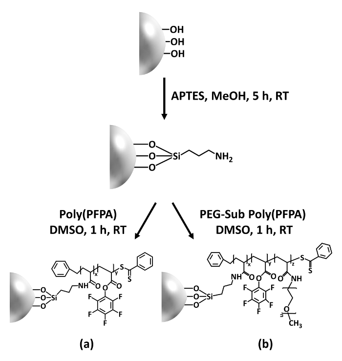
Figure 1: Schematic for the preparation of poly(PFPA) grafted SiO2 beads using APTES as a linker molecule. (a) Poly(PFPA) grafted beads. (b) Partially PEG-substituted poly(PFPA) grafted beads.
to:
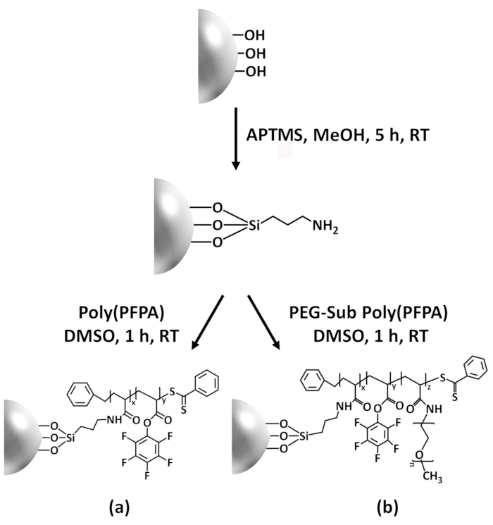
Figure 1: Schematic for the preparation of poly(PFPA) grafted SiO2 beads using APTMS as a linker molecule. (a) Poly(PFPA) grafted beads. (b) Partially PEG-substituted poly(PFPA) grafted beads.
Figure 2 was updated from:
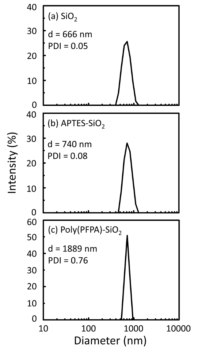
Figure 2: DLS measurements for (a) bare SiO2 beads (SiO2), (b) APTES functionalized SiO2 beads (APTES-SiO2), and (c) poly(PFPA) grafted SiO2 beads (poly(PFPA)-SiO2), dispersed in DMSO. The Z-average diameter (d) and polydispersity index (PDI) of each sample are reported.
to:
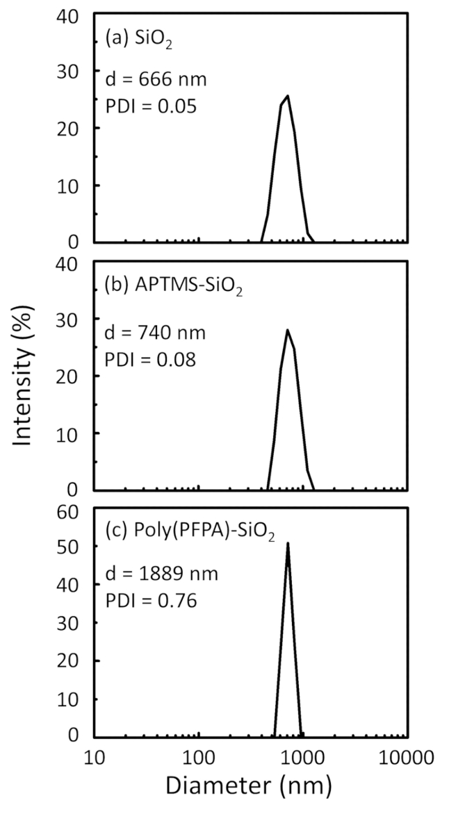
Figure 2: DLS measurements for (a) bare SiO2 beads (SiO2), (b) APTMS functionalized SiO2 beads (APTMS-SiO2), and (c) poly(PFPA) grafted SiO2 beads (poly(PFPA)-SiO2), dispersed in DMSO. The Z-average diameter (d) and polydispersity index (PDI) of each sample are reported.
Figure 3 was updated from:
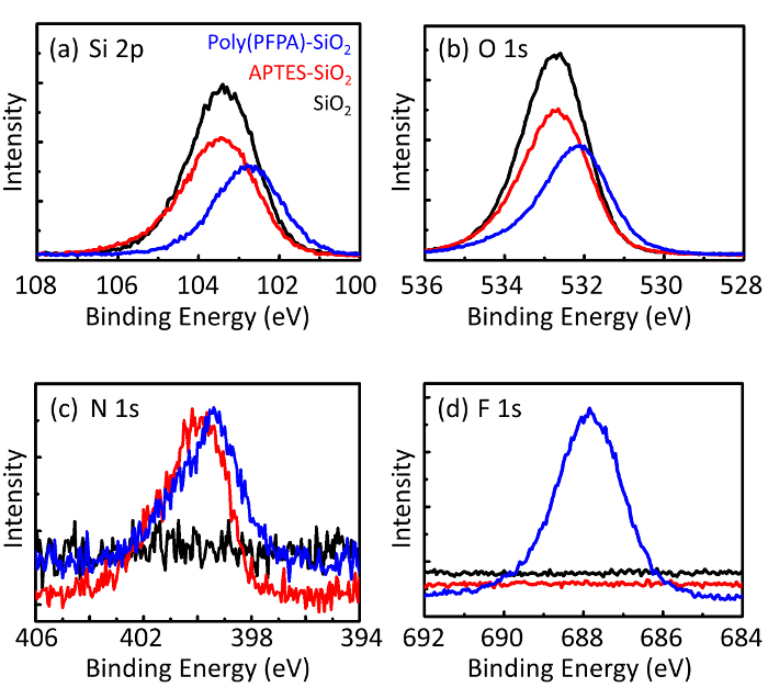
Figure 3: XPS spectra for bare SiO2 beads (SiO2), APTES functionalized SiO2 beads (APTES-SiO2), and poly(PFPA) grafted SiO2 beads (poly(PFPA)-SiO2). The peaks examined correspond to (a) Si 2p, (b) O 1s, (c) N 1s, and (d) F 1s.
to:
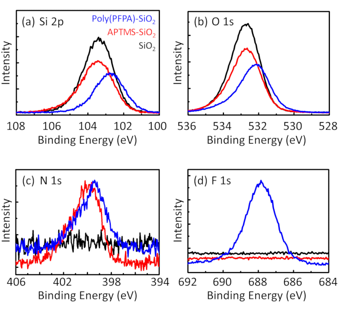
Figure 3: XPS spectra for bare SiO2 beads (SiO2), APTMS functionalized SiO2 beads (APTMS-SiO2), and poly(PFPA) grafted SiO2 beads (poly(PFPA)-SiO2). The peaks examined correspond to (a) Si 2p, (b) O 1s, (c) N 1s, and (d) F 1s.
The first and second paragraphs of the Discussion were updated from:
The synthesis of poly(PFPA) grafted SiO2 beads is illustrated in Figure 1. By employing APTES as a linker molecule, poly(PFPA) brushes covalently grafted to SiO2 substrate can be prepared via a simple two-step process. Although some of the PFP units are sacrificed for the reaction with APTES, a large number of the PFP units are expected to remain available for later reaction with either amino-PEG or antibodies. The PFP groups are known to form low energy surfaces so poly(PFPA) brushes do not solvate well in water28. For IP application, the antibodies need to be immobilized on the poly(PFPA) brushes, and this exchange reaction is done in aqueous buffer solution in order to preserve the activity of the antibodies. As reported in our previous publication, partial substitution of the PFP units with hydrophilic molecules such as amine-functionalized PEG can improve surface hydrophilicity, leading to increased antibody immobilization efficiency18. In this study, partially PEG substituted poly(PFPA) is also prepared, then grafted to the SiO2 surface using the same APTES linker molecule. Overall, the methods illustrated in Figure 1 allow the preparation of poly(PFPA) grafted surfaces with different degrees of PEG substitution. These polymer brushes with tunable surface properties provide an ideal platform for antibody immobilization and subsequent IP application.
The bead preparation process is monitored by both DLS and XPS. The DLS results for various functionalized SiO2 beads in DMSO are summarized in Figure 2. The bare SiO2 beads exhibit hydrodynamic diameter of 666 nm, in agreement with the manufacturer reported bead size (0.676 μm; SD = 0.03 μm). After APTES treatment, the bead diameter increases to 740 nm; and with poly(PFPA) treatment, the bead diameter further increases to 1889 nm. It is important to point out that the polydispersity index (PDI) for the poly(PFPA) grafted beads is rather large (PDI = 0.76), which is indicative of poor quality sample containing large aggregates. Although the DLS curve only shows one nano-sized peak, small amount of aggregates may be present in the suspension. The functionalized SiO2 beads are also examined by XPS to determine surface composition (Figure 3). Following APTES treatment, N 1s peak associated with the amine groups on APTES is detected. And, following poly(PFPA) treatment, F 1s peak associated with the PFP units on the polymer is detected. Together these data show the successful functionalization of the SiO2 surface, first with APTES, then with poly(PFPA).
to:
The synthesis of poly(PFPA) grafted SiO2 beads is illustrated in Figure 1. By employing APTMS as a linker molecule, poly(PFPA) brushes covalently grafted to SiO2 substrate can be prepared via a simple two-step process. Although some of the PFP units are sacrificed for the reaction with APTMS, a large number of the PFP units are expected to remain available for later reaction with either amino-PEG or antibodies. The PFP groups are known to form low energy surfaces so poly(PFPA) brushes do not solvate well in water28. For IP application, the antibodies need to be immobilized on the poly(PFPA) brushes, and this exchange reaction is done in aqueous buffer solution in order to preserve the activity of the antibodies. As reported in our previous publication, partial substitution of the PFP units with hydrophilic molecules such as amine-functionalized PEG can improve surface hydrophilicity, leading to increased antibody immobilization efficiency18. In this study, partially PEG substituted poly(PFPA) is also prepared, then grafted to the SiO2 surface using the same APTMS linker molecule. Overall, the methods illustrated in Figure 1 allow the preparation of poly(PFPA) grafted surfaces with different degrees of PEG substitution. These polymer brushes with tunable surface properties provide an ideal platform for antibody immobilization and subsequent IP application.
The bead preparation process is monitored by both DLS and XPS. The DLS results for various functionalized SiO2 beads in DMSO are summarized in Figure 2. The bare SiO2 beads exhibit hydrodynamic diameter of 666 nm, in agreement with the manufacturer reported bead size (0.676 μm; SD = 0.03 μm). After APTMS treatment, the bead diameter increases to 740 nm; and with poly(PFPA) treatment, the bead diameter further increases to 1889 nm. It is important to point out that the polydispersity index (PDI) for the poly(PFPA) grafted beads is rather large (PDI = 0.76), which is indicative of poor quality sample containing large aggregates. Although the DLS curve only shows one nano-sized peak, small amount of aggregates may be present in the suspension. The functionalized SiO2 beads are also examined by XPS to determine surface composition (Figure 3). Following APTMS treatment, N 1s peak associated with the amine groups on APTMS is detected. And, following poly(PFPA) treatment, F 1s peak associated with the PFP units on the polymer is detected. Together these data show the successful functionalization of the SiO2 surface, first with APTMS, then with poly(PFPA).
