This protocol describes both in vivo and ex vivo methods to fully visualize and characterize hyaloid vessels, a model of vascular regression in mouse eyes, using optical coherence tomography and fundus fluorescein angiography for the live imaging and ex vivo isolation and subsequent flat mount of hyaloid for quantitative analysis.
Method Article
Assessment and Characterization of Hyaloid Vessels in Mice
In This Article
Summary
Abstract
In the eye, the embryonic hyaloid vessels nourish the developing lens and retina and regress when the retinal vessels develop. Persistent or failed regression of hyaloid vessels can be seen in diseases such as persistent hyperplastic primary vitreous (PHPV), leading to an obstructed light path and impaired visual function. Understanding the mechanisms underlying the hyaloid vessel regression may lead to new molecular insights into the vascular regression process and potential new ways to manage diseases with persistent hyaloid vessels. Here we describe the procedures for imaging hyaloid in live mice with optical coherence tomography (OCT) and fundus fluorescein angiography (FFA) and a detailed technical protocol of isolating and flat-mounting hyaloid ex vivo for quantitative analysis. Low-density lipoprotein receptor-related protein 5 (LRP5) knockout mice were used as an experimental model of persistent hyaloid vessels, to illustrate the techniques. Together, these techniques may facilitate a thorough assessment of hyaloid vessels as an experimental model of vascular regression and studies on the mechanism of persistent hyaloid vessels.
Introduction
The blood supply in the eye is essential to ensure the normal development of the retina and surrounding ocular tissues and to equip a proper visual function. There are three vascular beds in the eye: the retinal vasculature, the choroid, and a transient embryonic circulatory network of hyaloid vessels. The development of the ocular vasculature requires spatial and temporal coordination throughout embryogenesis and tissue maturation. Among the three vascular beds, the hyaloid vasculature is the first functional blood supply system to provide nutrition and oxygen to the newly formed embryonic lens and the developing retina. Hyaloid vessels regress at the same time that the retina vasculatures develop and mature1. The regression of hyaloid vasculature is pivotal to allow a clear visual pathway for the development of visual function; hence, this vascular regression process is as important as the growth of retinal vasculature. Impaired hyaloid regression may lead to eye diseases. Moreover, the regression of hyaloid vessels provides a model system to investigate the cellular and molecular mechanisms involved in the regulation of vascular regression, which may have implications for the angiogenic regulation in other organs as well.
The hyaloid vasculature, derived from the hyaloid artery (HA), is composed of vasa hyaloidea propria (VHP), tunica vasculosa lentis (TVL), and pupillary membrane (PM). It provides nourishment to the developing retina, the primary vitreous, and the lens during embryonic development2. Arising from the HA, VHP branches anteriorly through the vitreous to the lens. The TVL cups the posterior surface of the lens capsule, and anastomoses to the PM, which connects to the anterior ciliary arteries, covering the anterior surface of the lens2,3, resulting in the formation of a network of vessels in the PM3,4,5. Interestingly, there are no veins in the hyaloid vasculature, and the system makes use of choroidal veins to accomplish venous drainage.
In the human embryo, the hyaloid vasculature is nearly complete at approximately the ninth week of gestation and starts to regress when the first retinal vessels appear, during the fourth month of gestation2. Beginning with atrophy of the VHP, regression of the capillary networks of the TVL, the PM, and lastly, the HA occurs subsequently2,3. Meanwhile, the primary vitreous retracts and the secondary vitreous starts to form, composed of the extracellular matrix components, including collagen fibers. By the sixth month of gestation, the primary vitreous is reduced to a small transparent canal extending from the optic nerve disc to the lens, called the Cloquet’s canal or hyaloid canal, and the secondary vitreous becomes the main component of the posterior segment2,3. The hyaloid circulation vanishes mostly at 35 to 36 weeks of gestation, just before birth3.
Unlike humans, in whom hyaloid vasculature is completely regressed at birth, the mouse hyaloid vascular system starts to regress after birth. As the mouse retina is born avascular and retinal vessels develop postnatally, hyaloid vessels regress concurrently from postnatal day (P) 4 and are mostly completely regressed by P216 (Figure 1). The PM disappears first between P10 and P12, and the VHP disappears between P12 and P16, while a small number of TVL and HA cells remain even at P16, and by P21 the hyaloid vascular system regression is almost complete6. In the meantime, retinal vasculature begins developing after birth. The superficial layer of vascular plexus fully extends to the peripheral retina at P7–P8, the deep layer (located in the outer plexiform layer) develops from P7–P12, and finally, the intermediate plexus in the inner plexiform layer develops between P12 and P157. As the retinal vasculature develops, it gradually replaces the function of concomitantly regressing hyaloid vessels, providing nutrition and oxygen to the developing eye. The postnatal occurrence of hyaloid vessel regression in mice provides an easily accessible experimental model to observe and study the hyaloid vasculature, as well as the molecular basis governing vascular regression processes under both physiological and pathological conditions8.
Failure of hyaloid regression can be seen in diseases such as PHPV, which is a rare congenital developmental anomaly of the eye resulting from a failed or incomplete regression of the embryological, primary vitreous and hyaloid vasculature9. The mechanisms regulating the regression process of hyaloid vasculature are complicated and broadly studied. One major molecular pathway essential for the normal regression of hyaloid vessels is the Wnt signaling pathway10, as genetic mutations in this pathway affecting both Wnt ligand and receptors have been linked with PHPV in humans9. Experimental studies identified a Wnt ligand, Wnt7b, which is produced by macrophages around hyaloid vessels in the developing eye to mediate this regression process. Wnt7b activates Wnt signaling by binding with the receptors frizzled4 (FZD4)/LRP5 in adjacent endothelial cells to initiate cell apoptosis, leading to the regression of hyaloid vessels10. As a result, Wnt7b-deficient mice show a persistence of hyaloid vessels10. Similarly, a nonconventional Wnt ligand, Norrin (encoded by the Ndp gene), also binds to FZD4/LRP5 to induce the hyaloid vessel regression during development. Ndpy/-, Lrp5-/-, and Fzd4-/- mice all display postponed hyaloid vessel regression, supporting a critical regulatory role of Wnt signaling11,12,13,14,15,16. Moreover, another Wnt coreceptor LRP6 overlaps with LRP5 in their function on modulating the Wnt signaling pathway in hyaloid vascular endothelial cells17. Other factors that may also contribute to hyaloid regression include the hypoxia-inducible factor18,19, vascular endothelial growth factor20,21, collagen-1822,23, Arf24, angiopoietin-225, and bone morphogenetic protein-426. In this paper, we use Lrp5-/- mice as a model of persistent hyaloid vessels to demonstrate the techniques of assessing and characterizing hyaloid vasculature through both in vivo and ex vivo methods.
The visualization of hyaloid vasculature in vivo and ex vivo is essential for studying the mechanisms of hyaloid vessel regression. Current methods to observe hyaloid vasculature mainly focus on visualizing and analyzing the VHP and HA, through OCT and FFA images, eye cross sections, and the hyaloid flat mount. OCT and FFA are powerful in vivo imaging tools, allowing longitudinal observation in live animals after they have opened their eyes. Moreover, isolated hyaloid flat mount provides visualization of the whole hyaloid vasculature and a means to achieve an accurate quantification of the vessel numbers. Yet the delicate and fragile nature of hyaloid vessels and the resulting technical difficulties of its isolation may have limited its use in eye research somewhat10,17,27. In this paper, we provide a detailed protocol of the visualization of hyaloid vessels, combining both in vivo live retinal imaging and ex vivo isolated hyaloid flat mount to enhance the feasibility of these techniques. This protocol has been adapted with modification and expansion from previous publications on the in vivo method of live fundus and OCT imaging28 and the ex vivo method of isolated hyaloid flat mount11.
Protocol
All animals were treated in accordance with the Association for Research in Vision and Ophthalmology (ARVO) Statement for the Use of Animals in Ophthalmic and Vision Research for animal experiments, following the Guidelines of the National Institutes of Health (NIH) regarding the care and use of animals for experimental procedures and the regulations set forth by the Institutional Animal Care and Use Committee (IACUC) at Boston Children’s Hospital. Lrp5-/- mice (stock no. 005823; Jackson Laboratory) and its wild-type (WT) control C57BL/6J mice (stock no. 000664; Jackson Laboratory) were used for this study.
1. Part I: In vivo imaging of hyaloid vessels using a rodent retinal imaging system
- Preparation of the mice anesthesia and pupil dilation
- Select the mice.
- Use mice that are older than P12 (after they have opened their eyes) for retinal imaging; both sexes are suitable.
- As desired, image mutant mice with delayed hyaloid regression (and their WT controls) throughout adulthood. WT mice older than 3 weeks may not exhibit much detectable remaining hyaloid vessels, hence P12–P21 is ideal for imaging remaining visible WT hyaloid.
- Prepare a ketamine/xylazine mixture for anesthesia.
- Take 2.3 mL of ketamine stock solution (100 mg/mL) and 0.7 mL of xylazine stock solution (20 mg/mL) and add 20 mL of sterile saline (0.9% sodium chloride) to make a working solution of a ketamine/xylazine mixture (10 mg/mL ketamine and 0.6 mg/mL xylazine).
- Anesthetize the mice by intraperitoneally injecting ketamine/xylazine mixture in a volume of 8x the mouse’s body weight (e.g., a 20 g mouse needs 160 μL of ketamine/xylazine working solution mixture to achieve a working dose of ketamine [80 mg/kg body weight] and xylazine [4.8 mg/kg body weight] mixture).
- Apply one drop of pupil-dilating drug solution (see Table of Materials) to each eye to dilate the mouse’s pupils immediately following anesthesia.
- Assess the level of anesthesia by pedal reflex (firm hindlimb toe pinch). Wait until the mouse is sufficiently anesthetized (no pedal reflex) and its pupils are widely dilated.
- Select the mice.
- Optical coherence tomography imaging of hyaloid vessels
- Moisturize the mouse’s corneas with artificial tears, and then, place the mouse on the positioning stage.
- Gently contact the mouse’s cornea with the lens of the OCT probe. Adjust the mouse and probe so that the optic nerve head is in the center of the visual field in the fundus image, to guide the OCT imaging.
NOTE: For more details on the general setup of a rodent retinal imaging system (see Table of Materials) and adjusting the mouse’s position, please refer to Gong et al.28. - Adjust the focus to achieve the optimal images of OCT.
- Adjust the angle of the indicated line in the OCT imaging software (see Table of Materials) to reveal persistent hyaloid vessels and, then, take images.
- Fundus fluorescein angiography imaging of hyaloid vessels
- Prepare a fluorescein solution: add 9 mL of sterile phosphate-buffered saline (PBS) to 1 mL of fluorescein solution (stock concentration: 100 mg/mL). The concentration of the final working solution is 10 mg/mL.
- Intraperitoneally inject the fluorescein working solution (5 μL/g body weight).
- Moisturize the mouse’s cornea with artificial tears, and then, place the mouse on the positioning stage.
- Position the lens of the Micron IV retinal imaging microscope to make direct gentle contact with the mouse cornea. Adjust the alignment slightly to position the optic nerve head in the center of the view field.
- Change the microscope filter to a green fluorescent channel.
- Focus on persistent hyaloid vessels to take images.
- Take multiple images after 1 min, 3 min, 5 min, and 10 min (no more than 10 min) post-injection to determine the best time point (signal-to-background ratio) for observing hyaloid vessels. Complete the FFA procedure within 10 min, after which the fluorescein may become too diffused and make vessels invisible.
- Recovery of the mice from anesthesia
- After the procedures, keep the mice on a warm heating pad.
- Wait until the mice are mobile again to return them to the mouse cage.
2. Part II: Ex vivo visualization of hyaloid vessels
- Preparation of fixed mouse eyes
- Select mice of the right age.
NOTE: Neonatal mice are typically sacrificed at P8 for hyaloid dissection. Both sexes are suitable. Mutant mice with a delayed hyaloid regression (and their respective WT controls) may be dissected and analyzed throughout adulthood. - Euthanize the mice by exposure to CO2.
- Enucleate the mouse’s eyes by blunt dissection.
- Open the eyelids wide with microsurgery forceps to allow access to the eyeball. Place curved microsurgery forceps under the globe in the orbit to grasp the optic nerve without squeezing the eyeball. Gently pull and remove the eyeball with the forceps.
- Alternatively, dissect the eyeball by using microsurgery scissors to carefully cut parallel to the globe from the four sides toward the back of orbit and separating the globe from surrounding connective tissues.
- Immerse the eyes in 4% paraformaldehyde in PBS buffer for 30 min at room temperature for fixation.
- Transfer the fixed eyeballs to ice-cold PBS buffer.
NOTE: The eyeballs may be stored in PBS at 4 °C for up to 1 week.
- Select mice of the right age.
- Embedding of hyaloid vessels with gelatin injection
- Prepare a 5% (w/v) gelatin solution.
- Weigh out 50 mg of gelatin.
- Dissolve the gelatin in 1 mL of distilled water.
- Incubate the gelatin solution in a 37 °C water bath until fully dissolved. Keep the solution in 37 °C water until use.
NOTE: A larger batch of solution may be prepared and stored at 4 °C as aliquots. Every time before use, the solution needs to be warmed in 37 °C water bath to achieve a clear consistency.
- Under a dissection microscope, inject 50 μL of gelatin solution into the vitreous body at the limbus; repeat the injection 3x at different sites to make a total of four injections, evenly spaced around the limbus (Figure 2A).
- Incubate the eyeballs in a 4 °C refrigerator or on ice for 30 min to solidify the injected gelatin in the vitreous space.
- Prepare a 5% (w/v) gelatin solution.
- Dissection and isolation of hyaloid vessels
NOTE: See Figure 2B-E.- Place the eyeballs in a Petri dish containing PBS to keep the tissue from drying. Make an incision with microsurgery scissors at the limbus and remove the cornea.
- Snip and remove the optic nerve.
- Under a dissection microscope, use two pairs of forceps to peel off and discard the sclera, choroid, and retinal pigment epithelium (RPE) layers and remove the iris.
- With the optic-nerve side of the retina facing up and the lens side down, inject 50 μL of PBS just beneath the retina cup to allow accumulation of the PBS buffer between the gelatinized vitreous body and the retina.
- Gently peel off and remove the retina cup and ciliary body from the vitreous body with microsurgery forceps.
- Using a transfer pipette, transfer the gelatin cup containing the hyaloid tissue immersed in PBS to a microscope slide
- Turn the rest of the tissue (lens/hyaloid) over, so the lens side is facing up.
- Lift the lens and gently loosen the connection between the lens/TVL and VHP, and then, cut the HA with microsurgery scissors to remove the lens-TVL. Keep the VHP-part of the hyaloid cup for the flat mount.
- Flat mounting and staining of hyaloid vessels
- Gently rinse the hyaloid cup with PBS on the slide to remove all dissection debris.
- Make sure the hyaloid cup is floating on the slide in adequate PBS solution.
- Gently arrange and adjust the position of the gelatinized hyaloid cup with microsurgery forceps, with the rim of the cup facing down on the slide, to achieve optimal appearance after melting.
- Place the slide with the hyaloid cup immersed in PBS onto a slide warmer at 37 °C, wait until the gelatin melts and the hyaloid flattens, and remove the slide when it is barely dry (do not over-dry).
- (Optional) Immunostain the hyaloid vessels
- Make blocking and penetrating buffer (e.g., 5% bovine serum albumin [BSA] and 0.1% Triton X-100 in PBS buffer). Add 50 μL of blocking buffer onto a flat-mounted hyaloid isolation and incubate it at room temperature for 30 min.
- Apply 50 μL of primary antibody (e.g., CD31) (1:100 dilution in blocking buffer) onto a flat mount of hyaloid isolation, and then, incubate it in a wet box at 4 °C overnight.
- Gently rinse 3x, for 5 min each time, with PBS.
- Apply 50 μL of secondary antibody (e.g., goat anti-rabbit secondary antibody-Alexa 488, 1:100 dilution) onto the hyaloid flat mount and incubate it at room temperature for 1 h.
- Gently rinse 3x, for 5 min each time, with PBS.
- Add a drop of anti-fade mounting medium with DAPI (4’, 6-diamidino-2-phenylindole) to stain the nuclei in the hyaloid vessels.
- Gently place a coverslip on the hyaloid to complete the flat mount.
- Imaging and quantification of flat-mounted hyaloid vessels
- Image the DAPI staining of hyaloid vessels with a fluorescent microscope and make sure the central HA is visible (exclude the samples without the HA).
- Identify vessel branches directly derived from the HA and manually count the number of vessel branches for quantification.
- Visualization of hyaloid vessels in the cross section of eyes
- Embed fixed eyeballs into optimal cutting temperature compound in a suitable tissue mold, and then, freeze the embedded eyes on dry ice or store them in a -20 °C freezer.
- Use a cryostat microtome to make a cross section of the embedded eyes into sections of 12 μm thickness at -20 °C.
- Collect the eye cross sections onto a microscope slide room temperature by gently touching the tissue sections, which will adhere to the slide.
- Air-dry to dehydrate the tissue sections for ~30 min, which can then be stored in a -20 °C freezer until ready for staining.
- Rinse the slide 1x with PBS to remove the remaining optimal cutting temperature compound on the slide.
- (Optional) Immerse the slide into an appropriate fixative buffer (e.g., 4% paraformaldehyde in PBS) for 15 min at room temperature for additional fixation, if needed.
- Permeabilize the tissue with 0.1% Triton X-100 in PBS for 30 min at room temperature.
- Prepare isolectin-IB4 staining buffer for blood vessel staining.
- Dissolve and reconstitute isolectin-IB4 (500 μg) powder with 50 mL of PBS in a 50 mL centrifuge tube.
- Slowly add 50 μL of 1 M CaCl2 solution, drop by drop, into the 50 mL of PBS/isolectin-IB4 mixture. This step needs to be performed slowly to avoid precipitation. The final concentration of CaCl2 is 1 μM. The presence of Ca2+ is required for isolectin-IB4 staining.
- Apply 30 μL of isolectin-IB4 staining buffer onto the eye cross sections on the slide and incubate it in a wet box at 4 °C overnight.
- Rinse 3x, for 5 min each time, with PBS.
- Add a drop of anti-fade mounting medium with DAPI to stain the nuclei in the sections.
- Gently place a coverslip on the tissue sections to complete the flat mount.
- Check the tissue under a microscope to visualize the presence of hyaloid vessels within the eyecup.
Results
In vivo imaging of hyaloid vessels in live mice
Figure 3A reveals cross-sectional views of OCT images for the retina and hyaloid tissues in 3-month-old WT and Lrp5-/- mice, an animal model with persistent hyaloid. The WT eye shows the absence of hyaloid tissue, whereas the Lrp5-/- eye shows two persistent hyaloid vessels derived from the optic nerve head. Figure 3B displays FFA images of persistent hyaloid vessels (green) in the fluorescent field in 6-week-old Lrp5-/- mice. The WT mouse shows no remnant of hyaloid vessels, and the Lrp5-/- mouse shows eight branches of hyaloid vessels in the vitreous body.
Ex vivo visualization of hyaloid vessels
Figure 4A,B demonstrates isolated hyaloid vessels as visualized in flat mounts, where vascular cells and associated macrophages were stained by their nuclei with DAPI staining (blue). The HA is at the center of each image, and hyaloid vessels are revealed by the DAPI-derived discontinuous lines. Each line represents one vessel of VHP. In Lrp5-/- mice, higher numbers of remaining hyaloid vessels were observed at P8 in flat mounts (Figure 4A). The WT mice had an average of 12 branches of hyaloid vessels at P8, whereas the age-matched Lrp5-/- pups showed around 25 branches of hyaloid vessels, demonstrating a significantly impaired regression of hyaloid vasculature (Figure 4B). In addition, delayed and incomplete retina vascular development, another characteristic often associated with persistent hyaloid vessels, was also observed in the Lrp5-/- pups (Figure 4C,D). Figure 5 shows the remaining hyaloid vessels in cross sections of Lrp5-/- eyes at P8, whereas the WT eyes do not display hyaloid vessels.
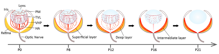
Figure 1: Schematic diagram depicting the developmental regression of hyaloid vasculature in mouse eyes. Hyaloid vessels and branches, including VHP, TVL, and PM, are derived from the HA, and occupy much of the space between the lens and the immature retina at birth (P0). Hyaloid involution in mice starts with the regression of PM capillaries as early as P4. At P8, PM, VHP, and TVL layers are continuously regressing, coinciding with the complete formation of the superficial layer of the retinal vessels. By P12, the involution of the PM layer is complete, whereas the atrophy of VHP and TVL is still in progress. In the meantime, a deep layer of retinal vessels starts to form during P7–P12. By P16, the regression of the hyaloid system is partially completed (with the remaining TVL vessels and the HA left), and the intermediate layer of retinal vascular plexus continues to develop. The retinal vasculature is fully mature by P21 and takes over the role of nourishing retinal tissues from the hyaloid vessels, which are now mostly regressed. At P21, the vitreous, in the absence of hyaloid vessels, shows a clear visual pathway. Dashed red lines represent the regressing vessels. VHP = vasa hyaloidea propria; TVL = tunica vasculosa lentis; PM = pupillary membrane; HA = hyaloid artery. Please click here to view a larger version of this figure.
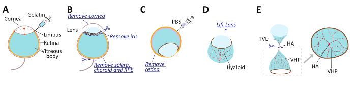
Figure 2: Schematic diagram depicting the sequential procedure of hyaloid vessel isolation for ex vivo flat-mounting visualization. (A) Injection of gelatin into the enucleated eye through four injecting points (red dots) at the limbus, to solidify the vitreous body. (B) Removal of the cornea and optic nerve, careful dissection of the iris, and peeling off the sclera-choroid-RPE complex. (C) Flipping the remaining retinal cup with the lens to make the retina side face up; then, injecting PBS between the retina and the vitreous body to separate them, followed by peeling off the retinal layer. (D) Flipping upside down again the rest of the tissue, containing the lens with the surrounding hyaloid. Lifting the lens slightly to loosen the connection of TVL and VHP. (E) Cutting at the HA between the TVL and the VHP, and the removal of the lens and TVL. Flat-mounting VHP layer. VHP = vasa hyaloidea propria; TVL = tunica vasculosa lentis; PM = pupillary membrane; HA = hyaloid artery. Please click here to view a larger version of this figure.
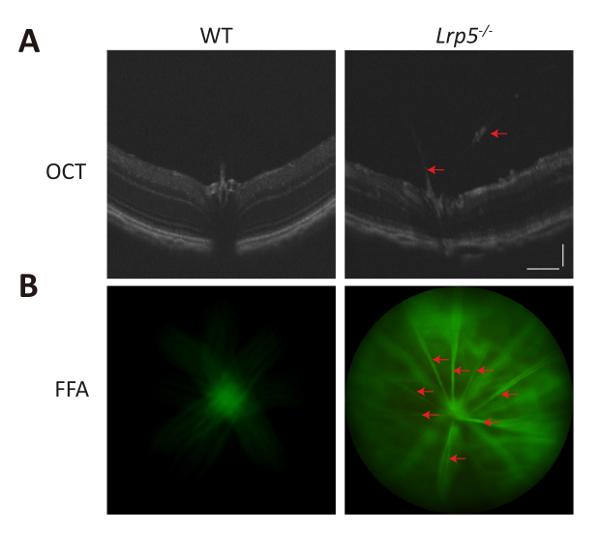
Figure 3: In vivo imaging of hyaloid vessels with optical coherence tomography (OCT) and fundus fluorescence angiography (FFA). A: Representative OCT images of WT and Lrp5-/- mice at 3-month-old showing persistent hyaloid vessels in the vitreous space in Lrp5-/- eyes. B: Representative FFA images of WT and Lrp5-/- mice at 6-weeks-old. Mice were injected with 1mg fluorescein sodium in 0.1ml saline per mouse after anesthesia, and images were taken 5 min after injection, focused on hyaloid vessels in Lrp5-/- mice or vitreous body in WT (in the absence of hyaloid vessels). Persistent hyaloid vessels were visualized in Lrp5-/- but not WT eyes. Red arrows indicate hyaloid vessels. Both scale bars: 100 μm (A). Please click here to view a larger version of this figure.
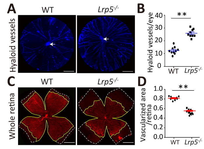
Figure 4: Visualization of delayed hyaloid vessel regression and delayed retinal vasculature development in Lrp5-/- mice. (A) Representative images of flat-mounted hyaloid vessels stained with DAPI (blue) in WT and Lrp5-/- eyes at P8. Arrows (white) indicate the hyaloid artery. (B) Quantification of the numbers of hyaloid vessels branching from the hyaloid artery in WT and Lrp5-/- eyes. (C) Representative flat-mounted retinas stained with isolectin-IB4 (red) for the vasculature in WT and Lrp5-/- eyes at P8. White dashed lines indicate the retina edge. Yellow lines indicate the vascularized area edge. (D) Quantification of vascular coverage of the superficial retinal vascular plexus in WT and Lrp5-/- eyes. n = 8–12/group. The scale bars in panels A and C = 1 mm. Data are shown as mean ± SEM. Two-tailed Student’s t-test was used for statistical analysis. **P < 0.01. This figure is modified with permission from Wang et al.27. Please click here to view a larger version of this figure.
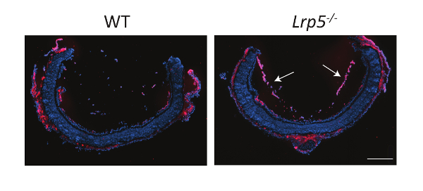
Figure 5: Visualization of hyaloid vessels in cross sections of Lrp5-/- eyes. Representative images of cross sections of eyes isolated from WT and Lrp5-/- mice at P8. The eyes were enucleated and embedded in optimal cutting temperature, and sections were cut using cryostat. The cross sections were stained with DAPI (blue) to show the nuclei and isolectin-IB4 (red) to show the blood vessels. White arrows indicate hyaloid vessels. The scale bar = 500 μm. This figure is adapted with permission from Chen et al.16. Please click here to view a larger version of this figure.
Discussion
Techniques to assess and characterize hyaloid vessels are intuitive and necessary procedures to observe the hyaloid vessel regression in animal models, to allow studies on the mechanisms underlying the vascular regression during development. While the in vivo retinal imaging allows the longitudinal observation of hyaloid regression in the same animal, access to a rodent fundus imaging system for OCT and FFA may be a limiting factor. In addition, in vivo imaging in live mice is not feasible before they open their eyes. Therefore, this methodology is not applicable during eye development in the neonatal stage. On the other hand, while imaging cross sections of isolated eyes may be used for any age of the mouse and has a low technical barrier, it is not quantitative when there are only a few visible hyaloid vessels, and the chance of obtaining an ideal image depends largely on the angle of sectioning (Figure 5). In comparison, the isolation of hyaloid vessels for flat mount visualization allows the complete imaging of the whole hyaloid vessel and accurate quantification, yet technical challenges may exist due to the delicate and fragile nature of hyaloid vessels. We hope the protocol detailed in this paper helps overcome these challenges.
Several critical steps in the hyaloid isolation protocol include the injection of the gelatin solution, the removal of the retina, and the final flat mounting. The technical difficulty of handling hyaloid vasculature lies in its delicate nature, the almost liquid-like state of vitreous where hyaloid vessels reside, and its close connection with the adjacent lens and the retina. Injecting gelatin solution intravitreally is key to solidifying the vitreous body containing hyaloid vessels and, thereby, making their texture firmer for easier dissection and handling. Gelatin is a gelling agent forming transparent elastic thermoreversible gels. By injecting the liquid form of gelatin (at room temperature or at 37 °C) into the vitreous space and cooling it to a lower temperature (4 °C), the vitreous body transforms into a firmer gel cup. Performing multiple gelatin injections into the eye allows the full dispersion of a sufficient amount of the gelatin into the vitreous space, to form a uniform and round gelatinized hyaloid tissue cup. Another technical difficulty lies in removing the retina without damaging hyaloid tissue. Unlike the choroid, which is relatively easy to dissect apart, the gelatinized vitreous body containing hyaloid vessels is challenging to separate from the nearby retina without damaging the hyaloid. We found that injecting PBS into the space between the vitreous body and the retina generated a liquid buffer layer, making it much easier to separate these two tissues. This is somewhat similar to the hydrodissection technique used in cataract surgery to separate the capsule and the cataract cortex. The final flat mounting is the last technically difficult step of the protocol. Warming the gelatinized hyaloid tissue in a PBS droplet on a slide warmer converts it back to a more liquid-like form to allow easier flat mounting. Proper arrangement and orientation of the hyaloid gel cup is still essential to ensure its even flattening after melting and drying.
The protocol of isolating hyaloid vessels may be further modified in several ways. The concentration of gelatin solution we used is 5%, yet higher or lower concentrations may also work, depending on the preference of the researcher to achieve a firmer or softer texture for handling. The DAPI staining of cellular nuclei may also be modified with isolectin-IB4 or other endothelial cell or macrophage markers, to better distinguish vascular endothelial cells from macrophages. A word of caution: the flat-mounted hyaloid vessels are very delicate and only loosely adhered to the slide; hence, the hyaloid slides need to be handled very gently and carefully if rinsing is needed during staining. This isolation protocol also has its limitations, including the complexity of performing the fine dissection and the difficulty of the long-term preservation of the samples due to the nature of the fragile tissue. Nevertheless, isolating and flat-mounting hyaloid vessels is still the most comprehensive and intuitive way to study hyaloid vasculature, to promote ocular and angiogenesis studies10,17,27,29,30,31.
The hyaloid vasculature provides an excellent experimental model of studying developmental vessel growth and regression, relevant for research fields in ophthalmology, angiogenesis, and programmed cell death, with which this paper aims to assist. Future modifications of the isolation protocol may be directed at improving the feasibility of a technical dissection procedure. Overall, the combined in vivo imaging and ex vivo isolation of hyaloid vessels is an advantageous method to allow the full assessment and characterization of hyaloid vessels in mice as a useful model of vascular regression.
Disclosures
The authors have nothing to disclose.
Acknowledgements
This work was supported by National Institutes of Health (NIH) grants (R01 EY024963 and EY028100) to J.C. Z.W. was supported by a Knights Templar Eye Foundation Career Starter Grant. The hyaloid isolation procedure described in this study was adapted with modification from protocols generously shared by Drs. Richard Lang, Toshihide Kurihara, and Lois Smith, to whom the authors are thankful.
Materials
| Name | Company | Catalog Number | Comments |
|---|---|---|---|
| AK-Fluor (fluorescein injection, USP) | Akorn | 17478-253-10 | |
| Anti-CD31 antibody | Abcam | ab28364 | |
| Antifade mounting medium | Thermo Fisher | S2828 | |
| Antifade Mounting Medium with DAPI | Vector Laboratories | H-1200 | |
| Artificial tear eyedrop | Systane | N/A | |
| Bovine serum albumin (BSA) | Sigma-Aldrich | A2058 | |
| C57BL/6J mice | The Jackson Laboratory | Stock NO: 000664 | |
| Calcium chloride (CaCl2) | Sigma-Aldrich | C1016 | |
| Cryostat | Leica | CM3050S | |
| Cryostat | Leica | CM3050 S | |
| Cyclopentolate hydrochloride and phenylephrine hydrochloride eyedrop | Cyclomydril | N/A | |
| Gelatin | Sigma-Aldrich | G9382 | |
| Goat anti-Rabbit IgG (H+L) Cross-Adsorbed Secondary Antibody, Alexa Fluor 488 | ThermoFisher Scientific | A-11008 | |
| Heating board | Lab-Line Instruments Inc. | N/A | |
| Isolectin GS-IB4, 594 conjugate | ThermoFisher Scientific | I21413 | |
| Ketamine hydrochloride injection | KetaVed | NDC 50989-996-06 | |
| Lrp5-/- mice | The Jackson Laboratory | Stock NO. 005823 | Developed by Deltagen Inc., San Mateo, CA |
| Micron IV and OCT | Phoenix Research Labs | N/A | Imaging software: InSight |
| Microscope | Zeiss | discovery v8 | |
| Microsurgery forceps | Scanlan International | 4004-05 | |
| Microsurgery scissors | Scanlan International | 6006-44 | |
| Optimal cutting temperature compound | Tissue-Tek | 4583 | |
| Optimal cutting temperature compound | Agar Scientific | AGR1180 | |
| Paraformaldehyde (16%) | Electron Microscopy Sciences | 15710 | |
| Peel-A-Way disposable embedding molds (tissue molds) | Fisher Scientific | 12-20 | |
| Phosphate-buffered saline (PBS) buffer (10X) | Teknova | P0496 | |
| Slide cover glass | Premiere | 94-2222-10 | |
| Superfrost microscope slides | Fisherbrand | 12-550-15 | |
| Triton X-100 | Sigma-Aldrich | X100 | |
| Xylazine sterile solution | Akorn: AnaSed | NDC: 59399-110-20 |
References
- Lutty, G. A., McLeod, D. S. Development of the hyaloid, choroidal and retinal vasculatures in the fetal human eye. Progress in Retinal and Eye Research. 62, 58-76 (2018).
- Anand-Apte, B., Hollyfield, J. Developmental anatomy of the retinal and choroidal vasculature. The Retina and its Disorders. Besharse, J., Bok, D. , Academic Press. Oxford, UK. (2011).
- Hobbs, R. P., Hartnett, M. E. Chapter 2: The hyaloidal vasculature and its role in development. Pediatric Retina: Second Edition. Hartnett, M. E. , Lippincott Williams & Wilkins. Philadelphia. (2013).
- Fruttiger, M. Development of the retinal vasculature. Angiogenesis. 10 (2), 77-88 (2007).
- Saint-Geniez, M., D'Amore, P. A. Development and pathology of the hyaloid, choroidal and retinal vasculature. The International Journal of Developmental Biology. 48 (8-9), 1045-1058 (2004).
- Ito, M., Yoshioka, M. Regression of the hyaloid vessels and pupillary membrane of the mouse. Anatomy and Embryology. 200 (4), 403-411 (1999).
- Stahl, A., et al. The mouse retina as an angiogenesis model. Investigative Ophthalmology & Visual Science. 51 (6), 2813-2826 (2010).
- Liu, C. H., Wang, Z., Sun, Y., Chen, J. Animal models of ocular angiogenesis: from development to pathologies. FASEB Journal. 31 (11), 4665-4681 (2017).
- Shastry, B. S. Persistent hyperplastic primary vitreous: congenital malformation of the eye. Clinical & Experimental Ophthalmology. 37 (9), 884-890 (2009).
- Lobov, I. B., et al. WNT7b mediates macrophage-induced programmed cell death in patterning of the vasculature. Nature. 437 (7057), 417-421 (2005).
- Kato, M., et al. Cbfa1-independent decrease in osteoblast proliferation, osteopenia, and persistent embryonic eye vascularization in mice deficient in Lrp5, a Wnt coreceptor. The Journal of Cell Biology. 157 (2), 303-314 (2002).
- Xia, C. H., et al. A model for familial exudative vitreoretinopathy caused by LPR5 mutations. Human Molecular Genetics. 17 (11), 1605-1612 (2008).
- Xu, Q., et al. Vascular development in the retina and inner ear: control by Norrin and Frizzled-4, a high-affinity ligand-receptor pair. Cell. 116 (6), 883-895 (2004).
- Ye, X., et al. Norrin, frizzled-4, and Lrp5 signaling in endothelial cells controls a genetic program for retinal vascularization. Cell. 139 (2), 285-298 (2009).
- Ohlmann, A. V., Adamek, E., Ohlmann, A., Lutjen-Drecoll, E. Norrie gene product is necessary for regression of hyaloid vessels. Investigative Ophthalmology & Visual Science. 45 (7), 2384-2390 (2004).
- Chen, J., et al. Retinal expression of Wnt-pathway mediated genes in low-density lipoprotein receptor-related protein 5 (Lrp5) knockout mice. PLoS One. 7 (1), 30203(2012).
- Nayak, G., et al. Developmental vascular regression is regulated by a Wnt/beta-catenin, MYC and CDKN1A pathway that controls cell proliferation and cell death. Development. 145 (12), (2018).
- Kurihara, T., et al. Astrocyte pVHL and HIF-alpha isoforms are required for embryonic-to-adult vascular transition in the eye. The Journal of Cell Biology. 195 (4), 689-701 (2011).
- Huang, T. Q., et al. Deletion of HIF-1alpha partially rescues the abnormal hyaloid vascular system in Cited2 conditional knockout mouse eyes. Molecular Vision. 18, 1260-1270 (2012).
- Yoshikawa, Y., et al. Developmental regression of hyaloid vasculature is triggered by neurons. The Journal of Experimental Medicine. 213 (7), 1175-1183 (2016).
- Garcia, C. M., et al. The function of VEGF-A in lens development: formation of the hyaloid capillary network and protection against transient nuclear cataracts. Experimental Eye Research. 88 (2), 270-276 (2009).
- Hurskainen, M., et al. Abnormal maturation of the retinal vasculature in type XVIII collagen/endostatin deficient mice and changes in retinal glial cells due to lack of collagen types XV and XVIII. FASEB journal. 19 (11), 1564-1566 (2005).
- Fukai, N., et al. Lack of collagen XVIII/endostatin results in eye abnormalities. The EMBO Journal. 21 (7), 1535-1544 (2002).
- McKeller, R. N., et al. The Arf tumor suppressor gene promotes hyaloid vascular regression during mouse eye development. Proceedings of the National Academy of Sciences of the United States of America. 99 (6), 3848-3853 (2002).
- Hackett, S. F., Wiegand, S., Yancopoulos, G., Campochiaro, P. A. Angiopoietin-2 plays an important role in retinal angiogenesis. Journal of Cellular Physiology. 192 (2), 182-187 (2002).
- Chang, B., et al. Haploinsufficient Bmp4 ocular phenotypes include anterior segment dysgenesis with elevated intraocular pressure. BMC Genetics. 2, 18(2001).
- Wang, Z., et al. Pharmacologic Activation of Wnt Signaling by Lithium Normalizes Retinal Vasculature in a Murine Model of Familial Exudative Vitreoretinopathy. The American Journal of Pathology. 186 (10), 2588-2600 (2016).
- Gong, Y., et al. Optimization of an Image-Guided Laser-Induced Choroidal Neovascularization Model in Mice. PLoS One. 10 (7), 0132643(2015).
- Kishimoto, A., et al. Histochemical characteristics of regressing vessels in the hyaloid vascular system of neonatal mice: Novel implication for vascular atrophy. Experimental Eye Research. 172, 1-9 (2018).
- Lang, R. A., Bishop, J. M. Macrophages are required for cell death and tissue remodeling in the developing mouse eye. Cell. 74 (3), 453-462 (1993).
- Riazifar, H., et al. Phenotypic and functional characterization of Bst+/- mouse retina. Disease Models & Mechanisms. 8 (8), 969-976 (2015).
Reprints and Permissions
