

Summary
Here we present a method to validate tail vein injections in rats by utilizing near-infrared fluorescence imaging data from dyes incorporated into agents or biological probes. The tail is imaged before and after the injection, the fluorescent signal is quantified, and an assessment of the injection quality is made.
Abstract
Intravenous (IV) administration of agents into the tail vein of rats can be both difficult and inconsistent. Optimizing tail vein injections is a key part of many experimental procedures where reagents need to be introduced directly into the bloodstream. Unwittingly, the injection can be subcutaneous, possibly altering the scientific outcomes. Utilizing a nanoemulsion-based biological probe with an incorporated near-infrared fluorescent (NIRF) dye, this method offers the capability of imaging a successful tail vein injection in vivo. With the use of a NIRF imager, images are taken before and after the injection of the agent. An acceptable IV injection is then qualitatively or quantitatively determined based on the intensity of the NIRF signal at the site of injection.
Introduction
The route of administration of agents into small animals serves as a critical point of many experiments. It determines where the agent is to be delivered and, subsequently, what will happen to the agent thereafter. Although other routes can be used for agent administration1, the intravenous route of delivery is a preferred route for certain agents. IV injection allows agents to be directly injected into the bloodstream, bypassing first-pass tissue effects and the need for extraneous solute absorption1. This also allows for targeting cells in the bloodstream2,3 and direct delivery to all tissues within the circulatory system. In rodents, several veins can be considered, including the jugular, the saphenous, and the tail vein.
In this method, a NIRF dye containing a biological probe—in this case, a nanoemulsion (Figure 1A)3,4,5,6—is injected into the lateral tail vein of rats. This particular NIRF-containing nanoemulsion has been used previously to image and track neuroinflammation in vivo and ex vivo7,8 in a rat model9 of neuropathic pain2,3,4,5,10,11. Imaging is conducted before and after the injection with a preclinical NIR fluorescence imager (see Table of Materials). This serves as a tool to validate the quality of the agent administration. Imaging prior to the tail vein injection serves as a basis for obtaining a baseline image.
Increasingly in animal studies, intravenously administered nanoemulsions are being utilized as biological probes and targeting agents12,13,14,15. It is a proven challenge to administer an agent via the tail vein16,17—be it a drug, a viral vector, or another probe—and to ensure that the entire contents of the injection have successfully entered the bloodstream and not the surrounding tissues17. Therefore, a method of visualizing and evaluating the quality of a successful injection is beneficial.
Typically, a heat lamp or warm water is used to warm the tail, which causes dilation of the vein, permitting its visualization prior to injection. While this ensures easier entry into the vein, there is not a quantitative way to discern whether the compound has entered the bloodstream in its entirety18,19,20,21. This becomes more difficult still in strains of animals where the vein contrasts faintly with the skin, such as in black mice. Typically, the investigator can gauge a failed injection by experiencing resistance during the injection and, in some cases, visualizing a bulge on the tail, indicating a subcutaneous leakage of the agent22,23.
In this study, NIRF imaging of the nanoemulsion injected into the lateral tail vein of live rats is performed on a small-animal NIRF imaging system (see Table of Materials). Rats are fed a special purified diet (see Table of Materials) to reduce nonspecific gut fluorescence. Simultaneous image acquisition of white light and 800 nm fluorescence is captured using the NIRF imager and associated software. The relative fluorescence intensity is measured on the tail at the pre-injection and post-injection states. The fluorescence intensity for the region of interest (ROI) at the site of injection is recorded and divided by the area of the ROI. Qualitative assessments can be made on which injections are acceptable. Optionally, further quantitative analysis can be performed by setting thresholds for acceptable injections and assigning ROI measurements into groups, at which point statistical significance can be calculated.
By utilizing this validation strategy following tail vein injections, the standard of a research study improves due to increased consistency of agent administration. This method of assessing the quality of tail vein injection can be easily customized for different injectable agents to include infra-red fluorescent probes provided commercially by several companies.
Protocol
All protocols were performed in accordance with the guidelines in the Guide for the Care and Use of Laboratory Animals of the National Institutes of Health and Institutional Animal Care and Use Committee (IACUC) at Duquesne University.
1. Preparation and Anesthesia
NOTE: Aseptic techniques are used for the entirety of the procedure. Only new sterile materials and autoclaved sterile instruments are to be used. Personal protective equipment (sterile gloves, hair bonnet, surgical mask, scrubs) needs to be worn to avoid contamination.
- Adult male Sprague-Dawley rats weighing 250–300 g were used in this protocol. Acclimate the rats to standard living conditions, keep them on a 12 h light/12 h dark cycle, and provide food and water ad libitum. House the animal socially, keep them on paper bedding, and provide a special diet (see Table of Materials) to avoid autofluorescence during imaging.
- With the use of a properly placed heating pad, anesthetize the animal under an initial 5% isoflurane in 20% oxygen, followed by a maintenance level of not less than 1.5% isoflurane and not more than 3%, unless the animal wakes up or retains feeling.
- Confirm proper anesthesia via a lack of response to tail pinches. Monitor the blood flow as well via vital signs throughout the procedure.
2. Pre-injection Image Aquisition
- Image the animal in a preclinical NIR fluorescence imager by positioning the animal laterally to expose the injection site on the lateral tail to establish a baseline of fluorescence in the tail (Figure 1C,E). Perform a simultaneous image acquisition of both a white light (body view) and near infrared channels using the NIRF imager and associated software, with linked lookup tables (LUT).
- Following imaging, move the animal back to the surgical table, and place it under anesthesia for the tail vein injection.
NOTE: Continue monitoring the animal’s vital signs and recheck proper anesthetization via tail pinch.
3. Tail Vein Injection with NIRF-containing Agent
- With the animal in the prone position, orient the tail with the dorsal side facing up. Dilate the tail vasculature in warm water for a minimum of 1 min. Orient the tail vein so the lateral side (either right or left) is turned 30° (clockwise or counterclockwise) to expose the right or left tail vein (Figure 1B).
- Once a lateral tail vein has been located (which appears dark-colored upon dilation), disinfect the entire tail with alcohol pads, repeating 2x.
- At an appropriate dosage based on the study design, begin injections in the distal coccygeal vertebrae region of the tail and moving more proximal if proper needle placement fails.
- Insert a 25–27 G sterile needle, bevel up, into the lateral tail vein, with the tail at a 180° angle, inserting the needle parallel to the lifted tail. Observe blood flashback in the rim of the needle to ensure correct placement. If no flashback is apparent, slowly move the needle tip (without removing it from the tail) to find vein insertion. If placed subcutaneously, no blood flashback will occur.
- Insert the syringe with the injectable materials into the rim of the needle. When proper placement is achieved, the injectable fluid will not incur resistance upon injection. The injection will advance smoothly and easily. Once injected, remove the needle and the syringe, apply pressure with sterile gauze for at least 1 min to ensure clotting, and mark the spot of injection with a pen on the tail, ensuring it is visible on the white light image.
NOTE: No hematoma or lesion will be visible at the site of injection. - If the needle tip moves during the syringe insertion, remove the needle and retry the needle entry procedure more proximal on the ipsilateral tail vein. Do not reuse the same needle if a different reentry point is tried.
NOTE: Alternatively, the injection can be performed with an IV catheter with a blood-flow indicator (see Table of Materials). This has the benefit of visual confirmation of the catheter during venipuncture. Insert the catheter, bevel side up, at the angle previously described. Observe prompt flashback in the entire length of the blood-flow indicator to ensure correct placement. Slight back pressure can be used to pull blood into the syringe to confirm proper placement in the vessel before injecting. Again, no resistance will be felt.
4. Post-injection Image Acquisition
- Perform quality assessment following tail vein injection in a preclinical NIR fluorescence imager in the same orientation as the baseline pre-injection image. Ensure the animal is still properly anesthetized — and will be so for the duration of imaging — prior to placing it in the imager.
NOTE: An imaging system containing a drawer housingwith anesthesia connections and a mask for the animal should be used if available. - Orient the animal on its lateral side to expose the injection site (as marked) on the lateral tail. Check to see if a NIRF signal is present only at the site of injection. This indicates a successful tail vein injection (Figure 1D).
NOTE: If the signal is dispersed throughout the entire tail, it is considered to be subcutaneous and, hence unsuccessful (Figure 1F). Figure 2 shows additional examples of failed injections.
5. Image Quantification
NOTE: Image quantification can be performed with the imaging software that accompanies the NIR imager, if this function is available. Alternatively, other commercially available image analysis software may be used24.
- In the post-injection image, draw a region-of-interest around the area of fluorescence at the injection site2,6.
- Measure the area and relative fluorescence intensity and record as area/intensity. Compare post-injection and baseline pre-injection images either qualitatively or quantitively by using appropriate statistical analysis(dependent on study groups and conditions).
NOTE: The researcher can decide on thresholds that discriminate good from bad injections or assign a percentage of quality to the injection.
Representative Results
Rats were injected with NIRF-containing nanoemulsion into the lateral tail vein, and pre- and post-injection images were taken with the small-animal imager (Table of Materials) as described in the protocol. Post-injection images are qualitatively assessed for injection quality and placed into ‘good injection’ (n = 7) and ‘bad injection’ (n = 4) groups. Qualitative assessment was carried out by observing the post-injection area fluorescence intensity. In an optimal injection, the NIRF signal is confined to the site of injection. No signal will be seen if the injection is successful because the agent has been fully displaced into the bloodstream. A bad-quality injection displays a NIRF signal that is dispersed along the length of the tail.
Images were analyzed with the accompanying NIRF imager software. Regions-of-interest were drawn at the site of pre-injection images (Figure 1C, E) and around the area of fluorescence in post-injection images (Figure 1D, F). Images where fluorescence was visible throughout the length of the tail were deemed unacceptable and removed from the analysis (Figure 2). Measurements of the area and fluorescence intensity were recorded. Values for area/fluorescence intensity were calculated and plotted (Figure 1G). A significant difference (unpaired t-test) in fluorescence intensity between pre- and post-injection images was observed in the ‘bad injection’ group (Figure 1G) (p = 0.0024).
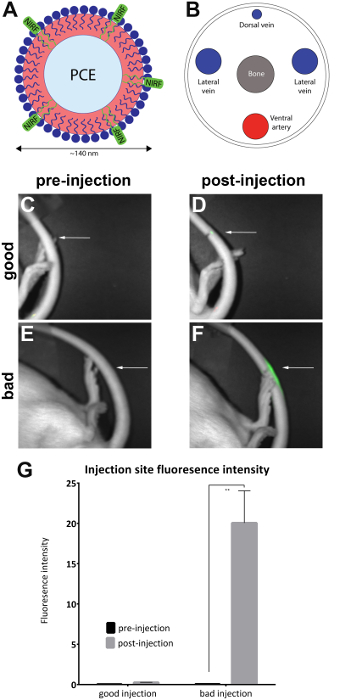
Figure 1: NIRF based nanoemulsion and images of tail vein. (A) A nanoemulsion-based biological probe containing NIRF dye was injected into (B) the lateral tail vein and imaged in a NIRF imager. (C and D) Pre- and postinjection images of a good injection. (E and F) Pre- and postinjection images of a bad injection. White arrows indicate the point of injection. It is possible to qualitatively assess the success of a good injection compared to a bad injection by assessing the extent of the NIRF signal at the site of injection. Unacceptable injections display fluorescence throughout the length of the tail and were removed from the analysis (Figure 2). (G) The images can also be analyzed to reveal a quantitative measure of fluorescence intensity, with thresholds for injection quality assigned by the investigator. The error bars on the graph reflect the SEM. For the ‘good injection’ group, n = 7. For the ‘bad injection’ group, n = 4. There is a statistical difference in fluorescence intensity in the ‘bad injection’ group when comparing pre- and postinjection images (unpaired t-test; p = 0.0024). Please click here to view a larger version of this figure.

Figure 2: Examples of bad injections. (A) Fluorescent signal seen in part of the tail. (B) Fluorescent signal seen over the full length of the tail. (C) Fluorescent signal dispersed heavily in the entire tail and caudal area of the animal’s body. Please click here to view a larger version of this figure.
Discussion
Research laboratories incur significant costs as a result of the misadministration of testing agents. Tail vein injections are a difficult technique to master to attain consistent success rate, with the most experienced of technologists often incurring misadministration errors. There is no reliable way to confirm a successful injection. This protocol offers a solution to this problem by giving researchers a qualitative and quantitative method to validate the success of a murine tail vein injection. Here, a NIRF-labelled nanoemulsion7,8,25 incorporates the agent of choice (in this case, a drug) and is imaged at the site of injection in a NIRF small-animal imager. There is also the option to develop a non-nanoemulsion-based agent and use the same principle of NIRF imaging by incorporating commercially available infra-red dyes. Additionally, ready-to-use imaging agents with a variety of applications, such as tumor imaging, metabolic imaging, cell trafficking, and apoptosis are also commercially available. An injection is performed either by using a sterile needle or, alternatively, an IV catheter; this depends on the preference of the researcher. In addition, automated tail vein injectors26 have been used to assist in this process and are compatible with this methodology. However, this technology has not yet become commercially available.
There are important steps in the tail vein injection method that ensure a higher rate of correct agent administration. First, the tail should be cleaned with ethanol to remove any dirt or debris, allowing researchers to better visualize the vein. Dilating the vein by submerging the tail in warm water is also a very important step in the method, as it allows a greater surface area for injection. Injecting at a more distal point on the tail vein allows for some error, in the event that multiple attempts are required. Injection should be attempted at a more proximal position in the tail as the tail vein increases in size as the caudal aspect of the animal’s body is approached. In addition, the contralateral tail vein can be used if needle placement fails in more than three to five sites on the ipsilateral tail vein.
A successful administration of a test agent results in little to no NIRF signal at the point of injection. If no resistance is felt during the administration of the injection and there is little to no fluorescence at the tail, then the injection can be recorded as successful. If resistance is felt during injection and there is a trail of NIRF signal along some length of the tail, then the injection is recorded as unsuccessful and is likely partly subcutaneous. Fluorescence images are taken pre- and post-injection, and the quality of the injection is assessed by observing qualitatively or analyzing quantitatively the fluorescence signal at the site of injection. The software accompanying the NIR fluorescence imager is often capable of performing this analysis.
The method can be adapted in several ways. It is applicable to tail vein injection in both mice and rats. Most small-animal NIR fluorescence imagers will be capable of accommodating murine rodents. Levels of anesthesia need to be adjusted depending on the weight of the animal, in accordance with the research laboratory’s IACUC protocol. Another possible modification is the preparation of a non-nanoemulsion-based probe either by incorporating an infrared dye into the researcher’s formulated agent or by purchasing a ready-to-use imaging agent, tailored to a specific biological application.
If a rat is relatively large, it can often be difficult to position it in the small-animal imager. It is thus recommended that a test image is taken with the animal in the drawer before injecting, and a field of view ascertained where the tail is visible. It is helpful to tape the tail to the drawer of the imager, to ensure it does not move during imaging.
Alternative methods seeking to assess the quality of tail vein injections in small animals are limited to the utilization of labeling reagents that do not interfere with concurrent experimental procedures and require euthanasia of the animals postinjection12,13. Some reagents may impact study outcomes and the therapeutic assessment of the animals involved, so care in experimental design is recommended.
This method can, in the future, be refined with advances in small-animal imaging technology, as well as improvements in infrared fluorescent probes. Biological probes with an incorporated infrared dye, designed for a variety of different applications, can be used at the agent administration stage of a study design to validate the quality of an injection, as outlined in this method2,3,27,28,29,30,31,32.
Disclosures
The authors have nothing to disclose.
Acknowledgments
J.A.P. and J.M.J. jointly designed the experimental approach for evaluating Nanoemulsions in the Chronic Constriction Injury rat model for effects on neuropathic pain. J.M.J. conceived and designed the overall macrophage-targeted drug delivery approach with nanoemulsions, the nanoemulsion composition, and processes for fabrication. J.M.J. produced the nanoemulsion, which was further fabricated by L.L. under the guidance of J.M.J. The stability of the nanoemulsion was assessed by J.M.J., L.L., and S.P. Animal care, surgery, behavior, tail vein injections, and NIRF imaging were carried out jointly by M.S. and A.M.S. under the guidance of J.A.P. The manuscript was written and prepared by M.S., and the protocol was written by A.M.S.
NIR optical imaging was performed on the Small Animal Imaging System at Duquesne University (supported by the Pittsburgh Tissue Engineering Initiative Seed Grant). J.M.J. acknowledges support from the DOD award number FA8650-17-2-6836, NIDA award number 1R21DA039621-01, NIBIB award number R21EB023104-02 and AFMSA Award number FA8650-17-2-6836. J.A.P. and J.M.J acknowledge support from the Pittsburgh Tissue Engineering Initiative Seed Grant. J.A.P. also acknowledges the Hunkele Dreaded Disease Award, the Samuel and Emma Winters Foundation, the Charles Henry Leach II Fund, and the Commonwealth Universal Research Enhancement Award. J.A.P. and J.M.J. acknowledge support from the Duquesne University Inaugural Provost’s Interdisciplinary Research Consortia Grant, which supports the Chronic Pain Research Consortium.
Materials
| Name | Company | Catalog Number | Comments |
| 100% Oxygen air tank | AirGas Heathcare | n/a | For ventilation of animal. |
| 70% Ethanol | Multiple sources | n/a | |
| Alcohol Pads | Henry Schein | 112-6131 | |
| Artificial Tears | Henry Schein | 100-2634 | This protects the rats eyes while it is anesthetized. |
| Beaker | Multiple sources | n/a | This holds warm water to dilate the tail veins. |
| Distilled water | Multiple sources | n/a | |
| Exhaust Fans | Hazard Technologies | n/a | For ventilation of lab, if it is not built in. |
| Face Mask | Multiple sources | n/a | |
| Gas Chamber with tubing and face mask | Multiple sources | n/a | |
| Gauze Pads | Henry Schein | 100-2634 | |
| Hair Bonnet | Multiple sources | n/a | |
| Heating Lamp | Multiple sources | n/a | |
| Heating Pad | Multiple sources | n/a | |
| Isoflurane | Southmedic Inc. | ND66794-013-25 | |
| Padded Bench Cloth | Box Board Products Inc. | 026755100I | |
| Pearl Small Animal Imager | Li-COR Biosciences | ||
| Pearl Trilogy Small Animal Imaging System | LI-COR Biosciences | n/a | Quote available via manufacturers web site. Other manufacturers such as Perkin Elmer (VisEn Medical FMT) offer preclinical NIR fluoresence imagers. |
| Scrubs, lab coat, shoe covers | Multiple sources | n/a | |
| Sharps container | Multiple sources | n/a | |
| special diet | Research Diets, Inc, New Brunswick, NJ | ||
| Sprague-Dawley rats | Hilltop Animals, Springdale, PA | ||
| Sterile injection cap | Multiple sources | n/a | |
| Sterile needle, 27 G | Multiple sources | n/a | |
| SURFLO IV Catheter, 24 G, yellow | TERUMO | SR+OX2419C1 | This is an alternative to using a sterile needle. It provides additional indication of correct venous insertion. |
| Surgical gloves | Multiple sources | n/a | |
| Surgical Tape | Multiple sources | n/a |
References
- Turner, P. V., Brabb, T., Pekow, C., Vasbinder, M. A. Administration of substances to laboratory animals: routes of administration and factors to consider. Journal of the American Association for Laboratory Animal Science: JAALAS. 50 (5), 600-613 (2011).
- Janjic, J. M., et al. Low-dose NSAIDs reduce pain via macrophage targeted nanoemulsion delivery to neuroinflammation of the sciatic nerve in rat. Journal of Neuroimmunology. 318, 72-79 (2018).
- Patel, S. K., Beaino, W., Anderson, C. J., Janjic, J. M. Theranostic nanoemulsions for macrophage COX-2 inhibition in a murine inflammation model. Clinical Immunology. 160 (1), 59-70 (2015).
- Patel, S. K., Janjic, J. M. Macrophage targeted theranostics as personalized nanomedicine strategies for inflammatory diseases. Theranostics. 5 (2), 150-172 (2015).
- Patel, S. K., Zhang, Y., Pollock, J. A., Janjic, J. M. Cyclooxgenase-2 inhibiting perfluoropoly (ethylene glycol) ether theranostic nanoemulsions-in vitro study. PLOS ONE. 8 (2), 55802 (2013).
- Liu, L., Bagia, C., Janjic, J. M. The First Scale-Up Production of Theranostic Nanoemulsions. BioResearch Open Access. 4 (1), 218-228 (2015).
- Patel, S. K., Patrick, M. J., Pollock, J. A., Janjic, J. M. Two-color fluorescent (near-infrared and visible) triphasic perfluorocarbon nanoemuslions. Journal of Biomedical Optics. 18 (10), 101312 (2013).
- O'Hanlon, C. E., Amede, K. G., O'Hare, M. R., Janjic, J. M. NIR-labeled perfluoropolyether nanoemulsions for drug delivery and imaging. Journal of Fluorine Chemistry. 137, 27-33 (2012).
- Bennett, G. J., Xie, Y. K. A peripheral mononeuropathy in rat that produces disorders of pain sensation like those seen in man. Pain. , (1988).
- Vasudeva, K., et al. In vivo and systems biology studies implicate IL-18 as a central mediator in chronic pain. Journal of Neuroimmunology. 283, 43-49 (2015).
- Vasudeva, K., et al. Imaging neuroinflammation in vivo in a neuropathic pain rat model with near-infrared fluorescence and (1)(9)F magnetic resonance. PLOS ONE. 9 (2), 90589 (2014).
- Cheng, Y., Liu, M., Hu, H., Liu, D., Zhou, S. Development, Optimization, and Characterization of PEGylated Nanoemulsion of Prostaglandin E1 for Long Circulation. AAPS PharmSciTech. 17 (2), 409-417 (2016).
- Fofaria, N. M., Qhattal, H. S., Liu, X., Srivastava, S. K. Nanoemulsion formulations for anti-cancer agent piplartine--Characterization, toxicological, pharmacokinetics and efficacy studies. International Journal of Pharmaceutics. 498 (1-2), 12-22 (2016).
- Ganta, S., et al. EGFR Targeted Theranostic Nanoemulsion for Image-Guided Ovarian Cancer Therapy. Pharmaceutical Research. 32 (8), 2753-2763 (2015).
- Shah, L., Kulkarni, P., Ferris, C., Amiji, M. M. Analgesic efficacy and safety of DALDA peptide analog delivery to the brain using oil-in-water nanoemulsion formulation. Pharmaceutical Research. 31 (10), 2724-2734 (2014).
- Maruyama, H., et al. High-level expression of naked DNA delivered to rat liver via tail vein injection. Journal of Gene Medicine. , (2002).
- Hibbitt, O. C., et al. Delivery and long-term expression of a 135 kb LDLR genomic DNA locus in vivo by hydrodynamic tail vein injection. Journal of Gene Medicine. , (2007).
- Sebestyén, M. G., et al. Mechanism of plasmid delivery by hydrodynamic tail vein injection. I. Hepatocyte uptake of various molecules. Journal of Gene Medicine. , (2006).
- Budker, V. G., et al. Mechanism of plasmid delivery by hydrodynamic tain vein injection. II. Morphological studies. Journal of Gene Medicine. , (2006).
- Lecocq, M., et al. Uptake by mouse liver and intracellular fate of plasmid DNA after a rapid tail vein injection of a small or a large volume. Journal of Gene Medicine. , (2003).
- Park, S., Park, H. -M., Sun, S. -H. Single-dose Intravenous Injection Toxicity of Water-soluble Danggui Pharmacopuncture (WDP) in Sprague-Dawley Rats. Journal of Pharmacopuncture. 21 (2), 104-111 (2018).
- Zhang, X., et al. Activatable fluorescence detection of epidermal growth factor receptor positive mediastinal lymph nodes in murine lung cancer model. PLOS ONE. 13 (6), 0198224 (2018).
- Liu, G., et al. Tracking of transplanted human umbilical cord-derived mesenchymal stem cells labeled with fluorescent probe in a mouse model of acute lung injury. International Journal of Molecular Medicine. 41 (5), 2527-2534 (2018).
- Schindelin, J., et al. Fiji: an open-source platform for biological-image analysis. Nature Methods. 9 (7), 676-682 (2012).
- Janjic, J. M., Srinivas, M., Kadayakkara, D. K., Ahrens, E. T. Self-delivering nanoemulsions for dual fluorine-19 MRI and fluorescence detection. Journal of the American Chemical Society. 130 (9), 2832-2841 (2008).
- Chang, Y. -C., et al. An Automated Mouse Tail Vascular Access System by Vision and Pressure Feedback. IEEE/ASME Transactions on Mechatronics. 20 (4), 1616-1623 (2015).
- Chen, Q., et al. Theranostic imaging of liver cancer using targeted optical/MRI dual-modal probes. Oncotarget. 8 (20), 32741-32751 (2017).
- Tansi, F. L., et al. Activatable bispecific liposomes bearing fibroblast activation protein directed single chain fragment/Trastuzumab deliver encapsulated cargo into the nuclei of tumor cells and the tumor microenvironment simultaneously. Acta Biomaterialia. 54, 281-293 (2017).
- Li, S., Johnson, J., Peck, A., Xie, Q. Near infrared fluorescent imaging of brain tumor with IR780 dye incorporated phospholipid nanoparticles. Journal of Translational Medicine. , (2017).
- Gao, M., Yu, F., Lv, C., Choo, J., Chen, L. Fluorescent chemical probes for accurate tumor diagnosis and targeting therapy. Chemical Society Reviews. , (2017).
- Wang, R., Han, X., You, J., Yu, F., Chen, L. Ratiometric Near-Infrared Fluorescent Probe for Synergistic Detection of Monoamine Oxidase B and Its Contribution to Oxidative Stress in Cell and Mice Aging Models. Analytical Chemistry. 90 (6), 4054-4061 (2018).
- Han, X., Song, X., Yu, F., Chen, L. A Ratiometric Near-Infrared Fluorescent Probe for Quantification and Evaluation of Selenocysteine-Protective Effects in Acute Inflammation. Advanced Functional Materials. 27 (28), 1700769 (2017).
