We present a protocol for the extraction of murine cerebral pericytes. Based on an antibiotic-free enrichment oriented pericyte extraction, this protocol is a valuable tool for in vitro studies providing high purity and high yield, thus decreasing the number of experimental animals used.
Method Article
A High Output Method to Isolate Cerebral Pericytes from Mouse
In This Article
Summary
Abstract
In recent years, cerebral pericytes have become the focus of extensive research in vascular biology and pathology. The importance of pericytes in blood brain barrier formation and physiology is now demonstrated but its molecular basis remains largely unknown. As the pathophysiological role of cerebral pericytes in neurological disorders is intriguing and of great importance, the in vitro models are not only sufficiently appropriate but also able to incorporate different techniques for these studies. Several methods have been proposed as in vitro models for the extraction of cerebral pericytes, although an antibiotic-free protocol with high output is desirable. Most importantly, a method that has increased output per extraction reduces the usage of more animals.
Here, we propose a simple and efficient method for extracting cerebral pericytes with sufficiently high output. The mouse brain tissue homogenate is mixed with a BSA-dextran solution for the separation of the tissue debris and microvascular pellet. We propose a three-step separation followed by filtration to obtain a microvessel rich filtrate. With this method, the quantity of microvascular fragments obtained from 10 mice is sufficient to seed 9 wells (9.6 cm2 each) of a 6-well plate. Most interestingly with this protocol, the user can obtain 27 pericyte rich wells (9.6 cm2 each) in passage 2. The purity of the pericyte cultures are confirmed with the expression of classical pericyte markers: NG2, PDGFR-β and CD146. This method demonstrates an efficient and feasible in vitro tool for physiological and pathophysiological studies on pericytes.
Introduction
Cerebral pericytes are an essential component of the neurovascular unit (NVU), which comprises a functional unit with the cerebral endothelial cells of the blood brain barrier (BBB), glial cells, extracellular matrix and neurons. Pericytes are a vital part in regulated functioning of the central nervous system (CNS) as they serve as one of the interfaces for the exchange of molecular and cellular information.
Cerebral pericytes are embedded in the abluminal side of the brain microvessels, and are essential for establishing1 and maintaining2 the BBB physiology. Several recent works have also highlighted the role of cerebral pericytes in angiogenesis3 and vessel maturation4, endothelial morphogenesis5 and survival6, and in controlling the brain cholesterol metabolism7. Importantly, the dysregulation in any of these processes are etiological hallmarks of neurodegenerative diseases.
Indeed, pericytes are a functional necessity for normal BBB functioning and its protection against the progression of several neurological diseases. Degenerating physiology and loss of pericytes are common denominators in the progression of Alzheimer's disease8, neuronal loss during white matter dysfunction9, multiple sclerosis10, septic encephalopathy11, acute phase ischemic stroke12 and in other neurological disorders. Pericytes are also instrumental in tumor metastasis13. Interestingly, pericytes have also been shown to exhibit a rescuing role after neurological trauma and disorders: in remyelination in brain1, ischemic stroke, spinal cord injury14 and promoting angiogenesis15. The susceptibility of pericytes to reinforce the pathophysiological manifestation of neurological trauma and disorders makes them a potential therapeutic target16.
In vitro research models of pericytes in the BBB are important tools to conduct extensive studies. These models provide a platform for more elaborate studies by representing working models of the BBB and more. For instance, these models can be used to understand the cellular physiology within pericytes and among other cell types of the NVU. Also, in vitro models are firsthand investigation tools for testing the pharmacological influence of new drugs and molecules on pericytes. These models can also be used to understand the pathophysiological role of pericytes in relation to neurological disorders. Nevertheless, the development of in vitro models requires increased output to enable experimental freedom. These models should be easy and quick, and reduce the number of experimental animals used. In addition, the ability to develop such models into a double and triple cell culture models is desirable.
There are many protocols that have been developed. The protocols proposed by Tigges et al.17, Chen et al.18, Thomsen et al.19, Yamazaki et al.20, and Crouch and Doetsch21 are commendable approaches that satisfy most of the necessities. All of these methods yield effective results, but the dependency on a large number of experimental animals remains a common denominator for these protocols. Therefore, it becomes mandatory to develop a high output method that can isolate and purify pericytes with maximum possible efficiency. In this protocol, the purity of the cells obtained after a second passage is verified with several pericytes markers. We checked for Platelet-Derived Growth Factor Receptor-β (PDGFR- β), which is used as a classical marker of pericytes17, and for NG2 (neuron-glial antigen 2), which is a marker of pericyte mediated vascular morphogenesis22 and vascularization23. We also checked for cluster of differentiation 146 (CD 146), which has been reported as one of the molecules expressed in the pericytes17,18.
Here, we present a protocol for the extraction of primary pericytes from mice (wild type or transgenics) that will satisfy all the aforementioned requirements with high output. We employ an antibiotic and immunopanning free selection-based method of proliferation for the primary cerebral pericytes, which will prove itself an efficient model for conducting in vitro studies.
Protocol
All experiments were performed following the Institute's guidelines for the animal use and handling. In accordance with the French legislation, the animal facility at the University of Artois has been approved by the local authorities (reference: B62-498-5). In compliance with the European Union Legislation (Directive 2010/63/EU), all the procedures were approved by the local animal care and use committee (Comité d'Ethique en Expérimentation Animale du Nord-Pas-De-Calais, reference: C2EA 75) and the French Ministry of Research (reference: 2015090115412152).
1. Preparation of solutions
- Prepare 500 mL of Washing Buffer A (WBA): 10 mM HEPES solution in Hank's Balanced Salt Solution (HBSS). Store at 4 °C.
- Prepare 500 mL of Washing Buffer B (WBB): 0.1% Bovine Serum Albumin (BSA) with 10 mM HEPES solution in HBSS. Store at 4 °C.
- Prepare 300 mL of 30% dextran solution in WBA by mixing the solution overnight at room temperature. Autoclave the solution at 110 °C for 30 min before use. After autoclaving, let the solution rest at room temperature for 2-3 h. Store the solution at 4 °C.
- Prepare 100 mL of 0.1% BSA solution in cold dextran solution. Shake vigorously for 3-4 min and store at 4 °C.
- Prepare complete Dulbecco's Modified Eagle Medium (DMEM) culture media by dissolving 20% calf serum, 2 mM glutamine, 50 µg/mL gentamycin, 1% vitamins, 2% amino acids Basal Medium Eagle (BME) in Basal DMEM media and store at 4 °C. Add 1 ng/mL Basic fibroblast growth factor (bFGF) prior to use.
- Prepare complete pericyte media by adding pericyte growth supplements (provided with the pericyte media) and 20% Fetal Calf Serum (FCS) in pericyte culture basal media and store at 4 °C.
2. Brain tissue recovery and removal of meninges
- For consistency, use mice of similar age and same gender in every batch of extraction. Use a pathogen-free animal shelter and provide ad libitum access to water. To ensure efficiency and minimal use of animals, avoid loss of tissue material.
- Euthanize C57BL/6J, 4-6 weeks old, male mice (Janvier labs, Le Genest-Saint-Isle, France).
- Quickly excise the brain tissue in sterile conditions, avoid any damage to the tissue. Carefully place the tissue in 40 mL of cold phosphate buffered saline (PBS).
- Transfer the brain tissue in cold PBS to a Petri dish (100 mm x 15 mm).
- Place the brain tissue on a sterile dry lint-free wipe and with curved tip forceps, remove the cerebellum, striatum and occipital nerves.
- Remove all the visible meninges with a cotton swab. Place the brain tissue upside down and open the lobes with a cotton swab using outward light strokes. Remove all the visible blood vessels.
- Place the meninges free brain tissue in a Petri dish (100 mm x 15 mm) with 15 mL of cold WBB.
3. Homogenization
- Transfer the tissue to a Dounce tissue grinder mortar tube and add 3-4 mL of WBB with forceps.
- Mince the tissue with a 'loose' pestle 55 times. Rinse the 'loose' pestle with WBB. Now mince the slurry with a "tight" pestle 25 times.
- Divide the slurry equally into two 50 mL tubes and add 1.5x volume of cold 30% BSA-dextran, vigorously shaking the tubes to mix the slurry.
4. Isolation of the vascular fraction
- After vigorously shaking the tubes, centrifuge the tubes for 25 min at 3,000 x g and 4 °C.
- Transfer the supernatant (along with the top myelin layer) to 2 new tubes, and centrifuge the tubes for 25 min at 3,000 x g and 4 °C. Preserve the pellets from the first centrifugation by adding 3 mL of cold WBB (keep the pellet at 4 °C).
- Repeat step 4.2 and carefully preserve the pellets from 2nd centrifugation.
- Discard the dextran and the myelin with tissue debris from the tubes from step 4.3. Preserve the pellets in cold WBB.
- Pool the contents of tube 1 and tube 2 from step 4.1 and make up to final volume of 10 mL with cold WBB.
- Repeat this step for tubes from step 4.2 and step 4.3.
NOTE: Finally, there are 3 tubes from 3 centrifugations. - Dissociate the pellet with 6 up and down strokes using a 10 mL pipette, until no visible clumps of the pellets are remaining.
- With the help of a vacuum filter assembly and the nylon mesh filter, filter the cell suspensions of each tube.
NOTE: This filtration step is important in order to remove longer/larger vessels via the mesh filter. - Recover and resuspend the capillaries by scraping the filter with the help of a flat tip forceps or a scraper in WBB at room temperature. Filter the suspension with a fresh filter to recover more capillaries
- Divide the filtrate equally into two tubes and centrifuge for 7 min at 1,000 x g and RT.
NOTE: During this centrifugation step, prepare the enzymatic solution. Determine the volume of WBB required in accordance with the number of animals used (see Table of Materials). Add 1x of DNase 1 and 1x of Tosyl-L-lysyl-chloromethane hydrochloride (TLCK) (see Table of Materials) in WBB and pre-warm to 37 °C. - Collect the pellets from step 4.10 in one tube with pre-warmed WBB with enzymes. Add pre-warmed 1x collagenase/dispase. Place the tube in the shaking table water bath at 37 °C for precisely 33 min.
- Stop the enzyme reaction by adding 30 mL of cold WBB. Centrifuge the suspension for 7 min at 1,000 x g and RT.
- Discard the supernatant carefully and dissociate the pellet in WBB with 6 up and down strokes using a 10 mL pipette. This step should be less rigorous and comparatively faster.
- Centrifuge the suspension for 7 min at 1,000 x g and RT.
NOTE: During this step, discard the coating from the culture dishes and rinse them once with DMEM at room temperature. Coat cell culture dishes for at least 1 h at RT. - Discard the supernatant from the tube obtained at step 4.14, and dissociate the pellet in new complete DMEM media, plate the cells (Day 0, P0) in 9 wells of 6-well plates (1 well of 9.6 cm2 each).
5. Proliferation of cerebral pericytes
- Maintain the cell cultures at 37 °C and 5% CO2 in a sterile incubator. Replace the culture media after 24 h (day 1) of plating the cells by carefully removing the debris. After day 1, change the culture media in every 48 h.
- Observe the cell culture for at least 7-8 days. By this time, cellular growths on the top of endothelial unilayer should be observable.
- Passage the cells on day 8-10 (depending on the confluency) in pericyte culture medium to passage 1 (P1) on gelatin coated culture plates. Change the culture media in every 2 days. Observe the cells for 6-7 days. Cells are consecutively split again to passage 2 (P2) on day 17 [and passage 3 (P3) on day 24 only if required], grown in pericyte medium in gelatin coated plates.
NOTE: Cells shall be ready for experiments/observation at nearly 80-90% confluency.
Results
This protocol (Figure 1) efficiently yields 9 wells (of 6-well plates) at the time of seeding at P0 (Figure 2A (P0: Day 1)).
From P0 to P2, there are specific morphological characteristics by which endothelial cells (indicated by white arrows) and s gradual increase in pericytes (indicated by black arrows) can be observed. In P0, the elongated endothelial cells developing from microvessels are in abundance (Figure 2A, P0: Day 3), while the abundance of such elongated cells is reduced in P1 and absent in P2. On the contrary, the pericytes appear as quadrilateral cells which are abundant in P2 (Figure 2A, P2: Day 18).
To confirm the purity of the pericyte culture in P2, we checked the expression of NG2, CD146 and PDGFR-beta as pericyte markers using quantitative PCR (Figure 2B), immunocytochemistry (Figure 2C), and western blot (Figure 3). Pericytes in P2 express higher levels of CD146, NG2 and PDGFR-β when compared to the expression in total mouse brain (Ms Br) extract. As a control, expression of endothelial markers Occludin and CD31, astrocytes marker Glial Fibrillary Acidic Protein (GFAP) and microglia marker CD11b were also observed absent in P2.
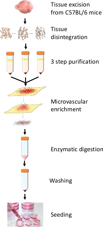
Figure 1: Summary of the protocol. This outline represents critical steps for pericyte extraction which begins with tissue disintegration with glass pestle grinder followed by 3-step separation in dextran and filtration. This protocol employs a 33 min enzyme digestion step. Please click here to view a larger version of this figure.

Figure 2: Cells morphology and markers expression. (A) Phase contrast images of pericytes in P0, P1 and P2 stages of proliferation. Abundant endothelial cells in P0 are indicated by white arrows. Their number decreased in P1 and they have disappeared in P2. Pericytes are indicated by black arrows. (B) Analysis of CD146, NG2 and PDGFR-β expression by PCR in pericytes in P2 with pericytes in P1 and mouse brain (Ms Br) samples. (C) Representative images of pericytes in P2 exhibiting positive immunostaining of CD146, PDGFR-β and, NG2. Scale bar: 50 μm (20x magnification) and 20 μm (40x magnification). Please click here to view a larger version of this figure.
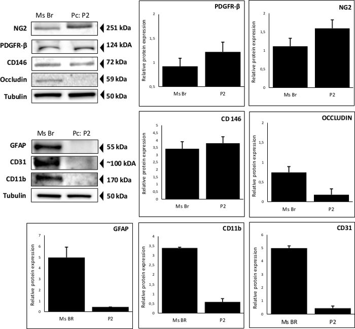
Figure 3: Representative purity of the cell cultures. Analysis of CD146, PDGFR-β, NG2, Occludin, GFAP, CD31, CD11b and Tubulin expression by western blotting in pericytes in P2 and mouse brain (Ms Br) samples. Please click here to view a larger version of this figure.
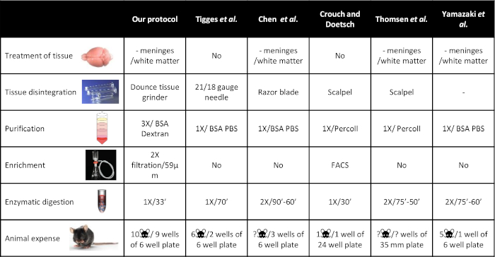
Figure 4: Representative comparison of different methods of tissue disintegration, purification, enrichment and enzymatic digestion. Each published protocol is summarized with indications on the number of animals used for each protocol. Outputs are also indicated with respect to the number of wells obtained upon seeding. Please click here to view a larger version of this figure.
Discussion
Cerebral pericytes are an integral part of the NVU and play an active role in induction and maintenance of the BBB24. Similarly, the role of these cells in the different neurodegenerative disorders and vascular pathologies is intriguing. Hence, an efficient high output primary pericyte cell model will provide an efficient platform for in vitro studies.
There are various protocols that have been proposed for the isolation of primary pericytes (Figure 4). Tigges et al.17 suggested a method including cortical tissue with meninges. This approach is tenderization of tissue from 6 mice (a 37 °C, 70 min digestion with papain/DNase enzymes) followed by a disintegration step via 21 G and 18 G needles. This protocol suggests a one-step separation (centrifugation in 22% BSA/PBS solution) that yields at least 2 collagen I coated wells of a 6-well plate. The cells are maintained in endothelial cell growth medium (ECGM) until passage 3 and later in pericyte growth medium (PGM) for passaging cells to promote pericyte proliferation. In another similar approach, Chen et al.18 proposed tissue dissociation by dicing the tissue with a sterilized razor blade and tissue digestion with collagenase/DNase for 90 min at 37 °C. Following one-step separation (centrifugation in 22% BSA) of the cells, the myelin layer is removed and the pellet is washed twice in ECGM. The microvessels are plated in 3 wells of a collagen I coated 6-well plates. After reaching confluence, the cells are passaged twice and later maintained in PGM. In the end, if cells are passed in ratio of 3, we can obtain 27 wells of 6-well plates only at the use of 10 mice in the beginning of the protocol.
In Thomsen et al., the authors suggest isolation of cerebral pericytes via a two-step enzyme digestion19. Meninges and white matter are removed, and brain samples are cut into small pieces. The tissue pieces undergo the first enzyme reaction in collagenase/DNase I for 75 min at 37 °C, following one step of separation in 20% BSA. The pellet is collected and further digested in collagenase/dispase/DNase I for 50 min at 37 °C. This step is followed by microvessel separation in a 33% Percoll gradient and further washed once. The microvessels are seeded on collagen IV/fibronectin coated 35 mm dishes. The proliferation of pericytes is favored by 10% FCS and gentamicin sulphate in DMEM for 10 days. In another two-step enzyme digestion approach, Yamazaki et al. suggest mincing of the excised tissue in cold DMEM20. In the first enzyme reaction, samples are treated with collagenase/DNase I for 75 min at 37 °C. Following one step centrifugation, the pellet is again washed once and a second enzyme reaction is initiated in collagenase/dispase for 60 min at 37 °C. Following a one-step separation, the pellet is resuspended and centrifuged in 22% BSA solution. Finally, the microvascular pellet is resuspended and plated in a 6-well plate. For 5 mouse brains, 1 well of a 6-well plate can be plated. To obtain the pericytes, endothelial cultures are passaged thrice while maintained in mouse brain endothelial cell (mBEC) medium II. Crouch and Doetsch20 suggest pericyte purification method by FACS. Tissue samples from cortex and ventricular–subventricular zone of mouse brain are micro-dissected and minced thoroughly with a scalpel. After collagenase/dispase enzyme incubation for 30 min at 37 °C, the digested tissue is separated from myelin and debris centrifuged in a 22% v/v Percoll solution. The cell suspension is then incubated in fluorescently conjugated antibodies for FACS analysis and sorting. The sorted cells are plated in collagen coated wells of 24 well plate. It is suggested that one cortex yields enough cells for one plating in 1 well of 24-well plate.
Even if productive, these methods come with several limitations, from the usage of high number of animals for single batch isolation to a very limited amount of output.
During the development of this proposed protocol, we were successful in obtaining high output: 9 wells of a 6-well plate from as few as 10 mice. To this end, removal of meninges ensures the one step removal of large vessels from the tissue. The Dounce tissue grinder is more appropriate for soft tissues such as the brain. It also ensures sample reduction with the loose pestle, homogenization with the tight pestle, and prevents unnecessary cellular damage. One of the main objectives in primary cell culture protocols is the minimal waste of tissue and extended retrieval of cerebral vasculature. In the presented protocol, this is achieved by repetitive centrifugation of the dextran-BSA infused tissue homogenate. A three-step centrifugation approach helps to recover large quantities of vasculature from the tissue homogenate. This provides a 3x enhanced recovery of microvessels. Following separation, filtration is the next essential step, which favors the exclusion of smooth muscle cell associated large vessels. As mentioned before a combination of different enzymes has been proposed for enzymatic digestion. While DNase and collagenase/dispase are used to reduce clumps of cells and isolate single cells respectively, it is very important to prevent cell death in such an invasive environment and this is prevented by TLCK, which thereby increases the final yield. Initially, the first passage is allowed to grow an endothelial monolayer, which later supports the growth of attached pericytes on the unilayer. Since the survival of primary endothelial cells is reduced upon passaging, it enhances the probability for pericyte retrieval. Moreover, this protocol employs another passaging that ensures avoidance of endothelial cell contamination. It should be noted that with a higher number of cells from P2, the dependency on further passaging of the cells is reduced. In addition, it reduces the possibility of pericyte growth being overtaken by smooth muscle cells, which proliferate on much higher rate.
In order to achieve a higher output, there are several steps that are critical and should be accurately performed with respect to temperature and time. The mixing of tissue homogenate into BSA-dextran should be fast. The pellet dissociation after the centrifugation steps should be quick to prevent cell death. Moreover, 33 min of enzymatic digestion should be done with precision and care. One of the limitations for this protocol is the 7-8 day duration that endothelial unilayer is allowed to grow and further facilitate the growth of pericytes. Evidently, the isolation of microvessels is btter, the growth of unilayer is faster, and hence there are an increased number of pericytes. It is recommended not to use less than 10 mice in each extraction to ensure an adequate number of microvascular fractions to support the pericyte growth further. If the aforementioned points are followed carefully, the desired cell density for cerebral pericyte culture can be easily achieved.
In vitro models provide a feasible platform for the development of derivative models to obtain more information on the pathophysiological relevance and communication among the other cells of the NVU during neurological disorders. Isolated pericytes can be incorporated in a bi- cellular culture (with endothelial or glial cells) and tri-cellular culture (endothelial and glial cells) models. The development of these models has not been discussed here. To conclude, this protocol provides one approach for the isolation of primary cells with higher output and a better platform for in vitro research related to the cerebral pericyte biology.
Disclosures
The authors have nothing to disclose.
Acknowledgements
LT and FG were granted by Agence Nationale de la Recherche (ANR, ANR-15-JPWG-0010) in the framework of the EU Joint Programme – Neurodegenerative Disease Research (JPND) for project SNOWBALL.
Materials
| Name | Company | Catalog Number | Comments |
|---|---|---|---|
| Amino acids BME | Sigma | B-6766 | Store at 4 °C. |
| Basal DMEM media | Invitrogen | 316000083 | Store at 4 °C. |
| Basic fibroblast growth factor | Sigma | F-0291 | Store at -20 °C. |
| BSA | Sigma | A-8412 | Store at 4 °C. |
| Collagenase dispase | Sigma | 10269638001 | Prepare a 10x stock solution in sterile PBS-CMF. Filter the solution with a 0.22 μm syringe filter and store at -20 °C. Note: For the enzyme digestion step of the protocol, for every set of 10 mice for extraction, 300 µL of 10x collagenase dispase is required. |
| Dextran | Sigma | 31398 | |
| DNase I | Sigma | 11284932001 | Prepare a 1000X stock solution by dissolving 100 mg in 10 ml sterile water and store at -20°C. |
| Gelatin | Sigma | G-2500 | Prepare the working coating by making a 0.2% gelatin solution in sterile PBS-CMF (8 g/L NaCl, 0.2 g/L KCl, 0.2 g/L KH2PO4, 2.86 g/L NaHPO4 (12 H2O), pH 7.4). Autoclave the solution for minimum 20 minutes at 120 °C and store at room temperature. Culture dishes to be coated for at least 4 hours at 4 °C. |
| Gentamycin | Biochrom AG | A-2712 | Store at 4 °C. |
| Glutamine | Merck | I.00289 | Store at -20 °C. |
| HBSS | Sigma | H-8264 | Store at 4 °C. |
| HEPES | Sigma | H-0887 | Store at 4 °C. |
| Matrigel | BD Biocoat | 354230 | Prepare a working coating solution of Matrigel by diluting stock in cold DMEM at 1:48 ratio with its final concentration to be 85 µg/cm2. Cell culture dishes should be coated at least for 1 hour at room temperature. |
| Pericyte Medium-mouse | Sciencell research laboratories | 1231 | Store at 4 °C. |
| Tosyl Lysin Chloromethyl Ketone | Sigma | T-7254 | Prepare a 1000X stock solution in WBA by dissolving 16 mg in 10.88 mL of WBA to make a 4 mM solution and store at 4 °C. |
| Vitamins | Sigma | B-6891 | Store at -20 °C. |
| Equipment Requirements | |||
| Filtration tools | Sefar, Nylon mesh, 60-micron porosity | ||
| Laboratory equipment | Swing bucket rotor centrifuge | ||
| Water bath with agitator | |||
| Laminar Flow Hood : BSL2 | |||
| Glassware (all components to be heat sterilized) | Dounce Tissue Grinder With Glass Pestle | ||
| Pestle I: 0.0035 - 0.0065 inches | |||
| Pestle II: 0.0010 - 0.0030 inches | |||
| Vacuum filter assembly with coarse porosity fritted glass filter support base | |||
| Surgical dissection tools (all components to be heat sterilized) | Forceps, scissors, Bunsen burner, cotton swabs, gauge |
References
- Azevedo, P. O., et al. Pericytes modulate myelination in the central nervous system. Journal of Cellular Physiology. 233 (8), 5523-5529 (2018).
- Hall, C. N., et al. Capillary pericytes regulate cerebral blood flow in health and disease. Nature. 508 (7494), 55-60 (2014).
- Gianni-Barrera, R., et al. PDGF-BB regulates splitting angiogenesis in skeletal muscle by limiting VEGF-induced endothelial proliferation. Angiogenesis. 21 (4), 883-900 (2018).
- Teichert, M., et al. Pericyte-expressed Tie2 controls angiogenesis and vessel maturation. Nature Communications. 8, 16106(2017).
- Dave, J. M., Mirabella, T., Weatherbee, S. D., Greif, D. M. Pericyte ALK5/TIMP3 Axis Contributes to Endothelial Morphogenesis in the Developing Brain. Developmental Cell. 44 (6), 665-678 (2018).
- Franco, M., Roswall, P., Cortez, E., Hanahan, D., Pietras, K. Pericytes promote endothelial cell survival through induction of autocrine VEGF-A signaling and Bcl-w expression. Blood. 118 (10), 2906-2917 (2011).
- Saint-Pol, J., et al. Brain Pericytes ABCA1 Expression Mediates Cholesterol Efflux but not Cellular Amyloid-beta Peptide Accumulation. Journal of Alzheimer's Disease. 30 (3), 489-503 (2012).
- Sagare, A. P., et al. Pericyte loss influences Alzheimer-like neurodegeneration in mice. Nature Communications. 4, 2932(2013).
- Montagne, A., et al. Pericyte degeneration causes white matter dysfunction in the mouse central nervous system. Nature Medicine. 24 (3), 326-337 (2018).
- Claudio, L., Raine, C. S., Brosnan, C. F. Evidence of persistent blood-brain barrier abnormalities in chronic-progressive multiple sclerosis. Acta Neuropathologica. 90 (3), 228-238 (1995).
- Nishioku, T., et al. Detachment of brain pericytes from the basal lamina is involved in disruption of the blood-brain barrier caused by lipopolysaccharide-induced sepsis in mice. Cellular and Molecular Neurobiology. 29 (3), 309-316 (2009).
- Yang, S., et al. Diverse Functions and Mechanisms of Pericytes in Ischemic Stroke. Current Neuropharmacology. 15 (6), 892-905 (2017).
- Yang, Y., et al. The PDGF-BB-SOX7 axis-modulated IL-33 in pericytes and stromal cells promotes metastasis through tumour-associated macrophages. Nature Communications. 7, 11385(2016).
- Hesp, Z. C., et al. Proliferating NG2-Cell-Dependent Angiogenesis and Scar Formation Alter Axon Growth and Functional Recovery After Spinal Cord Injury in Mice. Journal of Neuroscience. 38 (6), 1366-1382 (2018).
- Guo, P., et al. Platelet-derived growth factor-B enhances glioma angiogenesis by stimulating vascular endothelial growth factor expression in tumor endothelia and by promoting pericyte recruitment. American Journal of Pathology. 162 (4), 1083-1093 (2003).
- Cheng, J., et al. Targeting pericytes for therapeutic approaches to neurological disorders. Acta Neuropathologica. 136 (4), 507-523 (2018).
- Tigges, U., Welser-Alves, J. V., Boroujerdi, A., Milner, R. A novel and simple method for culturing pericytes from mouse brain. Microvascular Research. 84 (1), 74-80 (2012).
- Chen, J., et al. CD146 coordinates brain endothelial cell-pericyte communication for blood-brain barrier development. Proceedings of the National Academy of Sciences of the United States of America. 114 (36), 7622-7631 (2017).
- Thomsen, M. S., Birkelund, S., Burkhart, A., Stensballe, A., Moos, T. Synthesis and deposition of basement membrane proteins by primary brain capillary endothelial cells in a murine model of the blood-brain barrier. Journal of Neurochemistry. 140 (5), 741-754 (2017).
- Yamazaki, Y., et al. Vascular Cell Senescence Contributes to Blood-Brain Barrier Breakdown. Stroke. 47 (4), 1068-1077 (2016).
- Crouch, E. E., Doetsch, F. FACS isolation of endothelial cells and pericytes from mouse brain microregions. Nature Protocols. 13 (4), 738-751 (2018).
- Ozerdem, U., Grako, K. A., Dahlin-Huppe, K., Monosov, E., Stallcup, W. B. NG2 proteoglycan is expressed exclusively by mural cells during vascular morphogenesis. Developmental Dynamics. 222 (2), 218-227 (2001).
- Stallcup, W. B., You, W. K., Kucharova, K., Cejudo-Martin, P., Yotsumoto, F. NG2 Proteoglycan-Dependent Contributions of Pericytes and Macrophages to Brain Tumor Vascularization and Progression. Microcirculation. 23 (2), 122-133 (2016).
- Armulik, A., et al. Pericytes regulate the blood-brain barrier. Nature. 468 (7323), 557-561 (2010).
Reprints and Permissions
