In the clinical context, patients with localized pancreatic cancer will undergo pancreatectomy followed by adjuvant treatment. This protocol reported here aims to establish a safe and effective method of modelling this clinical scenario in nude mice, through orthotopic implantation of pancreatic cancer followed by distal pancreatectomy and splenectomy.
Method Article
An Orthotopic Resectional Mouse Model of Pancreatic Cancer
In This Article
Summary
Abstract
There is a lack of satisfactory animal models to study adjuvant and/or neoadjuvant therapy in patients being considered for surgery of pancreatic cancer (PC). To address this deficiency, we describe a mouse model involving orthotopic implantation of PC followed by distal pancreatectomy and splenectomy. The model has been demonstrated to be safe and suitably flexible for the study of various therapeutic approaches in adjuvant and neo adjuvant settings.
In this model, a pancreatic tumor is first generated by implanting a mixture of human pancreatic cancer cells (luciferase-tagged AsPC-1) and human cancer associated pancreatic stellate cells into the distal pancreas of Balb/c athymic nude mice. After three weeks, the cancer is resected by re-laparotomy, distal pancreatectomy and splenectomy. In this model, bioluminescence imaging can be used to follow the progress of cancer development and effects of resection/treatments. Following resection, adjuvant therapy can be given. Alternatively, neoadjuvant treatment can be given prior to resection.
Representative data from 45 mice are presented. All mice underwent successful distal pancreatectomy/splenectomy with no issues of hemostasis. A macroscopic proximal pancreatic margin greater than 5 mm was achieved in 43 (96%) mice. The technical success rate of pancreatic resection was 100%, with 0% early mortality and morbidity. None of the animals died during the week after resection.
In summary, we describe a robust and reproducible technique for a surgical resection model of pancreatic cancer in mice which mimics the clinical scenario. The model may be useful for the testing of both adjuvant and neoadjuvant treatments.
Introduction
Pancreatic ductal adenocarcinoma (pancreatic cancer [PC]) is associated with a poor prognosis1. Surgical resection remains the only potentially curative treatment for PC and should be considered for patients presenting with early stage disease. Unfortunately, even with R0 resection (i.e., resection margins free of tumor), the recurrence rate (local or from undetected metastatic disease) is high2,3. Therefore, systemic adjuvant therapy is indicated in almost all patients who undergo resection4. Furthermore, while neoadjuvant therapy is now recommended only for borderline-resectable cancers, its indications are expanding such that its routine use is the focus of much clinical research5,6,7,8. In order to develop novel therapeutic approaches for PC involving resection, these approaches need to be first assessed in pre-clinical models that accurately recapitulate clinical settings.
Orthotopic mouse models of PC have been frequently used in the past to test drug treatments9,10. Many of these were produced by injection of cancer cells alone into mouse pancreas, resulting in tumors that lacked the prominent stroma that is characteristic of PC. More recently, co-injection orthotopic models, such as the one we first developed by injecting a mixture of human PC and human pancreatic stellate cells (PSCs, the primary producers of the collagenous stroma in PC), have come into regular use11,12. The tumors produced by such co-injection of cancer and stromal cells exhibit (i) both the cancer elements and the characteristic stromal (desmoplastic) component of PC, and (ii) enhanced cancer cell proliferation and metastasis11. Thus, this model closely resembles human PC. While a number of resectional models of orthotopic PC have been described13,14,15,16, none have reflected the clinical realities of pancreatic resection in humans as accurate as this model, and therefore have been suboptimal for testing adjuvant or neoadjuvant treatments.
The aims of the mouse model presented were to demonstrate how to: (i) successfully implant orthotopic pancreatic cancer while minimizing inadvertent peritoneal dissemination and (ii) subsequently completely resect the cancer. The paper highlight tips and potential pitfalls of this technique.
Protocol
All procedures were approved by the Animal Care and Ethics Committee of the University of New South Wales (17/109A). Female athymic Balb/c nude mice, aged 8-10 weeks weighing 16-19 g, were used for this protocol. Mice were housed in micro-isolator cages and fed commercially available pelleted food and water ad libitum.
1. Orthotopic pancreatic cancer implantation
- Prepare the cells for implantation. First, calculate the number of cells required for the procedure (1 x 10⁶ luciferase-tagged AsPC-1 cells and 1 x 10⁶ cancer-associated human pancreatic stellate cells [CAhPSCs] are required for each animal).
- Maintain these cells in a humidified temperature-controlled CO2 incubator and perform routine mycoplasma testing. Culture medium used for AsPC-1 and CAhPSCs are RPMI 1640 (with 300 mg/L L-glutamine, 20% v/v foetal bovine serum, 1% v/v penicillin/streptomycin) and IMDM (with 4 mM L-glutamine, 10% v/v foetal bovine serum, 1% v/v penicillin/streptomycin).
- Use standard cell culture techniques to trypsinize the cells into a cell suspension. Neutralize the trypsin using the respective complete culture medium at a volume twice that of the trypsin solution used.
- Wash these cells twice with phosphate buffered saline (PBS) and resuspend into a mixture containing 1 x 10⁶ AsPC-1 cells and 1 x 10⁶ CAhPSCs in a 50 μL cell suspension.
- Keep this suspension on ice until use.
- Prepare a class II biosafety cabinet for the procedure. Use a heating mat overlaid by a sterile plastic drape. For magnification during the procedure, use a pair of 2.5x to 3.5x magnification surgical loupes.
- Prepare purse-string swabs by cutting a hole, 1 cm in diameter, into a gauze swab. Secure this hole with a purse-string suture. Any fine braided suture can be used for this (e.g., 5/0 polyglycolic acid suture). Braided suture material is recommended as it allows the loose knot to stay in place after tightening. This is illustrated in Figure 1a.
- Anesthetize the mouse with 80 mg/kg of ketamine and 10 mg/kg of xylazine by intraperitoneal injection.
- Administer 5 mg/kg enrofloxacin antibiotic prophylaxis, 2.5 mg/kg flunixin analgesia and 1 mL of 0.9% saline subcutaneously.
- Once anesthetized, place the mouse on the sterile field in a supine position and apply povidone-iodine followed by 70% ethanol for skin preparation.
- Make a longitudinal incision in the skin of the left cranial quadrant of the abdomen, and then enter the abdomen by incising the muscular layer between forceps.
- Load a 29 G insulin syringe with 50 μL of cell suspension–this equates to 1 x 10⁶ CAhPSCs and 1 x 10⁶ luciferase-tagged AsPC-1 cells per injectate. Mount it on the injection device. The design and function of this injection device is explained in detail in Figure 1b and its legend.
- Place the purse-string swab over the laparotomy incision and then exteriorize the spleen and pancreatic tail through the opening of this swab. Tighten the purse-string to gently encircle the body of the pancreas, exposing the pancreatic tail for injection. It is important to be tight enough that the gauze contacts the pancreas circumferentially while at the same time not constricting it.
- Using a pair of forceps, grasp the tail of the pancreas and gently place lateral tension on it. Puncture the ventral peritoneal surface with the needle at a shallow angle and then inject the cell suspension into the pancreas in a slow and controlled fashion (over 10−15 s) with the injection device.
- During the injection process, carefully observe for leakage—both around the injection site (from reflux) and on the other side of the pancreatic lobule (in case of through-and-through penetration). If visible leakage occurs, stop the injection and note the volume of leakage by checking the volume of remaining injectate in the syringe. If the leakage is of small volume (<10 μL), and then absorb any leakage with gauze and reposition the needle into a different pancreatic lobule to complete the injection.
- Replace the spleen and pancreas and close the abdominal wall with 5/0 polyglycolic acid suture in a continuous fashion. Close the skin with clips.
- Monitor the mouse in a warmed cage until recovered from the anaesthetic. Once awake and alert, move the mouse back to its cage.
2. Cancer resection surgery: Distal pancreatectomy and splenectomy
- The timing of resection in relation to implantation can vary depending on the experimental protocol. In general, allow the tumors to grow at least for 3 weeks prior to resection, but optimize this empirically for the particular implanted cancer cell line.
- On the day prior to the resection surgery, perform bioluminescence imaging on the animals to confirm the presence of a localized primary tumor. Note that this imaging study is simply used to exclude mice with obvious extra-pancreatic disease from resection. Neither size nor radiant flux should be used as thresholds for determining eligibility for resection.
- Weigh mice and inject with D-luciferin intraperitoneally (150 mg/kg).
- Determine the timing of the imaging step in relation to luciferin injection for each experiment by the performance of a luciferin kinetic curve. The period of time where the radiant flux is above 90% of its maximum represents the optimal time for bioluminescence imaging (in this experiment, 18 to 26 minutes post-injection)
- Induce anaesthesia and maintain using isoflurane (4% and 3% with oxygen, respectively) and perform imaging using a bioluminescent imaging device (e.g., IVIS Lumina II). Use automatic exposure and binning settings (this can, however, be optimized for the expected radiant flux).
- Prepare the class II biosafety cabinet for procedure. Use a heating mat overlaid by a sterile plastic drape. For magnification during dissection, use a pair of 2.5x to 3.5x magnification surgical loupes.
- Anesthetize the mouse with 80 mg/kg of ketamine and 10 mg/kg of xylazine by intraperitoneal injection.
- Administer 5 mg/kg enrofloxacin antibiotic prophylaxis, 2.5 mg/kg flunixin analgesia and 1 mL of 0.9% saline subcutaneously.
- Place the mouse on the sterile field in a supine position and apply povidone-iodine followed by 70% ethanol for skin preparation.
- Make a longitudinal incision in the skin of the left cranial quadrant of the abdomen, preferably through the previous incision site.
- Bluntly dissect the skin off the underlying muscular abdominal wall, and then place an Alm self-retaining retractor to hold the skin wound open.
- Incise the muscular layer between forceps just to one side of the suture line of the previous operation, and then extend the incision to excise the entire previous suture line.
- Exteriorize the spleen and distal pancreas and retract it cranially. At the caudal aspect of the pancreas, the colon may be found attached by filmy adhesions. If this is found, bluntly dissect the colon off.
- Carefully pass a pair of forceps dorsal to the body of the pancreas and splenic vessels and open this space. This frees up a segment of pancreas for subsequent ligation.
- Ligate the body of the pancreas proximal to the tumor with a titanium ligation clip, and then transect the pancreas distal to this with cautery. An alternative way to control the pancreatic stump is to ligate it in continuity with 5/0 polyglycolic acid suture before transection.
- Retract the pancreas caudally and cauterize the gastrosplenic vessels between the cranial pole of the spleen and the stomach.
- Remove the specimen and confirm haemostasis.
- Close the abdominal wall with 5/0 polyglycolic acid suture in a continuous fashion. Close the skin with clips.
3. Postoperative management
- In the immediate post anaesthetic period (for both of the above procedures), monitor the mouse in a warmed cage until recovered from the anaesthetic. Once awake and alert, move the mouse back to its cage. In the postoperative period, monitor the animals for pain and signs of distress. Administer 0.05 mg/kg buprenorphine by subcutaneous injection and closely observe the animals for 12 hours.
- Subsequently, monitor mice daily for weight, food intake and activity. Examine incision sites and palpate for tumor size. Remove skin clips on the seventh postoperative day.
- Euthanize the mouse if humane endpoints are reached. These humane endpoints include: loss of body weight >20%, features of untreatable distress (including hunched posture, lack of movement or grooming) and tumor size greater than 1 cm3 as estimated by external palpation.
Results
Fifty-nine consecutive mice underwent implantation surgery. Gross leakage occurred in eight (14%) mice. The degree of leakage at the time of injection is estimated as described above in the protocol section. After three weeks to allow these implanted tumors to grow, pre-resection bioluminescence imaging was performed to exclude mice with gross metastatic disease prior to resection. Forty-five (76%) mice underwent surgical resection.
All 45 (100%) mice underwent successful distal pancreatectomy/splenectomy with no issues of haemostasis. A macroscopic proximal pancreatic margin greater than 5 mm was achieved in 43 (96%) mice.
At the time of resection, local metastasis was found in 9/45 (20%) mice – mostly in the suture line (discontinuous with the primary tumor) with three of the nine showing additional isolated nodules on the greater curve of the stomach and one showing a subcapsular nodule on the liver. The primary pancreatic tumor was adherent to the suture line in five (11%) mice and to the liver in one (2%) mouse. These adherent structures were excised en bloc.
The mean (SEM) surgery time (induction to closure) was 22 (0.9) minutes. None of the animals died within 1 week after resection.
One-week post resection, mice underwent bioluminescence imaging to detect residual disease. The ratio of the maximum radiance over the ventral surface of the mouse was compared to that of the background. Thirty-two (71%) mice had a maximum radiance ratio (mouse:background) of <10, indicating minimal or no residual disease.
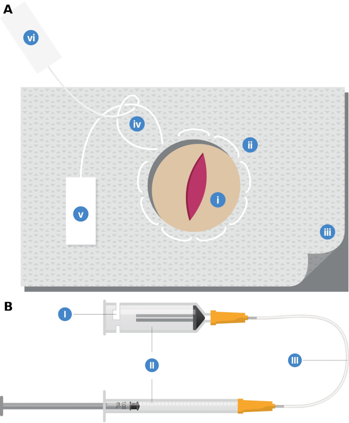
Figure 1: Custom-made devices to facilitate tumor implantation. (a) Purse-string gauze swab: (i) Central hole, approximately 1 cm in diameter, through which the pancreatic tail will be placed at the time of injection; (ii) Purse-string suture around the hole; (iii) Double-layered gauze; (iv) Single throw knot; (v) One limb of the suture material is secured to the gauze with sterilising indicator tape; (vi) A handle, made from indicator tape, is fashioned on the other end of the suture material. (b) Injection device: (I) Actuating syringe. Slots cut through the body of this syringe allows the injection syringe (with the cell suspension injectate; not shown) to be mounted on this syringe body; (II) Controller syringe. This is filled with water. Depression of the plunger on the smaller controller syringe by the surgical assistant causes displacement of the larger actuating syringe plunger. The displacement of the actuating plunger is smaller, but with a mechanical advantage which allows the injection to overcome the resistance associated with the injection syringe mechanism as well as the tissue’s resistance to expansion by the injectate. This allows for precise and smooth injection of 50 μL over 10–15 seconds; (III) Polytetrafluoroethylene (PTFE) connection tubing with internal diameter of 0.5 mm. Please click here to view a larger version of this figure.
Discussion
A resectional orthotopic mouse model of pancreatic cancer is important because it allows for the testing of adjuvant and neoadjuvant treatments. This is particularly important in pancreatic cancer where surgery remains the most effective treatment but is associated with high risk of recurrence. This paper describes a method which will reliably produce a pancreatic cancer which is potentially curable with resection, replicating the clinical scenario where neoadjuvant/ adjuvant therapy is required.
Significance with respect to existing methods
Despite the importance of adjuvant and neoadjuvant therapies in pancreatic cancer, there are few well-described orthotopic resectional mouse models in the literature. These described resectional models varied in their fidelity of replication of the clinical situation in humans. These previous models can be broadly classified into: (i) tumor excision only, with fluorescence guidance; (ii) subtotal pancreatic resection with no splenectomy; (iii) distal pancreatectomy/splenectomy.
Tumor excision with fluorescence guidance has been described in the greatest number of reports15,17,18,19,20,21. Many of these papers originated from the same research group. Unfortunately, in humans, local excision of the tumor alone (enucleation) is not performed for pancreatic adenocarcinoma (PC) due to the high likelihood of local recurrence, as well as the inability to assess lymph node status22,23. Therefore, the use of a non-clinically relevant comparator group (non-fluorescence guided enucleation) clouds the reporting of the oncological outcomes in papers describing this technique. Not surprisingly, the non-fluorescence enucleation groups invariably had excessive rates of local recurrence15,20,21. In contrast, Torgenson et al.14 described a similar fluorescence-guided resection technique, and reported a reasonably low recurrence rate of 58% (at eight weeks post-resection). Overall, these studies appear to demonstrate the utility of fluorescence guidance for visualization of residual disease during surgery. However, this is not yet the standard of care in humans, which is a limitation in terms of its use in a mouse model aiming to replicate the clinical scenario. Of course, this may change if fluorescence-guided surgery were to be widely adopted in clinical practice.
Another resection model was based on subtotal pancreatectomy without splenectomy for a tumor implanted into the body of the pancreas13,24. The clinical relevance of this is also called into question as the operation described was neither a pancreaticoduodenectomy nor distal pancreatectomy as performed in humans. Not surprisingly, these mice also suffered from high rates of tumor recurrence, both distant and local. Of particular note is that splenic recurrence was common, suggesting either inadequate resection or possible peritoneal tumor seeding at implantation24.
Ni et al.16 described a distal pancreatectomy/splenectomy model performed with fluorescence imaging guidance. Disappointingly, despite the use of a clinically relevant operation (with fluorescence guidance), the survival was very short (mean survival of 18 days), even in the distal pancreatectomy group. This degree of progressive disease appears to be even worse than palliative treatment models25,26,27, suggesting the possible presence of gross residual disease after resection. Most recently, Giri et al.28 reported a distal pancreatectomy and partial splenectomy mouse model. This study is notable in that it represents an immunocompetent mouse model of cancer. However, this study reported almost universal local and other intraperitoneal tumor recurrence, possibly indicating occult iatrogenic metastasis at implantation.
The use of mouse models where there is gross residual disease post resection for testing adjuvant treatments may be inappropriate. The issue is that treatment for gross residual disease cannot truly be classified as adjuvant treatment but rather should be considered to be treatment with palliative intent. In that case, such mouse models offer no advantage compared to non-resectional models with low volume disease.
Tips and pitfalls of critical steps
Tumor implantation procedure
In order to replicate the clinical scenario, there are distinct challenges in this model which relate to the implantation and resection procedures. For the implantation procedure, the major challenges which need to be overcome are successful implantation and prevention of leakage. These two issues are interrelated as failure of injection would result in gross leakage of the tumor cell suspension into the abdominal cavity. This would produce a mouse model with peritoneal metastasis, which will progress regardless of pancreatic resection. This reflects the well-known clinical scenario in humans where pancreatic resection in metastatic PC does not affect the patient outcome. This is the basis of the staging laparoscopy in humans29.
The success of implantation of the tumor can be seen intraoperatively as the successful generation of a “bubble” of cell suspension without obvious leakage. Of most importance in achieving a good result is the accurate placement of the needle within the pancreatic parenchyma. This could only be achieved by “stretching out” the pancreas so that the peritoneal surface is taut. Puncture should occur with the needle bevel facing upwards (ventrally). Once the needle punctures the peritoneal surface, it should be advanced while the needle tip is slightly lifted-up so that the beveled surface glides just beneath the peritoneum. This will prevent inadvertent through-and-through puncture of the pancreas, a common pitfall due to the small dimensions of mouse pancreatic lobules. Once the entire bevel is within the substance of the pancreas, the cell suspension is injected. Magnification of vision with surgical loupes is highly desirable to visualize accurately the depth of the needle penetration.
A number of techniques can be used to further minimize the risk of inadvertent leakage.
Selection of a large lobule for injection. Small lobules require higher pressures to inflate (following Laplace’s law), thereby increasing the risk of leakage around the needle at the puncture site.
Optimization of the speed of injection. The use of an injection device (Figure 1b) which allows the cell suspension to be injected over 10-15 seconds serves three purposes. First, it decreases the rate of change of pressure in the pancreas, giving the tissues time to deform and reduces the risk of reflux of the suspension. Second, it allows the injection process to be monitored and, if necessary, stopped and needle repositioned. Any leakage can be mopped up by a povidone-iodine-soaked gauze. Third, it frees the operator from needing to depress the plunger, allowing the operator to focus on keeping the needle tip within the pancreas while the assistant injects the cell suspension.
Use of a double-layered purse-string gauze. This gauze forms a collar around the pancreatic tail which will absorb any leakage of the cell suspension and therefore minimize contamination in the abdominal cavity.
Some studies in the literature have used an extracellular matrix mixture (Matrigel) which solidifies with time after injection13,15,24. This may reduce the risk of leakage post-injection. However, a potential disadvantage of this strategy is that Matrigel or other similar extracellular matrix solutions may exert non-physiological effects on PSCs30. For instance, Matrigel has been shown to render PSCs quiescent thereby potentially negating the effects of PSCs in the model31,32. An alternative to injection of cancer cells is the orthotopic implantation of tumor tissue (either directly from patients or from subcutaneous mouse models). However, these approaches have their own disadvantages. First, heterogeneity may arise from sampling error or from variations in the volume of tissue implanted. Such heterogeneity may reduce the power of subsequent treatment comparisons. Second, passaging of tumor tissue with a subcutaneous mouse model may lead to selection of sub-clones which have different biological behaviours to the original patient tumor.
Tumor resection procedure
In this model, we have utilized a distal pancreatectomy/splenectomy procedure akin to that performed in humans. The challenges relating to the resectional surgery depend on pathological and anatomical factors.
The key pathological factor is tumor dissemination. Low volume local spread can be resected at the time of pancreatic resection, although it may indicate the possibility of more distant peritoneal and other metastasis. We routinely excise the suture line from the first operation as it is a possible area of local recurrence. If the tumor is attached to surrounding structures, such as the abdominal wall or the left lobe of the liver, these can be resected en bloc. Anatomically, the key step is dissecting the plane dorsal to the body of the pancreas. The splenic vein can often be visualized behind the pancreas once the pancreas is exteriorized. This is a key landmark, as the embryological bloodless plane is immediately dorsal to this.
There are two other potential anatomical pitfalls in the model described here. The colon may be adherent to the caudal aspect of the pancreatic body. Failure to mobilize this structure away could lead to inadvertent colonic injury at the time of pancreatic division or ligation. The gastrosplenic vessels are small and may easily bleed if avulsed or inadequately cauterized. Furthermore, once avulsed, the bleeding point often retracts deep into the abdomen behind the greater curve of the stomach, making subsequent control of bleeding more challenging. Therefore, careful retraction of the spleen and cautery of the gastrosplenic vessels are required. One approach for successful hemostasis is to cauterize these vessels on the hilar aspect of the spleen which minimizes the risk of inadvertent thermal injury to surrounding hollow viscera.
We have found that using a titanium ligation clip, widely used in human surgery for ligation of vessels, is a rapid and effective way of controlling the pancreatic stump, with consequent reduction in total operative time compared to the use of ligatures. This was also used by Giri et al.28.
Limitations of the technique
There are limitations to this resectional model of the pancreas. One limitation relates to the time allowed to produce recurrence/metastasis. On the one hand, one needs to maximize the development of metastatic disease, but on the other hand, one needs to resect the tumor before it became locally advanced. The period between implantation and resection may therefore need to be adjusted for the particular clinical scenario one wishes to replicate. Another limitation relates to the inadvertent spillage and subsequent peritoneal metastasis of cancer cells which is discussed above.
A major challenge of adjuvant treatment models is dissecting the adjuvant treatment effect from the surgical treatment effect. Clearly, a well-designed study which is randomized, with a control group undergoing resection surgery is required. To further improve the assessment of the relative treatment effects, we suggest assessing tumor burden in vivo (for example, by using luciferase-tagged cancer cells and performing in vivo bioluminescence imaging). Despite the semi-quantitative nature of this assessment in orthotopic models (as the bioluminescence signal is attenuated by passage through the overlying tissues), this approach allows longitudinal assessment of tumor burden, including the assessment of the post-surgical residual disease.
Modifications and future applications
The implanted cell line and/or cell numbers with or without pancreatic stellate cells could be modified to reflect the target clinical scenario12. The duration between implantation and resection could also be modified to change the risk of metastasis formation. Other variations could include implantation of patient- or mice-derived xenografts or organoids33.
Neoadjuvant therapy can also be tested within the basic features of the model described here. It would simply require commencement of drug treatment prior to surgical resection34. Similarly, both neoadjuvant and adjuvant therapy could be studied in the same mice.
Finally, while we have described the use of athymic Balb/c nude mice which represents an immunodeficient model, an alternative immunocompetent model may involve KPC tumor cells implanted into C57B6 mice28. This may be a useful alternative for the testing of adjuvant/neoadjuvant immune therapies.
In summary, we describe a robust and reproducible technique for a surgical resection model of pancreatic cancer in mice which mimics the clinical scenario and does not require specialized equipment. This model may be useful for the testing of both adjuvant and neoadjuvant treatments.
Disclosures
The authors have nothing to disclose with respect to this project.
Acknowledgements
Authors have received support from the Avner Pancreatic Cancer Foundation.
Materials
| Name | Company | Catalog Number | Comments |
|---|---|---|---|
| Animals, Materials and Equipment for Implantation Procedure | |||
| AsPC-1 human pancreatic cancer cell line, luciferase tagged (luc+ gene from Promega PGL3 Basic plasmid) | American Type Culture Collection, Manassas, VA, USA | supplied by Professor Takashi Murakami, Saitama Medical University, Saitama, Japan | |
| Autoclip wound clips, 9 mm | Becton Dickson Pty Ltd, North Ryde, NSW, Australia | 500346 | |
| Basic Dressing Pack | Multigate Medical Products Pty Ltd, Villawood, NSW, Australia | ||
| Cancer associated human pancreatic stellate cells | Pancreatic Research Group cell bank | In house cell bank | |
| Cryogenic tubes, 1.0 mL | Thermo Fisher Scientific Australia Pty Ltd, Scoresby, VIC, Australia | 366656 | |
| Disposable stainless-steel scalpel blade with handle, size 15 | Livingstone International, Mascot, NSW, | SCP15 | |
| Foetal bovine serum (FBS) | Life Technologies Corporation, Tullamarine, VIC, Australia | 16000044 | |
| Gilles fine tooth forceps 12 cm | Generic stainless steel microsurgical instrument set | ||
| Heated mats to maintain body temperature during surgery and postoperative recovery | Generic | ||
| Homozygous athymic nude mice: Strain BALB/c-Fox1nu/Ausb, female | Australian Bioresources, Moss Vale, NSW, Australia | ||
| Iscove's modified Dulbecco's medium (IMDM) with 4mM L-glutamine and no phenol red | Life Technologies Corporation, Tullamarine, VIC, Australia | 21056023 | |
| Jewellers forceps 11.5 cm | Generic stainless steel microsurgical instrument set | ||
| Micro needle holder (round handle) 15 cm straight | Generic stainless steel microsurgical instrument set | ||
| Micro scissors (round handle) 15 cm straight | Generic stainless steel microsurgical instrument set | ||
| Penicillin 10,000 U/mL, streptomycin 10,000 μg/mL | Life Technologies Corporation, Tullamarine, VIC, Australia | 15140122 | |
| Polyglycolic acid suture, size USP 5/0 on 13mm half-circle round-bodied needle | Braun Australia Pty Ltd, Bella Vista, NSW, Australia | C1049407 | |
| Portable weighing scale | Precision balances, Bradford, MA, USA | ||
| Reflex clip applier and clip remover | World Precision Instruments, Sarasota, FL, USA | 500345 | |
| Roswell Park Memorial Institute (RPMI) 1640 with phenol red and 300 mg/L Lglutamine | Life Technologies Corporation, Tullamarine, VIC, Australia | 11875085 | |
| Round bodied vessel dilator 15 cm, 0.1 mm tip | Generic stainless steel microsurgical instrument set | ||
| Trypsin 0.05%, EDTA 0.02% | Life Technologies Corporation, Tullamarine, VIC, Australia | 25300054 | For pancreatic stellate cells |
| Trypsin 0.25%, EDTA 0.02% | Life Technologies Corporation, Tullamarine, VIC, Australia | 25200056 | For ASPC-1 cells |
| U-100 insulin syringes, 0.5 mL with 29 G (0.33 mm) × 13 mm needle | Terumo Medical Corporation, Elkton, MD, USA | ||
| Equipment for Resection Procedure | |||
| Alm self-retaining retractor | Generic stainless steel microsurgical instrument set | ||
| Autoclip wound clips 9 mm | Becton Dickson Pty Ltd, North Ryde, NSW | 500346 | |
| Basic Dressing Pack | Multigate Medical Products Pty Ltd, Villawood, NSW, Australia | 08-559NP | |
| Disposable stainless-steel scalpel blade with handle, size 15 | Livingstone International, Mascot, NSW, | SCP15 | |
| Gilles fine tooth forceps 12 cm | Generic stainless steel microsurgical instrument set | ||
| Hand-held high temperature fine tip cautery | Bovie Medical Corporation, Melville, NY, USA | AA01 | |
| Heated mats to maintain body temperature during surgery and postoperative recovery | Generic | ||
| IVIS Lumina II Bioluminescent Imaging Device | Caliper Life Sciences, Hopkinton, MA, USA | ||
| Jewellers forceps 11.5 cm | Generic stainless steel microsurgical instrument set | ||
| Micro needle holder (round handle) 15 cm straight | Generic stainless steel microsurgical instrument set | ||
| Micro scissors (round handle) 15 cm straight | Generic stainless steel microsurgical instrument set | ||
| Polyglycolic acid suture, size USP 5/0 on 13mm half-circle round-bodied needle | Braun Australia Pty Ltd, Bella Vista, NSW, Australia | C1049407 | |
| Portable weighing scale | Precision balances, Bradford, MA, USA | ||
| Reflex wound clip applier and clip remover | World Precision Instruments, Sarasota, FL, USA | 500345 | |
| Round bodied vessel dilator 15 cm, 0.1 mm tip | Generic stainless steel microsurgical instrument set | ||
| Titanium “Weck style” Ligaclip, small | HZMIM, Hangzhou, China | ||
| Titanium Ligaclip applier for open surgery, small | HZMIM, Hangzhou, China | ||
| Volatile anaesthetic machine, including vapouriser and induction chamber | Generic | Generic vapouriser and induction chamber | |
| Drugs for Procedures | |||
| 70% w/w ethanol solution | Sigma-Aldrich Pty Ltd, Castle Hill, NSW, Australia | Applied topically as surgical skin preparation | |
| Buprenorphine 0.3 mg/mL | Troy Laboratories Pty Ltd, Glendenning, NSW, Australia | Dose: 0.05 mg/kg s.c. | |
| D-Luciferin (1 U/g) | PerkinElmer, Inc., Waltham, MA, USA | 122799 | diluted in PBS to 15 mg/mL. Dose: 150 mg/kg i.p |
| Enrofloxacin 50 mg/mL | Troy Laboratories Pty Ltd, Glendenning, NSW, Australia | Dose: 5 mg/kg s.c. | |
| Flunixin 50 mg/mL | Norbrook Laboratories Australia, Tullamarine, VIC, Australia | Dose: 2.5 mg/kg s.c. | |
| Isoflurane | Zoetis Australia Pty Ltd., Rhodes, NSW, Australia | Dose (vapourised with oxygen): 4% induction, 3% maintenance | |
| Ketamine 100 mg/mL | Maylab, Slacks Creek, QLD, Australia | Dose: 80 mg/kg i.p. | |
| Povidone-Iodine 10% w/v solution | Perrigo Australia, Balcatta, WA, Australia | RIO00802F | Applied topically to the anterior abdomen as surgical skin preparation |
| Refresh eye ointment (liquid paraffin 42.5% w/w, soft white paraffin 57.3% w/w) | Allergan Australia Pty Ltd, Gordon, NSW, Australia | Applied to both eyes | |
| Sodium chloride 0.9% w/v | Braun Australia Pty Ltd, Bella Vista, NSW, Australia | 9481P | Dose: 900 μL s.c. |
| Water for injections BP | Pfizer Australia, Sydney, NSW, Australia | For dilution of drugs | |
| Xylazine 20 mg/mL | Troy Laboratories Pty Ltd, Glendenning, NSW, Australia | Dose: 10 mg/kg i.p. |
References
- Noone, A. M., et al. SEER Cancer Statistics Review, 1975-2015, National Cancer Institute. , Available from: https://seer.cancer.gov/csr/1975_2015/ (2018).
- Sugiura, T., et al. Margin status, recurrence pattern, and prognosis after resection of pancreatic cancer. Surgery. 154 (5), 1078-1086 (2013).
- Hishinuma, S., et al. Patterns of recurrence after curative resection of pancreatic cancer, based on autopsy findings. Journal of Gastrointestinal Surgery. 10 (4), 511-518 (2006).
- NCCN Clinical Practice Guidelines in Oncology - Pancreatic Adenocarcinoma (Version 3.2019). National Comprehensive Cancer Network. , Available from: https://www.nccn.org/professionals/physician_gls/pdf/pancreatic.pdf (2019).
- Breslin, T. M., et al. Neoadjuvant chemoradiotherapy for adenocarcinoma of the pancreas: treatment variables and survival duration. Annals of Surgical Oncology. 8 (2), 123-132 (2001).
- Mokdad, A. A., et al. Neoadjuvant Therapy Followed by Resection Versus Upfront Resection for Resectable Pancreatic Cancer: A Propensity Score Matched Analysis. Journal of Clinical Oncology. 35 (5), 515-522 (2017).
- Tachezy, M., et al. Sequential neoadjuvant chemoradiotherapy (CRT) followed by curative surgery vs. primary surgery alone for resectable, non-metastasized pancreatic adenocarcinoma: NEOPA- a randomized multicenter phase III study (NCT01900327, DRKS00003893, ISRCTN82191749). BMC Cancer. 14, 411(2014).
- Barbour, A. P., et al. The AGITG GAP Study: A Phase II Study of Perioperative Gemcitabine and Nab-Paclitaxel for Resectable Pancreas Cancer. Annals of Surgical Oncology. , (2020).
- Fu, X., Guadagni, F., Hoffman, R. M. A metastatic nude-mouse model of human pancreatic cancer constructed orthotopically with histologically intact patient specimens. Proceedings of the National Academy of Sciences of the United States of America. 89 (12), 5645-5649 (1992).
- Marincola, F., Taylor-Edwards, C., Drucker, B., Holder, W. D. Orthotopic and heterotopic xenotransplantation of human pancreatic cancer in nude mice. Current Surgery. 44 (4), 294-297 (1987).
- Vonlaufen, A., et al. Pancreatic stellate cells: partners in crime with pancreatic cancer cells. Cancer Research. 68 (7), 2085-2093 (2008).
- Xu, Z., et al. Role of pancreatic stellate cells in pancreatic cancer metastasis. American Journal of Pathology. 177 (5), 2585-2596 (2010).
- Tepel, J., et al. Adjuvant treatment of pancreatic carcinoma in a clinically adapted mouse resection model. Pancreatology. 6 (3), 240-247 (2006).
- Torgenson, M. J., et al. Natural history of pancreatic cancer recurrence following "curative" resection in athymic mice. Journal Surgical Research. 149 (1), 57-61 (2008).
- Metildi, C. A., et al. Fluorescence-guided surgery allows for more complete resection of pancreatic cancer, resulting in longer disease-free survival compared with standard surgery in orthotopic mouse models. Journal of the American College of Surgeons. 215 (1), 126-135 (2012).
- Ni, X., Yang, J., Li, M. Imaging-guided curative surgical resection of pancreatic cancer in a xenograft mouse model. Cancer Letters. 324 (2), 179-185 (2012).
- Hiroshima, Y., et al. Hand-held high-resolution fluorescence imaging system for fluorescence-guided surgery of patient and cell-line pancreatic tumors growing orthotopically in nude mice. Journal of Surgical Research. 187 (2), 510-517 (2014).
- Hiroshima, Y., et al. Metastatic recurrence in a pancreatic cancer patient derived orthotopic xenograft (PDOX) nude mouse model is inhibited by neoadjuvant chemotherapy in combination with fluorescence-guided surgery with an anti-CA 19-9-conjugated fluorophore. PLoS One. 9 (12), 114310(2014).
- Hiroshima, Y., et al. Fluorescence-guided surgery in combination with UVC irradiation cures metastatic human pancreatic cancer in orthotopic mouse models. PLoS One. 9 (6), 99977(2014).
- Metildi, C. A., et al. Ratiometric activatable cell-penetrating peptides label pancreatic cancer, enabling fluorescence-guided surgery, which reduces metastases and recurrence in orthotopic mouse models. Annals of Surgical Oncology. 22 (6), 2082-2087 (2015).
- Metildi, C. A., et al. Fluorescence-guided surgery with a fluorophore-conjugated antibody to carcinoembryonic antigen (CEA), that highlights the tumor, improves surgical resection and increases survival in orthotopic mouse models of human pancreatic cancer. Annals of Surgical Oncology. 21 (4), 1405-1411 (2014).
- NCCN Guidelines: Pancreatic Adenocarcinoma. National Comprehensive Cancer Network. , Available from: https://www.nccn.org/professionals/physician_gls/pdf/pancreatic.pdf (2019).
- Maithel, S. K., Allen, P. J. Blumgart's Surgery of the Liver, Biliary Tract and Pancreas, 2-Volume Set (Sixth Edition). Jarnagin, W. R. , Content Repository Only 1007-1023 (2017).
- Egberts, J. H., et al. Dexamethasone reduces tumor recurrence and metastasis after pancreatic tumor resection in SCID mice. Cancer Biology & Therapy. 7 (7), 1044-1050 (2008).
- Xu, Z. H., et al. Targeting the HGF/c-MET pathway in advanced pancreatic cancer: a key element of treatment that limits primary tumor growth and eliminates metastasis. British Journal of Cancer. , (2020).
- Pothula, S. P., et al. Targeting the HGF/c-MET pathway: stromal remodelling in pancreatic cancer. Oncotarget. 8 (44), 76722-76739 (2017).
- Pothula, S. P., et al. Hepatocyte growth factor inhibition: a novel therapeutic approach in pancreatic cancer. British Journal of Cancer. 114 (3), 269-280 (2016).
- Giri, B., et al. An Immunocompetent Model of Pancreatic Cancer Resection and Recurrence. Journal of Gastrointestinal Surgery. , (2020).
- Allen, V. B., Gurusamy, K. S., Takwoingi, Y., Kalia, A., Davidson, B. R. Diagnostic accuracy of laparoscopy following computed tomography (CT) scanning for assessing the resectability with curative intent in pancreatic and periampullary cancer. Cochrane Database of Systematic Reviews. (11), 009323(2013).
- Vaillant, F., Lindeman, G. J., Visvader, J. E. Jekyll or Hyde: does Matrigel provide a more or less physiological environment in mammary repopulating assays. Breast Cancer Research. 13 (3), 108(2011).
- Jesnowski, R., et al. Immortalization of pancreatic stellate cells as an in vitro model of pancreatic fibrosis: deactivation is induced by matrigel and N-acetylcysteine. Laboratory Investigation. 85 (10), 1276-1291 (2005).
- Phillips, P. A., et al. Cell migration: a novel aspect of pancreatic stellate cell biology. Gut. 52 (5), 677-682 (2003).
- Boj, S. F., et al. Organoid models of human and mouse ductal pancreatic cancer. Cell. 160 (1-2), 324-338 (2015).
- Egberts, J. H., et al. Superiority of extended neoadjuvant chemotherapy with gemcitabine in pancreatic cancer: a comparative analysis in a clinically adapted orthotopic xenotransplantation model in SCID beige mice. Cancer Biology & Therapy. 6 (8), 1227-1232 (2007).
Reprints and Permissions
