Here we present development of a mock circulation setup for multimodal therapy evaluation, pre-interventional planning, and physician-training on cardiovascular anatomies. With the application of patient-specific tomographic scans, this setup is ideal for therapeutic approaches, training, and education in individualized medicine.
Method Article
Development and Evaluation of 3D-Printed Cardiovascular Phantoms for Interventional Planning and Training
In This Article
Summary
Abstract
Catheter-based interventions are standard treatment options for cardiovascular pathologies. Therefore, patient-specific models could help training physicians' wire-skills as well as improving planning of interventional procedures. The aim of this study was to develop a manufacturing process of patient-specific 3D-printed models for cardiovascular interventions.
To create a 3D-printed elastic phantom, different 3D-printing materials were compared to porcine biological tissues (i.e., aortic tissue) in terms of mechanical characteristics. A fitting material was selected based on comparative tensile tests and specific material thicknesses were defined. Anonymized contrast-enhanced CT-datasets were collected retrospectively. Patient-specific volumetric models were extracted from these datasets and subsequently 3D-printed. A pulsatile flow loop was constructed to simulate the intraluminal blood flow during interventions. Models' suitability for clinical imaging was assessed by x-ray imaging, CT, 4D-MRI and (Doppler) ultrasonography. Contrast medium was used to enhance visibility in x-ray-based imaging. Different catheterization techniques were applied to evaluate the 3D-printed phantoms in physicians' training as well as for pre-interventional therapy planning.
Printed models showed a high printing resolution (~30 µm) and mechanical properties of the chosen material were comparable to physiological biomechanics. Physical and digital models showed high anatomical accuracy when compared to the underlying radiological dataset. Printed models were suitable for ultrasonic imaging as well as standard x-rays. Doppler ultrasonography and 4D-MRI displayed flow patterns and landmark characteristics (i.e., turbulence, wall shear stress) matching native data. In a catheter-based laboratory setting, patient-specific phantoms were easy to catheterize. Therapy planning and training of interventional procedures on challenging anatomies (e.g., congenital heart disease (CHD)) was possible.
Flexible patient-specific cardiovascular phantoms were 3D-printed, and the application of common clinical imaging techniques was possible. This new process is ideal as a training tool for catheter-based (electrophysiological) interventions and can be used in patient-specific therapy planning.
Introduction
Individualized therapies are gaining increasing importance in modern clinical practice. Essentially, they can be classified in two groups: genetic and morphologic approaches. For individualized therapies based on unique personal DNA, either genome sequencing or the quantification of gene expression levels is necessary1. One can find these methods in oncology, for example, or in metabolic disorder treatment2. The unique morphology (i.e., anatomy) of each individual plays an important role in interventional, surgical, and prosthetic medicine. The development of individualized prostheses and pre-interventional/-operative therapy planning represent central focusses of research groups today3,4,5.
Coming from industrial prototype production, 3D-printing is ideal for this field of personalized medicine6. 3D-printing is classified as an additive manufacturing method and normally based on a layer-by-layer deposition of material. Nowadays, a broad variety of 3D-printers with different printing techniques is available, enabling processing of polymeric, biologic, or metallic materials. Due to increasing printing speeds as well as the continuous widespread availability of 3D-printers, manufacturing costs are becoming progressively less expensive. Therefore, the use of 3D-printing for pre-interventional planning in daily routines has become economically feasible7.
The aim of this study was to establish a method for generating patient-specific or disease-specific phantoms, usable in individualized therapy planning in cardiovascular medicine. These phantoms should be compatible with common imaging methods, as well as for different therapeutic approaches. A further goal was the use of the individualized anatomies as training models for physicians.
Protocol
Ethical approval was considered by the ethical committee of the Ludwig-Maximilians-Universität München and was waived given that the radiological datasets used in this study were retrospectively collected and fully anonymized.
Please refer to the institute's MRI safety guidelines, especially regarding the used LVAD ventricle and metal components of the flow loop.
1. Data acquisition
- Prior to creating the anatomical phantoms, select a suitable radiological dataset, preferably from patients in cardiovascular disciplines. The virtual 3D-model can be derived from both, computed tomography (CT) or magnetic resonance imaging (MRI) datasets.
- Select the pixel size and slice thickness (ST) of the dataset to adapt to the size of the structures intended to be represented in the 3D-model. This experiment used an ST of 0.6 mm with a matrix size of 512 x 512 and a field of view of 500 mm leading to a pixel size of 0.98 mm. Ensure that the value of both pixel size and ST must lie below the size of the smallest feature that should be visible in the images and the 3D model, e.g., <0.3 mm for datasets of infants or representation of coronaries, <0.6 mm for the main cardiovascular structures of an adult patient.
- Perform standard acquisition for CT angiography (CTA) in dual-source spiral technique with a ST of 0.6 mm for adult patients. For adults, inject 80 mL of iodine contrast agent at a speed of 4 mL/s and start acquisition 11 s after bolus tracking in the ascending aorta at a threshold of 100 HU. The tube voltage and tube current are selected automatically by the scanner according to the patient's body type. Perform reconstruction in a soft tissue kernel using a high degree of iterative reconstruction.
NOTE: CTA acquisition parameters and protocols are highly dependent on the available CT scanner, patient size and patient circumference. The presented parameters are experience-based and should be taken as a starting point for adjustment rather than a fixed requirement. - For MR angiography (MRA), perform non contrast-enhanced (non-CE) MRA using an in-house modified sequence that utilizes a fully balanced gradient waveform, using both ECG- and respiratory triggering (TE 3.59, TR 407.40, matrix size 224x224). Achieve accelerated MRI data acquisition by using compressed sensing which combines parallel imaging, sparse sampling, and iterative reconstruction. As an example, acquisition times of about 5 min for the thoracic aorta are possible.
NOTE: Be sure to select a dataset that is free of movement artifacts. To reduce motion artifacts, perform image acquisition using prospective ECG triggering and additional respiratory triggering for non-CE MRA. Furthermore, when selecting a model for general use, ensure that there are no metallic implants as this can improve the quality of the finished model. - For the segmentation and 3D-printing of cardiovascular anatomies, use contrast-enhanced datasets. The use of native cardiovascular datasets makes the separation of hollow anatomic structures (e.g., vessels or ventricle) from blood difficult, due to comparable Hounsfield values of roughly 30 HU8.
NOTE: A higher Hounsfield value gradient between blood volume and surrounding soft tissue will allow for an easier separation in the segmentation process. If the gradient is very small, parts of the soft tissue will be displayed as part of the blood volume, resulting in a poor model quality and additional post processing. - When exporting the dataset, make sure to select a reasonably low slice thickness (roughly 0.3 - 0.6 mm for CTA and 0.8 - 1.0 mm for MRA), since the resolution and surface quality of the printed model depends greatly upon this parameter.
NOTE: If the slice thickness is too thin, the required computing power for modeling will increase substantially, which slows the process accordingly. On the other hand, excessive slice thickness can result in the loss of small details in the patients' anatomy.
2. 3D-model creation
NOTE: The creation of a 3D-model from a radiological dataset is called the segmentation process, and a special software is required. The segmentation of medical images bases itself upon Hounsfield units, to form 3-dimensional models9. This study uses a commercial segmentation and 3D-modeling software (see Table of Materials), but similar results can be achieved using available freeware. The following steps will be described for modeling from a contrast-enhanced CT dataset.
- After importing the dataset into the segmentation software, crop the dataset to limit the area of interest, i.e., heart and aortic arch. Achieved this by selecting the Crop Images tool and moving the edges of the ROI by clicking and moving the sides of the frame. This can be done in all three orientations. Therefore, a focus on the ROI, together with a decrease of file size is obtained, which enables higher computing speed, leading to reduced overall working time.
- Define a range of Hounsfield unit values (approx. 200-800 HU) by opening the Threshold tool, resulting in a combined mask of the contrast-enhanced blood volume and bone structures (Figure 1A, e.g., sternum, parts of the ribcage, and spine).
- Remove all bone parts which are undesirable in the final 3D-model by using the Split Mask tool which enables the marking and separation of multiple areas and overall slices, based on Hounsfield values and location.
- Following this separation, ensure that a mask containing the contrast-enhanced blood volume remains. This can be done, by scrolling through the coronal and axial planes and matching the created mask with the underlying dataset. From this mask, calculate a rendered 3D polygon surface-model (the so-called STL) (Figure 1B).
NOTE: Tool names might differ in other segmentation programs. - For further adaption and manipulation, transfer the 3D-model to a 3D-modeling software (see Table of Materials). To export the 3D-model, click on the Export-Tool and select the 3D-modeling software, or a fitting data format for the exported file. Subsequently, confirm your selection and the export process will be performed.
- Use the Trim tool to crop the blood volume to the specific area of interest (e.g., removing parts of the aorta or some of the heart cavities). Click the tool and draw a contour around the parts that need removal.
NOTE: Depending on the dataset quality and the accuracy of the segmentation, some minor surface repairs and modifications might be required at this point. Further design operations allow the manipulation of patient-specific models according to the purpose of use, e.g., in training. Some examples for engineering, according to the patients' anatomy, include scaling the entire model or single structures, to create or delete connections, combining parts of different models in one. Such features are particularly interesting for training models with congenital abnormalities, as CT and MRI images are rare in pediatrics, where the minimization of radiation and sedation is key. Therefore, the adaptation and modification of existing models is especially helpful for the 3D-printing of congenital heart defect models. - Click the Local Smoothing tool to adjust the surface of the segmented model manually and locally. Focus on removing rough polygon shapes, single peaks and rough edges created by the previous trimming operations.
- To allow the later connection of the model to a flow loop, include tubular parts with defined diameters adjusted to the available hose connectors and tube diameters (Figure 1C). Therefore, place a datum plane parallel to the opening cross-section of the vessels at a distance of roughly 10 mm.
- To place the plane, select the tool Create Datum Plane and use the preset 3-Point Plane. Next, click on three equally spaced points on the vessels cross-section to create the plane. Afterwards, input an offset of 10 mm in the command window and confirm the operation.
- Select the New Sketch tool from the menu and choose the previously created datum plane as location of the sketch. In the sketch, place a circle roughly on the centerline of the vessel and set the radius constraint to match the outer diameter of your hose connector (24 mm for aortic inlet, 8-10 mm for subclavian, carotid, and renal vessels, and 16-20 mm for the distal opening of the vessel).
- From the created sketch, use the Extrude tool to create a cylinder with a length of 10 mm. Orient the extrusion to move away from the vessel opening, to create a distance between the cylinder and the vessel cross-section of 10 mm. Then, use the Loft tool, to create a connection between the vessel ending and the geometrically defined cylinder. At this point, ensure a smooth transition between the two cross-sections, thereby avoiding turbulence and low flow areas in the final 3D flow model (Figure 1D).
NOTE: By following these steps, a 3D-model of the blood volume of the aorta and adherent arteries will be created. Furthermore, it will include the connectors required for subsequently connecting it to a flow loop. - To make a hollow blood space, use the Hollow tool in the software. In the command window, input the required wall thickness (in this experiment: 2.5 mm) Furthermore, the direction of the hollowing process has to be set to Outside. Afterwards, confirm selection and the hollowing process will be executed.
NOTE: This step allows the selection of a fixed wall thickness for the entire model. Since "hollowing" creates a defined wall thickness on all surfaces, a fully closed model will result. Therefore, the ends of all vessels will need to be trimmed once more using the step described in step 2.6 (Figure 1E). When using flexible 3D-printing materials, this step is essential to define the final bio-mechanic properties of the phantom. By increasing the wall thickness of the model, higher resilience and lower elasticity will logically result. If the mechanical properties of the native tissue and the 3D-printing material are not known, tensile tests must be performed at this point. Since the wall thickness is constant across the entire model, the desired mechanical properties should be recreated in the region of interest of the model. - Some processing software offer a "Wizard" to ensure the printability of the final model, which is highly recommended. This optional processing step will analyze the model's polygon mesh and mark overlaps, defects and small objects, which are not connected to the model. Usually, the wizard offers solutions to remove the found issues, resulting in a printable 3D-model (Figure 1F).
- Export the final model as .stl-file by selecting the Export option in the File tab.
NOTE: To confirm the accuracy of the designed 3D-model, some software enables the overlay of the final STL's contour and the underlying radiological dataset. This allows a visual comparison of the 3D-model to the native anatomy. Furthermore, a printer with a suitable spatial resolution of < 40 µm must be selected, to allow for an accurate print of the digital model.
3. 3D-printing and flow loop setup
- Upload the .stl-file to a 3D-printer, using the slicing software provided by the manufacturer, to produce a physical phantom of the anatomy. Ideally, one should use a printing layer height of ≤ 0.15 mm to ensure high resolution and good printing quality.
NOTE: There is a wide range of elastic printing materials and suitable 3D-printers available on the market. Different setups can be used to print the previously described digital models. However, resolution, post-processing and mechanical behavior might differ from the presented results. - After uploading the printing file from the slicing software to the 3D-printer, ensure that the amount of printing material and support material in the printer's cartridges is sufficient for the 3D-model and start the print.
- Following the printing process, remove the support material from the finished model. First, remove the support material manually by gently squeezing the model, followed by immersion in water or a respective solvent (depending on support material). Dry in an incubator set to 40 °C overnight.
NOTE: The removal of the support material can be a time-consuming step, depending on the complexity of the anatomical model. While the use of tools like spatulas, spoons and medical probes can slightly decrease the post-processing time, it also increases the danger of perforating the model's wall, rendering it useless for fluid testing. When using the Polyjet printing technology, the entire model will be encased by a support material. This is required to keep the uncured model material in place while it is cured using UV-light. In hollow tubular models, this will lead to a much higher demand for support material compared to actual model material. The model presented in Figure 2 uses roughly 200 g of model material and 2,000 g of support material. - Next, embed the model in 1% agar. This reduces movement artifacts during clinical imaging of the model. Secondly, agar offers a better haptic feedback during sonographic imaging, and a better force feedback during catheterization, as compared to submersion in water.
- Use a plastic box with at least 2 cm side margins around the model. Drill holes into the walls of the box to allow the tubes to be connected from the vessels to the pump and the reservoir.
- Prepare an agar solution by adding 1% w/v in water and bringing to a boil. After boiling and stirring the mixture, let it cool for 5 min and pour into the box to create a bed of at least 2 cm height, on which the model will be placed.
NOTE: If the model is placed directly onto the bottom of the box, the pulsatility of the fluid inside the model will create an asymmetric upward movement.
- While the agar bed sets, connect the model to non-compliant PVC tubes, using commercial hose connectors at every opening. A tube diameter of 3/8" is recommended for large vessels (e.g., aorta) and/or anatomical structures with high blood flow (e.g., ventricles). For smaller vessels a 1/8" tube is sufficient. Use zip ties to fix the connection between the hose connectors and the 3D-model and ensure there is no fluid leakage.
- Guide the PVC tubes through the drilled holes into the box and then place the model on top of the set agar bed. To prevent agar leaking from these holes, use heat proof modeling clay to seal it. Subsequently, fill the box with agar, covering the model by adding a 2 cm layer on top and leaving for an hour at room temperature for the agar to fully cool and set. This will require more of the agar mixture described in step 3.4.
NOTE: The agar once cured will be usable for about a week, if refrigerated. Once it visibly reduces in volume, it should be replaced by a fresh batch. - Connect a pulsating pneumatic ventricle pump to the model using the 3/8" tubing attached to the proximal opening. Connect the other tubes to the reservoir and subsequently, connect the reservoir to the inlet of the ventricle pump to create a closed flow loop. (Figure 2; e.g., ventricular assist device (VAD)-ventricle). The pump should have a stroke volume of 80 - 100 mL to ensure sufficient physiological flow in adult anatomies. For pediatric anatomies, smaller pumping chambers are available.
- The ventricle should be agitated by a piston pump with a stroke volume of 120 - 150 mL, to account for air compression in the connective tube system.
4. Clinical imaging
NOTE: To prevent artifacts in clinical imaging, it has to be ensured that there are no air pockets in the fluid circuit.
- CT imaging
- For CT imaging, place the entire flow loop within the CT scanner with the drive unit standing close by. Connect the contrast agent pump directly to the reservoir of the flow loop, so the flooding of the model with contrast agent can be simulated during scanning. This is especially useful for visualizing vascular pathologies.
- Perform CT as a dynamic scan over the whole model to visualize contrast agent inflow. Tube voltage is set at 100 kVp, tube current at 400 mAs. Collimation is 1.2 mm. Inject 100 mL of 1:10 diluted iodinated contrast agent into the model's reservoir, at a speed of 4 mL/s. Start the scan using bolus triggering in the leading tube, with a 100 HU threshold and 4 s delay.
- Sonography
- Put a small amount of ultrasonic gel on top of the agar block to reduce artifacts. Start the pump and use the ultrasonic head to locate the anatomical structure of interest for ultrasonic imaging (i.e., heart valves). Use 2D-echo mode to evaluate leaflet movement, as well as opening and closing behavior of the valve. Use color Doppler to evaluate blood flow across the valve and spectral Doppler to quantify the flow velocity following the heart valve.
- Catheterization/Interventions
- Insert an access port into the PVC tube directly below the 3D-model, to allow for an easier access of the anatomy with a cardiac catheter or guidewire. After starting the flow loop, check for leakage at the port entrance point. If necessary, use a two-component adhesive to seal the opening.
- Place the 3D-model on the patient table underneath the C-arm(s) of the X-ray machine. Use X-ray imaging to guide the catheter and guidewires through the anatomic structure. For balloon dilation or stentgraft placement use continuous X-ray mode to visualize the expansion of the device.
NOTE: Catheterization and intervention training on 3D-printed models allows for the interchangeable use of different anatomical and pathological models. This further increases the variety and realism of the training setting.
- 4D-MRI
- Use a 1.5 T scanner for MRI acquisition and ensure that the acquisition protocol consists of a non contrast-enhanced MRA as described above and the 4D-Flow sequence. For 4D-Flow acquire an isotropic dataset with 25 phases and a slice thickness of 1.2 mm (TE 2.300, TR 38.800, FA 7 °, matrix size 298 x 298). Set the velocity encoding at 100 cm/s. The in vitro measurements are performed using simulated ECG- and respiratory triggers.
- For 4D-Flow analysis the box with the embedded model and the VAD-ventricle are placed in the MRI scanner and covered with an 18-channel body coil. With regard to the magnetic field of the MRI scanner, the pneumatic drive unit has to be placed outside the scanner room; therefore, a longer connective tube system is usually required.
- Perform the 4D-Flow image analysis with a commercially available software. First, import the 4D-MRI dataset by selecting it from the flash drive. Next, perform semi-automated offset correction and correction of aliasing to improve image quality. Subsequently, the centreline of the vessel is automatically traced, and the software extracts the 3D volume.
- Finally, perform quantitative analysis of flow parameters by clicking on the individual tabs in the analysis window. Flow visualization, pathline visualization, and flow vector will be visualized without further input. For quantification of pressure and wall shear stress in the respective tab, place two planes by clicking on the button Add Plane. The planes will be automatically placed perpendicular to the vessel's centreline.
- Move the planes to the ROI by dragging them along the centreline, so one plane is placed at the beginning of the ROI and one at the end. In the diagram next to the 3D-model the pressure drop across the ROI and wall shear stress will be visualized and quantified.
Results
The described representative results focus on a few cardiovascular structures commonly used in planning, training, or testing settings. These were created using isotropic CT-datasets with a ST of 1.0 mm and a voxel size of 1.0 mm³. The aortic aneurysm models' wall thickness was set at 2.5 mm complying with comparative tensile testing results of the printing material (tensile strength: 0.62 ± 0.01 N/mm2; Fmax: 1. 55 ± 0.02 N; elongation: 9.01 ± 0.34 %) and porcine aortic samples (width: 1 mm; Fmax: 1.62 ± 0.83 N; elongation: 9.04 ± 2.76 %).
The presented 3D-printed models offer a wide range of possibilities in CT-imaging. The printed material can easily be distinguished from the surrounding agar and possible metallic implants (Figure 3A). Therefore, the use of a contrast agent is normally not required, except for generating dynamic imaging sequences. This can be especially useful for the evaluation of endovascular stentgrafts, since it allows for the visualization of possible prosthesis mismatches and subsequently appearing endoleaks.
As a staple in daily clinical work, sonographic imaging is a prime example for the application of 3D-printed models as training setup. It can be used for both the evaluation of heart valve dynamics, as well as investigation of the whole heart, particularly in pediatrics. Ultrasonic imaging of the 3D-printed model reveals a good permeability of the ultrasonic waves. Furthermore, it is possible to distinguish between the model's wall, the surrounding agar and thin dynamic objects, like heart valve leaflets (Figure 3B). The agar layer on top of the model provides realistic haptic feedback during the scanning process.
The usage of 4D-MRI in the flow analysis within the flow loop offers a wide range of possible applications in pre-interventional imaging. 4D-MRI sequence enables visualization of fluid flow, turbulences, and wall shear stress within the 3D-printed model. This allows for the analysis of flow patterns following artificial heart valves, which can lead to high wall shear stress and turbulence in the ascending aorta and aortic arch (Figure 3C). The impact of turbulence and high wall shear stress is specifically interesting for the analysis of aortic aneurysms. Thus, the 3D-models can help to better understand the occurrence of aneurysms in both the thoracic and abdominal aorta.
3D-printed cardiovascular models provide a realistic training environment for diagnostic and interventional cardiology. The simulation setup allows the trainees to practice the handling of guiding wires/catheters and maneuvering through the vessels and heart structures, intracardiac pressure measurements, balloon dilatation of stenotic vessels or valves, positioning and dilation of stents, as well as angiographic imaging (visualization of inner structures of the 3D-model, e.g., heart valves). The skills and tasks for both roles, first and second operator, as well as the communication amongst the two are included during the training. Modification of the 3D-printed models in the 3D-modeling software enables the adaptation of the model structure and size (infant to adult) to any training level and goals. Therefore, students as well as proficient practitioners benefit from the training to the same extent. Workshops for all training levels - medical students to pediatric cardiologists with years of experience - have successfully been carried out on 3D-models representing the most common congenital defects, which include patent ductus arteriosus (PDA), pulmonary valve stenosis (PS), aortic valve stenosis (AS), coarctation of the aorta (CoA) and atrial septal defect (ASD). The appearance of the 3D-models under X-ray imaging, as well as the haptic feedback from the manipulation of the instruments inside the model, were assessed as extremely realistic. Repetitive training on 3D-models leads to well-versed orientation in 3D, improved perception of haptic feedback and - most important for the patient - minimization of radiation exposure.
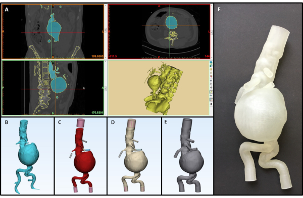
Figure 1: Design steps from a radiological dataset to a printed anatomical model (Pathology: infrarenal aortic aneurysm). (A) CT-dataset-based segmentation process (B) Rough 3D-model after segmentation (C) Smoothed model with added tubular connectors (D) Final model of the blood volume with connectors (E) Hollow model with defined wall thickness (F) 3D-printed flexible model. Please click here to view a larger version of this figure.
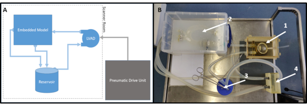
Figure 2: Setup of the flow loop. (A) Schematic model of the flow loop (B) Final flow loop setup with LVAD (1), embedded model (2), a reservoir (3) and a 3D-printed tube connector (optional) (4) Please click here to view a larger version of this figure.
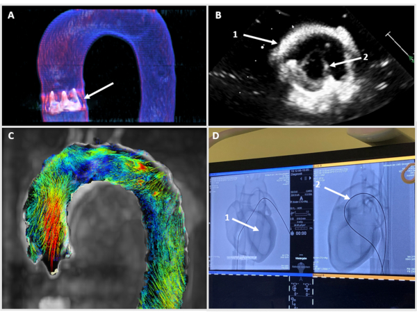
Figure 3: Clinical imaging techniques. (A) CT-reconstruction of a 3D-printed aortic arch with a biological surgical heart valve (B) Ultrasonic image of a 3D-printed aortic root (1) with an open biological surgical heart valve (2) (C) 4D-MRI flow visualization in the aortic arch (D) X-ray imaging of a 3D-printed pediatric heart (1) during a catheter intervention (2) Please click here to view a larger version of this figure.
Discussion
The presented workflow allows to establish individualized models and thereby perform pre-interventional therapy planning, as well as physician training on individualized anatomies. To achieve this, patient-specific tomographic data can be used for segmentation and 3D-printing of flexible cardiovascular phantoms. By implementation of these 3D-printed models in a mock circulation, different clinical situations can be realistically simulated.
Nowadays, many therapy planning procedures focus upon the digital simulation of different scenarios, in order to identify the most favorable outcome10,11. In contrast to these in-silico simulations, the described 3D-printed setup enables tactile feedback in training procedures; a material compliance close to the human original is possible in pulsatile perfusion. On the other hand, many published 3D-printed cardiovascular phantoms only use rigid material and therefore are limited to a mainly visual use12,13.
However, it must be understood that current 3D-printing techniques and materials remain the biggest limitation in reproducing biomechanical properties for the presented workflow14. While an exact recreation of the anatomical shape is possible, the mechanical behavior of the created models will still differ from native aortic tissue to an extent. To mimic different tissues with varying bio-mechanical properties in one phantom, so far as it is possible at all, can be accomplished only by a few sophisticated multi-material 3D-printers15. Creating tissue mimicking materials for 3D-printing remains a focus of scientific research; the development of novel materials will result in even more realistic results16,17. As long as, only commercially available printing material and/or one-component-printing is available, the mechanical properties of the phantom can be adjusted by means of variations of the wall thicknesses, as was conducted in this study. It is, therefore, not recommended only to duplicate the thickness of the tissue of interest from the underlying tomographic data. It is important to stress that there exists a wide range of different 3D-printers with different materials and varying mechanical properties on the market18. Comprehensive mechanical testing is, therefore, recommended, prior to 3D-printing. For printing of cardiovascular structures, (i.e., aortic or ventricular walls), different native tissue samples are required for reference. Following the described segmentation and printing workflow, the creation of flexible and anatomically accurate as well as engineered but realistic 3D-printed models of a wide range of cardiovascular anatomies is possible.
The cost-effectiveness of 3D-printed models depends significantly upon the material properties. In interventional training high durability of each model (even after balloon dilation) is necessary, to reduce overall costs. When looking at patient-specific therapy planning, one must take into account the beneficial effect of a printed model. A 3D-printed model will not prove cost-effective for a "standard" surgical patient, but might offer tremendous insight in patients with complex anatomies. Therefore, the costs of training models have to be weighed against their prospective benefits.
Until now, a few commercially available phantoms for clinical training exist on the market; some academic models have been published19,20. These models normally have pre-defined anatomies and usually prove difficult to employ in patient-specific settings. Furthermore, high acquisition costs complicate the widespread use of these tools in physicians' training. The presented customizable mock circulation can be created on a low budget if necessary. Tomographic, fluoroscopy and sonographic scanners, for acquisition of the patient-specific data as well as for the later use of the mock circulation, are standard equipment of any general or university hospital in developed countries. Segmentation of the cardiovascular anatomy and creation of the virtual 3D-model can be performed with the mentioned licensed software, but freeware is also available21. The freeware options offer excellent results when creating 3D-models from radiological datasets, although a high amount of initial work is required to adjust the software to individual needs. Furthermore, a subsequent editing of the digital 3D model requires an additional software, which is why a comprehensive software suite covering all these aspects is highly recommended for a quick and smooth workflow. If necessary, printing of the flexible phantoms can be done by contract 3D-manufacturing if there is no suitable 3D-printer on site. By anatomical reduction on the region of interest, the size of the 3D-printed phantom can be reduced, which comes with faster printing times and lower costs.
The most critical point of the process described above is the initial image acquisition. As a result, the higher the quality of the tomographic data, the more accurate will prove the final 3D-printed phantom. There are two major factors in obtaining suitable data from CT or MRI: Prevention of artifacts and spatial resolution. To prevent artifacts, ideally no metallic materials (e.g., implants) will be next to the region of interest, if no specific artifact reduction techniques are available22. In order to reduce motion artifacts, ECG- and respiratory triggering should be performed during image acquisition23,24. Spatial resolution depends on the imaging device; however, a slice thickness of 1.0 mm or less is necessary to obtain suitable 3D-printed phantoms without excessive digital postprocessing.
The above-mentioned modularity, cost-effectiveness as well as versatility predisposes the individualizable mock circulation for supplementary use in daily clinical routine. The presented method can be beneficial for a wide range of clinical and basic research fields. The use of realistic models is excellent for teaching young doctors and students the basics of sonography, as well as interventional techniques. Especially with interventions, such a model will render the technology more accessible and increase the overall knowledge base of doctors, long-term. CT and MRI imaging, especially when looking at hemodynamic flow patterns in the aortic vessels, can be a major addition both in basic science, as well as determining the outcome of surgical and transcatheter interventions.
Disclosures
The authors declare no conflict of interest.
Acknowledgements
This publication was supported by the German Heart Foundation/German Foundation of Heart Research.
Materials
| Name | Company | Catalog Number | Comments |
|---|---|---|---|
| 3-matic | Materialise AB | Software Version 15.0 - Commercial 3D-Modeling Software | |
| Affiniti 50 | Philips Medical Systems GmbH | Ultrasonic Imaging System | |
| Agilista W3200 | Keyence Co. | Polyjet 3D-Printer with a spatial resolution of 30µm | |
| AR-G1L | Keyence Co. | flexible 3D-Printing material | |
| Artis Zee | Siemens Healthcare GmbH | Angiographic X-ray Scanner | |
| cvi42 | CCI Inc. | Software Version 5.12 - 4D Flow Analysis Software | |
| Diagnostic Catheter, Multipurpose MPA 2 | Cordis, A Cardinal Health company | Catheter for pediatric training models, Sizes 4F for infants and 5F for children, young adults | |
| Excor Ventricular Assist Device | Berlin Heart GmbH | 80 -100ml stroke volume | |
| Imeron 400 Contrast Agent | Bracco Imaging | CT - Contrast Agent | |
| IntroGuide F | Angiokard Medizintechnik GmbH | Guidewire with J-tip; diameter: 0.035" length: 220cm | |
| Lunderquist Guidewire | Cook Medical Inc. | (T)EVAR interventional guidewire | |
| MAGNETOM Aera | Siemens Healthcare GmbH | MRI Scanner | |
| Magnevist Contrast Agent | Bayer Vital GmbH | MRI - Contrast Agent | |
| Mimics | Materialise AB | Software Version 23.0 - Commercial Segmentation Software | |
| Modeling Studio | Keyence Co. | 3D-Printer Slicing Software | |
| PVC tubing | |||
| Radifocus Guide Wire M | Terumo Europe NV | Straight guidewire; diameter: 0.035" length: 260cm | |
| Really useful box 9L | Really useful products Ltd. | ||
| Rotigarose - Standard Agar | Carl Roth GmbH | 3810.4 | |
| Solidworks | Dassault Systemes SE | Software Version 2019-2020; CAD Design Software | |
| SOMATOM Force | Siemens Healthcare GmbH | Computed Tomography Scanner | |
| syngo via | Siemens Healthcare GmbH | Radiological Imaging Software |
References
- Goetz, L. H., Schork, N. J. Personalized medicine: motivation, challenges, and progress. Fertility and Sterility. 109 (6), 952-963 (2018).
- Gwin, W. R., Disis, M. L., Ruiz-Garcia, E. Immuno-Oncology in the Era of Personalized Medicine. Advances in Experimental Medicine and Biology. 1168, 117-129 (2019).
- Spetzger, U., Frasca, M., König, S. A. Surgical planning, manufacturing and implantation of an individualized cervical fusion titanium cage using patient-specific data. European Spine Journal. 25 (7), 2239-2246 (2016).
- Gardner, S. J., Kim, J., Chetty, I. J. Modern radiation therapy planning and delivery. Hematology/Oncology Clinics of North America. 33 (6), 947-962 (2019).
- Haglin, J. M., et al. Patient-specific orthopaedic implants. Orthopaedic surgery. 8 (4), 417-424 (2016).
- Liaw, C. Y., Guvendiren, M. Current and emerging applications of 3D printing in medicine. Biofabrication. 9 (2), 024102(2017).
- Pugliese, L., et al. The clinical use of 3D printing in surgery. Updates in Surgery. 70 (3), 381-388 (2018).
- Kamalian, S., Lev, M. H., Gupta, R. Handbook of Clinical Neurology. Masdeu, J. C., Gonzalez, R. G. , Elsevier. 3-20 (2016).
- Bücking, T. M., et al. From medical imaging data to 3D printed anatomical models. PLoS One. 12 (5), 0178540(2017).
- Steinberg, E. L., Segev, E., Drexler, M., Ben-Tov, T., Nimrod, S. Preoperative planning of orthopedic procedures using digitalized software systems. Israel Medical Association Journal. 18 (6), 354-358 (2016).
- Hua, J., Aziz, S., Shum, J. W. Virtual surgical planning in oral and maxillofacial surgery. Oral and Maxillofacial Surgery Clinics of North America. 31 (4), 519-530 (2019).
- Schmauss, D., Haeberle, S., Hagl, C., Sodian, R. Three-dimensional printing in cardiac surgery and interventional cardiology: a single-centre experience. European Journal of Cardiothoracic Surgery. 47 (6), 1044-1052 (2015).
- Smelt, J. L. C., et al. Operative Planning in thoracic surgery: A pilot study comparing imaging techniques and three-dimensional printing. The Annals of Thoracic Surgery. 107 (2), 401-406 (2019).
- Masaeli, R., Zandsalimi, K., Rasoulianboroujeni, M., Tayebi, L. Challenges in three-dimensional printing of bone substitutes. Tissue Engineering Part B: Reviews. 25 (5), 387-397 (2019).
- Rafiee, M., Farahani, R. D., Therriault, D. Multi-material 3D and 4D printing: A survey. Advanced Science. 7 (12), 1902307(2020).
- Wang, S., et al. Development and testing of an ultrasound-compatible cardiac phantom for interventional procedure simulation using direct three-dimensional printing. 3D Printing and Additive Manufacturing. 7 (6), 269-278 (2020).
- D'Souza, W. D., et al. Tissue mimicking materials for a multi-imaging modality prostate phantom. Medical Physics. 28 (4), 688-700 (2001).
- Tejo-Otero, A., Buj-Corral, I., Fenollosa-Artés, F. 3D printing in medicine for preoperative surgical planning: A review. Annals of Biomedical Engineering. 48 (2), 536-555 (2020).
- Rotman, O. M., et al. Realistic vascular replicator for TAVR procedures. Cardiovascular Engineering and Technology. 9 (3), 339-350 (2018).
- Hussein, N., et al. Hands-on surgical simulation in congenital heart surgery: Literature review and future perspective. Seminars in Thoracic and Cardiovascular Surgery. 32 (1), 98-105 (2020).
- Fedorov, A., et al. 3D slicer as an image computing platform for the quantitative imaging network. Magnetic resonance imaging. 30 (9), 1323-1341 (2012).
- Katsura, M., Sato, J., Akahane, M., Kunimatsu, A., Abe, O. Current and novel techniques for metal artifact reduction at ct: practical guide for radiologists. Radiographics. 38 (2), 450-461 (2018).
- Pépin, A., Daouk, J., Bailly, P., Hapdey, S., Meyer, M. E. Management of respiratory motion in PET/computed tomography: the state of the art. Nuclear Medicine Communications. 35 (2), 113-122 (2014).
- Scott, A. D., Keegan, J., Firmin, D. N. Motion in cardiovascular MR imaging. Radiology. 250 (2), 331-351 (2009).
Reprints and Permissions
