This protocol describes a method for facilitating the collection of samples from radical prostatectomy specimens. The goal is to map, characterize, and micro-macro dissect tissue samples from the specimens based on anatomopathological criteria before storing them in a Biobank.
Method Article
Enhancing Prostate Tumor Biobanking Reliability with Improved Sampling Technique and Histological Characterization
Eduardo Giner Moreno*1 , Francisco Quereda-Flores*2 , Josselyn A. Hernández Chinchilla*1 , Raquel Amigo Moreno3 , María Dolores Perez Ruiz1 , Aileen Carcelén López3 , Jorge Rocher Benlloch3 , Pilar Molió Sanchís3 , Adrián Casares Calzada3 , María Ángeles Marqués-Torrejón4 , Conrado Martínez-Cadenas4 , José Luis Ruiz Cerdá2,5 , Alba Loras Monfort4,5 , David Ramos Soler1,6
1Department of Pathology, Hospital Universitario y Politécnico La Fe, 2Department of Urology, Hospital Universitario y Politécnico La Fe, 3Biobanco La Fe, Instituto de Investigación Sanitaria La Fe, 4Department of Medicine, Jaume I University of Castellon, 5Department of Surgery, University of Valencia, 6Department of Pathology, University of Valencia
* These authors contributed equally
Opens in a new tab
In This Article
Summary
Loading...
$$\rightleftharpoonup{xx}$$
$$\longleftharp{xx}$$,
$$\longrightharp{xx}$$,
Abstract
Loading...
$$\rightleftharpoonup{xx}$$
$$\longleftharp{xx}$$,
$$\longrightharp{xx}$$,
Acquiring fresh and well-characterized tumor tissue samples is critical for conducting high-quality "omics" studies. However, it can be particularly challenging in the context of prostate cancer (PC) due to the unique nature of this organ and the high heterogeneity associated with this tumor. On the other hand, histopathologically characterizing samples before their storage without causing significant tissue alterations is also an intriguing challenge. In this context, we present a new method for acquiring, mapping, characterizing, and micro-dissecting resected prostate tissue based on anatomopathological criteria.
Unlike previously published protocols, this method reduces the time required for histopathological analysis of the prostate specimen without compromising its structure, which is crucial for assessing surgical margins. Furthermore, it enables the delineation and micro-macro dissection of fresh prostate tissue samples, with a focus on histological tumor areas defined by pathological criteria such as Gleason score, precursor lesions (high-grade prostatic intraepithelial neoplasia - PIN), and inflammatory lesions (prostatitis). These samples are then stored in a Biobank for subsequent research analyses.
Introduction
Loading...
$$\rightleftharpoonup{xx}$$
$$\longleftharp{xx}$$,
$$\longrightharp{xx}$$,
Prostate cancer (PC) is the 2nd most frequent cancer in men and the 5th leading cause of death worldwide1. Patient treatment and prognosis depend on the staging and grading (Gleason score) of the tumor, as evidenced by the higher 5-year survival rates of localized and low-grade tumors (Gleason grade 6) (99%) compared to high Gleason grades and metastatic tumors (31%)2.
PC local relapse and treatment failure have been linked to the characteristic high genetic intratumor heterogeneity of this tumor type3. Additionally, PC is considered to be a multifocal disease with several tumor foci exhibiting different morphological, histological, and molecular characteristics4, which may originate independently or derive from a common tumor cell ancestor5. Previous studies have shown that tumor evolution differs among patients based on specific genetic drivers that can promote metastasis or confine the cell lineage to the prostate5. Therefore, molecular characterization of the different tumor foci is crucial not only for providing a more accurate diagnosis and prognosis but also for tailoring effective and personalized treatment for the patient.
In this context, biomedical research and integrative multi-omics approaches are offering unprecedented opportunities to classify cancers into different subtypes, identify diagnostic and prognostic biomarkers, and discover markers related to treatment response. Furthermore, these approaches contribute to a better understanding of the biology of this disease6,7. Biological samples, whether tissues or biofluids, can be analyzed using various multi-omics platforms (genomics, transcriptomics, proteomics, metabolomics, etc.) to uncover the biological features underlying cancer pathophysiology, thereby addressing current limitations related to genetic and phenotypic heterogeneity6. However, it's important to consider that the quality of data derived from omics studies depends on the quality of the samples collected from tumors, their accurate characterization, and subsequent processing and storage8.
In this context, obtaining fresh PC tissue for research presents a methodological challenge due to the difficulty of successful tumor sampling9. Previous methods involved random sampling following radical prostatectomy, yielding poor results10. However, more recent approaches incorporate targeted protocols based on both magnetic resonance imaging (MRI) and biopsy data, resulting in improved efficacy in tumor sample collection11.
On the other hand, histopathological characterization of samples prior to their storage without significant tissue alteration also poses an interesting challenge. Consequently, in many cases, the histopathological determination of samples is performed after their analysis (e.g., HR 1H NMR metabolomic analysis)12. This practice entails unnecessary expenses, time consumption, and the loss of a significant number of samples that are eventually excluded from the analysis (for example, samples that, following histopathological analysis, turn out not to be tumor samples). In other cases, the histopathological characterization of samples is performed before their analysis. In fact, some previous studies have attempted to standardize methods for providing representative high-quality research samples from radical prostatectomy specimens for genomics and metabolomics13,14. Nevertheless, sampling efficiency is significantly higher when performed from already histologically confirmed sections (88%) that disrupt tissue, compared to when performed from unconfirmed sections (45%)1.
Here, a new methodology is presented to overcome these limitations, aiming to obtain fresh and well-characterized PC samples before storage in the Biobank. This method has been developed through collaborative efforts between different clinical services (Urology, Pathology, and the La Fe Hospital Biobank). It's important to highlight that Biobanks play an essential role in the collection, processing, preservation, and storage of biological samples while ensuring the high quality of samples and data, as well as compliance with ethical and legal requirements8,15,16.
Access restricted. Please log in or start a trial to view this content.
Protocol
Loading...
$$\rightleftharpoonup{xx}$$
$$\longleftharp{xx}$$,
$$\longrightharp{xx}$$,
This method was developed through collaborative efforts involving different clinical services (Urology, Pathology, and the La Fe Hospital Biobank). The study was conducted in compliance with institutional, national, and international guidelines for human welfare, and it received approval from the Ethics Committee for Biomedical Research at the Instituto de Investigación Sanitaria Hospital Universitario y Politécnico La Fe (Valencia, Spain). All samples were stored at the La Fe Hospital Biobank (PT13/0010/0026). The overall procedure is detailed in Figure 1.
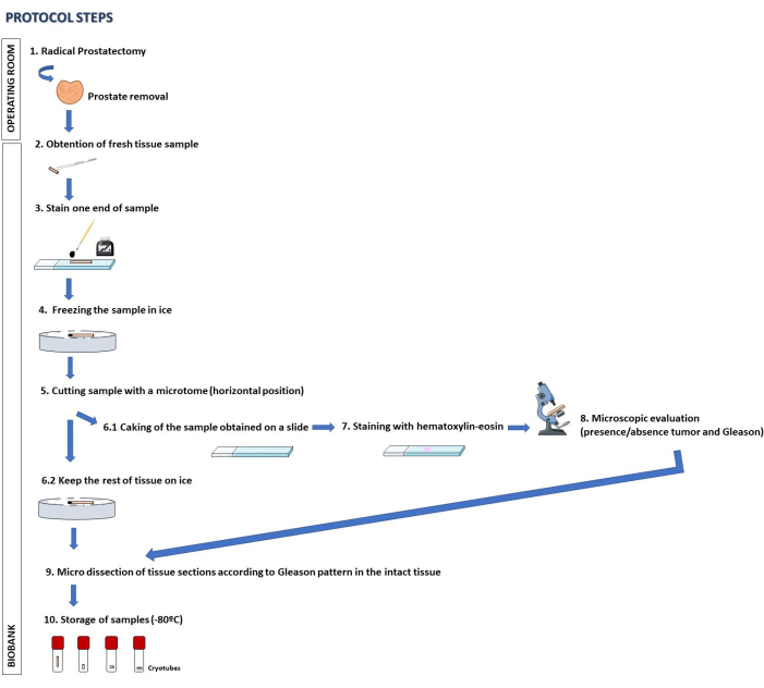
Figure 1: Overall procedure scheme. Schematic representation of the stepwise procedure described in the protocol section. Please click here to view a larger version of this figure.
1. Tumor targeting
- Before surgery, utilize multiparametric or biparametric Magnetic Resonance Imaging (mpMRI or bpMRI)17,18 to locate the tumor and map its position within the prostate gland. Combine the information obtained from MRI with findings from transrectal ultrasound or rectal examination if the latter yields discordant results.
- To enhance tumor localization, review the results of preoperative ultrasound-guided and MRI-guided transrectal prostate biopsies.
NOTE: In the diagnostic procedure for prostate cancer, biopsies are taken directly from the prostate via transrectal access. These biopsies include systematic sampling from the prostate (involving cylinders distributed all around the gland) and cylinders directly targeted to suspicious findings on MRI. To target the retrieval of these samples, transrectal ultrasound is used, which is combined with MRI findings in a cognitive manner ("cognitive fusion") or with "real MRI-ultrasound fusion software" when available.
2. Radical prostatectomy and prostate collection
- Perform a standardized robotic-assisted radical prostatectomy through a transperitoneal approach, following the steps outlined by Huynh et al.19.
- Start with abdominal access, insufflation, and camera port placement (see Table of Materials). Place the remaining five ports under direct vision. Set pneumoperitoneum to 12 mm.
- Release the bladder by incising the peritoneum and dissect the perirectal space.
- Open the pelvic fascia on each side.
- Perform a complete dissection of the anterior and lateral aspects of the prostate and bladder.
- Carry out the opening of the bladder neck, identify and section the proximal urethra to gain access to the posterior aspects of the prostate and bladder.
- Dissect the seminal vesicles and rectum space19.
- Dissect the lateral aspects of the prostate, sparing the neurovascular bundles.
- Ensure hemostasis of vessels in the retropubic space.
- Dissect the apex and section the distal urethra.
- Complete the vesicourethral anastomosis.
- Extract the prostate and perform the closure of the incision19.
- Collect the fresh prostate in a dry container. It is crucial not to fix or paraffin-embed the sample specimen.
- Transport the fresh surgical specimen, which includes the entire prostate, to the Biobank for sample processing.
3. Prostate processing and sample collection
- Select targeted prostate zones for sampling based on tumor location information retrieved from preoperative mpMRI, preoperative biopsy results, and direct prostatectomy specimen palpation. Record this information on a form that includes patient identification data, prostate areas finally sampled, and the number of samples collected.
NOTE: When MRI findings, preoperative biopsy results, and direct prostatectomy specimen palpation are concordant, target all the samples to that or those locations (between 4 and 6 cylinders). If there is any discordance, increase the number of cylinders and separately target all zones that are suspicious of being affected by any of those methods. - Use a disposable automatic needle (surgeon) to retrieve cylindrical samples from the selected areas (step 1.1). Then, identify (pathologist) the outer zone of the cylinder, clamp the cylinder with tissue forceps, place it on a slide, and mark the outer zone with tissue ink. Repeat this process with all cylinders taken.
- Place the collected cylinders horizontally on a sample holder disk (see Table of Materials) filled with cryostat embedding medium and transfer them to the microtome cooling platen for cutting.
- Once placed on the platen, attach the sample holder disk to the specimen chuck, and cut it with careful movements of the wheel following the next steps:
- Use the optimal cutting temperature compound (OCT compound, see Table of Materials) to embed the tissue sample before frozen sectioning on a microtome-cryostat.
- Afterward, cut the sample using a cryostat-microtome at -25 °C. Collect thin cylinder-shaped sections of tissue (10 µm) onto treated microscope slides. Ensure to collect only a few microns of tissue to ensure an adequate amount of intact tumor tissue for storage in the Biobank.
- Gather two or three thin sections per slide and manually stain them to control the final coloration. The technician's eye ensures optimal staining, and any necessary re-staining can be done. Follow these steps
- Fix slides in 96% ethanol, hydrate in distilled water, and stain with Harris hematoxylin (see Table of Materials) for 1 min to stain the nuclei. Remove excess hematoxylin by rinsing the slides with distilled water.
- Treat slides with 30% ammonium hydroxide to turn the hematoxylin blue. Remove excess solution with rinses of distilled water.
- Stain slides with eosin (see Table of Materials) for 30 s.
- Next, dehydrate the slides with increasing titrations of alcohol, clear with xylene (100%), and mount them with DPX (a mixture of distyrene, a plasticizer, and xylene, used as a synthetic resin mounting media that replaces xylene-balsam, see Table of Materials). During these steps, dip the slides into and remove them from the jar of Coplin solution.
- Finally, place a coverslip on top of the stained tissue and use a medium with a suitable refractive index (see Table of Materials) for viewing under a microscope.
4. Sample characterization
- Analyze the sample under a light microscope (pathologist), discriminating the areas of carcinoma and determining the Gleason score of the tumor. Document all these data and mark the precise tumor zones on the slides.
- During this step, label the slide with the precise location of the tumor and its respective Gleason Score20. Also, mark the location of PIN (Prostatic Intraepithelial Neoplasm) based on morphological identification.
5. Sample micro-macro-dissection and storage
- Clearly mark different histological components with distinct markers on the slide (pathologist) and directly correlate them with frozen tissue on the disk.
- Then, using a surgical blade, macro-micro dissect the areas of the sample marked on the slide within the OCT disc, dividing them into tumoral, peri-tumoral, PIN, and normal prostatic tissue.
- As the embedding medium begins to defrost, handle the tissue with forceps and place it into correctly labeled cryotubes.
NOTE: Carefully store each dissected fragment within the corresponding tube, following the histological diagnosis of the cryo-slices. - Store the labeled cryotubes with fully characterized histological samples in the Biobank at -80 °C for subsequent use in research studies.
NOTE: The steps described in this section can be performed by a trained technician under the supervision of the pathologist. The surgical specimen should be transported to the Biobank as soon as possible to prevent tissue and nucleic acid degradation. According to the reference guides, efforts must be made to avoid prolonged ischemia time (>30 min)21. The samples can be kept in the Biobank indefinitely as long as the sample quality principles are maintained.
Access restricted. Please log in or start a trial to view this content.
Results
Loading...
$$\rightleftharpoonup{xx}$$
$$\longleftharp{xx}$$,
$$\longrightharp{xx}$$,
The results reveal that this technique has made it possible to obtain tumor material in 61% of the cases studied (25 out of 41 cases) (Table 1).
Table 1: Histopathological data of study samples. Summary of histopathological data for the samples used in the study. The diagnostic cylinder refers to the prostate biopsy sample obtained for diagnostic purposes, while the processed cylinder corresponds to the cylinder obtained from the prostatectomy specimen for res...
Access restricted. Please log in or start a trial to view this content.
Discussion
Loading...
$$\rightleftharpoonup{xx}$$
$$\longleftharp{xx}$$,
$$\longrightharp{xx}$$,
In any research study, obtaining quality samples is an essential requirement to reduce systematic biases and obtain reliable results22. Therefore, control of preanalytical variables such as the temperature at which samples are processed and stored, the time elapsed from sample collection to storage, the use of sterilized materials, or the effects that the addition of preservatives or other additives can have on samples must be considered in any protocol involving biological samples. Not only is th...
Access restricted. Please log in or start a trial to view this content.
Disclosures
Loading...
$$\rightleftharpoonup{xx}$$
$$\longleftharp{xx}$$,
$$\longrightharp{xx}$$,
The authors declare no conflict of interest.
Acknowledgements
Loading...
$$\rightleftharpoonup{xx}$$
$$\longleftharp{xx}$$,
$$\longrightharp{xx}$$,
A.L. acknowledges a "Margarita Salas" postdoctoral contract (number 21-076), and MAM-T a 'Maria Zambrano' research contract (number MAZ/2021/03 UP2021-021). Both contracts have been funded by the European Union-Next generation EU.
Access restricted. Please log in or start a trial to view this content.
Materials
| Name | Company | Catalog Number | Comments |
|---|---|---|---|
| Cadiere forceps | Intuitive | PN1052082-US 10/2021 | Part number: 471049. 18 uses. |
| Conventional slides | Knittel Glass | 2021 | Ground/ Frosted end |
| Cryostat microtome | Thermo Fisher Scientific | --- | Criostato CryoStar NX50 |
| Cryotubes | Greiner Bio-One GmbH | Ref.: 122280. | CRYO S. PP, with screw cap, sterile. |
| Da Vinci surgical system | Intuitive | PN1052082-US 10/2021 | XI model |
| Dissection instruments | Bayer | --- | Two tweezers and a surgical blade |
| DPX Eukitt | Medizin- und Labortechnik GmbH | 6.00.01.0001.06.01.01 | |
| Eosin | Agilent | 157252 | |
| Fenestrated bipolar forceps | Intuitive | PN1052082-US 10/2021 | Part number: 471205. 14 lives. |
| Force bipolar | Intuitive | PN1052082-US 10/2021 | Part number: 471405. 12 uses. |
| Freezers | Thermo Scientific | MODEL 907. -80 ºC | |
| Hematoxylin | Agilent | 157251 | |
| Inmunohistochemistry Slides | Agilent-Dako | K802021-2 | |
| Large needle driver | Intuitive | PN1052082-US 10/2021 | Part number: 471006. 15 uses. |
| Maryland bipolar forceps | Intuitive | PN1052082-US 10/2021 | Part number: 471172. 14 uses. |
| Microscope | Olympus | --- | Olympus cx40 |
| Microtome blades | PFM Medical | a35 | |
| Monopolar curved scissors | Intuitive | PN1052082-US 10/2021 | Part number: 470179. 10 uses. |
| OCT compound | NEG-50 | LOT.117340 | |
| PlusSpeed S Single-use Biopsy Device with beveled tip | Peter Pflugbeil GmbH | PSS-1825-S | |
| ProGasp forceps | Intuitive | PN1052082-US 10/2021 | Part number: 471093. 18 uses. |
| Sample holder Disc | Davidson Cryo Chuck. BradleyProducts | 30 mm | |
| Tissue ink | Pelikan | 2021 | Ink 4001 brilliant black (301168) |
| Xylol | Quimipur | Ref. 169 |
References
Loading...
$$\rightleftharpoonup{xx}$$
$$\longleftharp{xx}$$,
$$\longrightharp{xx}$$,
- Rawla, P. Epidemiology of prostate cancer. World Journal of Oncology. 10 (2), 63-89 (2019).
- Epstein, J. I. The 2019 Genitourinary Pathology Society (GUPS) white paper on contemporary grading of prostate cancer. Archives of Pathology & Laboratory Medicine. 145 (4), 461-493 (2021).
- Zhang, W. Intratumor heterogeneity and clonal evolution revealed in castration-resistant prostate cancer by longitudinal genomic analysis. Translational Oncology. 16, 101311(2022).
- Haffner, M. C., et al. Genomic and phenotypic heterogeneity in prostate cancer. Nature Reviews Urology. 18 (2), 79-92 (2021).
- Woodcock, D. J. Prostate cancer evolution from multilineage primary to single lineage metastases with implications for liquid biopsy. Nature Communications. 11 (1), 5070(2020).
- Heo, Y. J., Hwa, C., Lee, G. H., Park, J. M., An, J. Y. Integrative multi-omics approaches in cancer research: from biological networks to clinical subtypes. Molecules and Cells. 44 (7), 433-443 (2021).
- Menyhárt, O., Győrffy, B. Multi-omics approaches in cancer research with applications in tumor subtyping, prognosis, and diagnosis. Computational and Structural Biotechnology Journal. 19, 949-960 (2021).
- Annaratone, L. Basic principles of biobanking: from biological samples to precision medicine for patients. Virchows Archiv. 479 (2), 233-246 (2021).
- King, C. R., Long, J. P. Prostate biopsy grading errors: A sampling problem. International Journal of Cancer. 90 (6), 326-330 (2000).
- Jayadevan, R., Zhou, S., Priester, A. M., Delfin, M., Marks, L. S. Use of MRI-ultrasound fusion to achieve targeted prostate biopsy. Journal of Visualized Experiments. 146, e59231(2019).
- Heavey, S., et al. Use of magnetic resonance imaging and biopsy data to guide sampling procedures for prostate cancer biobanking. Journal of Visualized Experiments. 152, 60216(2019).
- Panach Navarrete, J. Estudio metabolómico en tejido prostático y orina para el diagnóstico y pronóstico del cáncer de próstata. , https://roderic.uv.es/handle/10550/81853 (2022).
- Bertilsson, H., et al. A new method to provide a fresh frozen prostate slice suitable for gene expression study and MR spectroscopy. The Prostate. 71 (5), 461-469 (2011).
- Show SOP - Biospecimen Research Database. , https://brd.nci.nih.gov/brd/sop/show/522 (2022).
- Dagher, G. Quality matters: International standards for biobanking. Cell Proliferation. 55 (8), e13282(2022).
- Yüzbaşıoğlu, A., Özgüç, M. Biobanking: sample acquisition and quality assurance for "omics" research. New Biotechnology. 30 (3), 339-342 (2013).
- Carpagnano, F. A. Multiparametric MRI: local staging of prostate cancer. Current Radiology Reports. 8 (12), 27(2020).
- Christophe, C. Prostate cancer local staging using biparametric MRI: assessment and comparison with multiparametric MRI. European Journal of Radiology. 132, 109350(2020).
- Huynh, L. M., Ahlering, T. E. Robot-assisted radical prostatectomy: a step-by-step guide. Journal of Endourology. 32 (S1), S28-S32 (2018).
- van Leenders, G. J. L. H., et al. The 2019 International Society of Urological Pathology (ISUP) consensus conference on grading of prostatic carcinoma. The American Journal of Surgical Pathology. 44 (8), e87-e99 (2020).
- Liu, A., Collins, C. C., Diemer, S. M. Biobanking metastases and biopsy specimens for personalized medicine. Journal of Biorepository Science for Applied Medicine. 3, 57-67 (2015).
- Ellervik, C., Vaught, J. Preanalytical variables affecting the integrity of human biospecimens in biobanking. Clinical Chemistry. 61 (7), 914-934 (2015).
- Arellano, H. L., Castillo, C. O., Metrebián, B. E. Diagnostic agreement of the Gleason score in needle biopsy and radical prostatectomy and its clinical consequences. Rev Méd Chile. 132 (8), 971-978 (2004).
- Torres Caparó, D. A. Concordancia en los valores de gleason en biopsia prostática transrectal y en prostatectomia radical en pacientes con cáncer de próstata del Hospital Cirujano Mayor Santiago Távara entre enero 2010 - junio del 2018. , Universidad Ricardo Palma. https://repositorio.urp.edu.pe/handle/20.500.14138/1886 (2019).
- Chavolla-Canal, A. J., et al. Concordancia del puntaje de Gleason en biopsia transrectal de próstata vs prostatectomía radical. Revista Mexicana de Urología. 81 (2), 1-10 (2021).
- Fan, X. J., et al. Impact of cold ischemic time and freeze-thaw cycles on RNA, DNA and protein quality in colorectal cancer tissues biobanking. Journal of Cancer. 10 (20), 4978-4988 (2019).
- Vaswani, A., et al. Comparative liquid chromatography/tandem mass spectrometry lipidomics analysis of macaque heart tissue flash-frozen or embedded in optimal cutting temperature polymer (OCT): Practical considerations. Rapid communications in Mass Spectrometry: RCM. 35 (18), e9155(2021).
- Zhang, W., Sakashita, S., Taylor, P., Tsao, M. S., Moran, M. F. Comprehensive proteome analysis of fresh frozen and optimal cutting temperature (OCT) embedded primary non-small cell lung carcinoma by LC-MS/MS. Methods (San Diego, Calif). 81, 50-55 (2015).
- Boyd, A. E., Allegood, J., Lima, S. Preparation of human tissues embedded in optimal cutting temperature compound for mass spectrometry analysis. Journal of Visualized Experiments. 170, e62552(2021).
Access restricted. Please log in or start a trial to view this content.
Reprints and Permissions
Request permission to reuse the text or figures of this JoVE article
Request Permission