Repair of the intracarotid artery in a mouse model after injection returns blood flow to the artery without negatively impacting the distribution of the injected material. Injection site repair facilitates subsequent injections through the same artery and prevents cerebral ischemia in mouse strains that lack a complete Circle of Willis.
Method Article
Facilitating Repeat Intracarotid Injections in Mouse Models by a Novel Injection Site Repair Technique
In This Article
Summary
Abstract
Given recent advances in the delivery of novel antitumor therapeutics using endovascular selective intraarterial delivery methods in neuro-oncology, there is an urgent need to develop methods for intracarotid injections in mouse models, including methods to repair the carotid artery in mice after injection to allow for subsequent injections. We developed a method of intracarotid injection in a mouse model to deliver therapeutics into the internal carotid artery (ICA) with two alternative procedures.
During injection, the needle is inserted into the common carotid artery (CCA) after tying a suture around the external carotid artery (ECA) and injected therapeutics are delivered into the ICA. Following injection, the common carotid artery (CCA) can be ligated, which limits the number of intracarotid injections to one. The alternative procedure described in this article includes a modification where intracarotid artery injection is followed by injection site repair of the CCA, which restores blood flow within the CCA and avoids the complication of cerebral ischemia seen in some mouse models.
We also compared the delivery of bone marrow-derived human mesenchymal stem cells (BM-hMSCs) to intracranial tumors when delivered through intracarotid injection with and without injection site repair following the injection. Delivery of BM-hMSCs does not differ significantly between the methods. Our results demonstrate that injection site repair of the CCA allows for repeat injections through the same artery and does not impair the delivery and distribution of injected material, thus providing a model with greater flexibility that more closely emulates intracarotid injection in humans.
Introduction
Delivery of therapeutics to brain tumors is challenging due to the impermeability of the blood-brain barrier (BBB) and the blood-tumor barrier (BTB). Direct intratumoral injection of therapeutics to circumvent the BBB may be achieved through the use of an Ommaya reservoir-catheter, low-flow microinfusion for convection-enhanced delivery, or local injection into the resection cavity or adjacent tissue1. However, the total volume of tumor tissue that is reached with these methods is limited2,3,4. Intraarterial injections have been used previously to deliver therapeutic agents to brain tumors with the aim of reaching more of the tumor5,6,7,8 and in recent times, the advances in both intraarterial delivery techniques and novel therapeutic agents have demonstrated the benefit of using this approach in the treatment of brain tumors7,9. These advances include the development of microcatheters, endovascular selective intraarterial (ESIA) delivery with advanced imaging, the use of osmotic agents to disrupt the BBB and BTB, and the development of targeted biological therapies. Therefore, to conduct preclinical testing of novel therapeutic agents that are administered via intraarterial injections, appropriate translational research models are necessary9,10.
In mouse models of brain tumors, therapeutic agents delivered intraperitoneally or intravenously (through the tail vein) pass through the liver or the heart and lungs, respectively, before being distributed to the entire body, including the brain. These first-pass effects may trap and remove the agent, or dilute the agent before reaching the brain, and may present dose-limiting toxicities prior to achieving a therapeutic dose in the brain. In contrast, intracarotid artery injection enables focused delivery to the brain prior to circulation by bypassing first-pass metabolism and limiting off-target delivery. While intracarotid injection in mice is more labor-intensive, the specificity and reproducibility of the technique result in reduced animal numbers to complete investigations11,12.
In general, in previously described methods of intracarotid artery injection in mice, the common carotid artery is ligated after injection and the circulation to the brain is provided by the contralateral carotid artery and posterior cerebral circulation via the circle of Willis11,12. This method has the inherent limitation of only allowing for a maximum of a single injection into the internal or external carotid artery. It is also critical that the mouse strains used in experiments where the carotid artery is ligated have a complete Circle of Willis to prevent cerebral ischemia due to the ligated artery13. Occlusion of the carotid artery has also been demonstrated to reduce cerebral blood flow and limit the distribution of injected particles14. Additionally, the occlusion of the carotid artery in mice following injection does not emulate intracarotid artery injection in human patients.
Our group has previously used intracarotid artery injections to successfully deliver mesenchymal stem cells to the brain10,15,16,17,18,19. In this article, we describe this method of intracarotid artery injection in detail and include a modification of the method that we developed, in which the injection site is repaired without occluding the artery, avoiding the limitations posed by post-injection carotid artery ligation. In this method, the common carotid artery (CCA) is prepared for injection by placing two sutures, one at either end of the intended injection site, and the lower suture (below the injection site) is tightened. The external carotid artery (ECA) is sealed using another suture. The needle is inserted into the CCA, and therapeutics are delivered into the internal carotid artery (ICA). Following this, the upper suture on the CCA is tightened to prevent backflow from the ICA. At this step, the injected CCA can either be ligated or repaired. If the CCA is to be ligated, sutures are tightened and left in place. If the injection site is repaired, sutures are removed after repair, and blood flow is restored. The details of these alternate procedures are provided below.
Protocol
All the steps outlined below are in compliance with our protocol, which follows guidelines established by and was approved by the Institutional Animal Care and Use Committee at The University of Texas MD Anderson Cancer Center.
1. Preparation of the surgical table and mouse for surgical procedure
- Preparation of the surgical table and mouse for surgical procedure (refer to Figure 1A,B)
- Arrange the dissecting microscope and light source in front of the isoflurane vaporizer with an active scavenging system or place on a downdraft table. Place the electric heating pad on the dissecting microscope base (or preferably under the microscope if the base is solid and can transmit heat) and cover with a sterile surgical drape.
NOTE: All surgical tools and drapes should be sterilized by autoclaving prior to use and all disposables should be sterile and individually packaged. Sterile gloves should be used throughout the setup and procedure and changed when necessary to maintain sterility. - Prepare the bed (4-5" long x 2" wide) on the surgical drape using vinyl lab tape. Cut a 1.5 inches x 1.5 inches square of gauze, roll it tightly, and place the rolled gauze under a 3 inch piece of vinyl tape at the "head" of the bed to form a pillow (head tilt with the use of a pillow allows for greater extension of ventral neck area).
- Lay 4 inch pieces of surgical tape along the sides of the bed (on top of the vinyl tape). [If using isoflurane, place an anesthesia nose cone with weight (or tape) attached near the head of the bed. Adjust it for specific placement once the mouse is anesthetized and restrained.] See Figure 1A,B.
- Using a needle driver or heavy forceps, tear off small pieces of cotton from a cotton swab and roll into balls of various sizes from 0.5 to 1 mm in diameter (8-10 cotton pieces per mouse). Keep the cotton on the surgical drape near the head of the bed.
- Using sterile instruments, cut 6-0 suture thread into 1 cm pieces (3-4 pieces per mouse). Place the prepared suture lengths on a sterile drape. Prepare a 1 mL syringe for administration of buprenorphine or other suitable analgesic (as approved by the Institutional Animal Care and Use Committee [IACUC] protocol). Sterilize all surgical tools according to IACUC standards prior to placing the tools in the sterile field.
- Arrange the dissecting microscope and light source in front of the isoflurane vaporizer with an active scavenging system or place on a downdraft table. Place the electric heating pad on the dissecting microscope base (or preferably under the microscope if the base is solid and can transmit heat) and cover with a sterile surgical drape.
- Preparation of mouse for the surgical procedure (refer to Figure 1C-F)
- Anesthetize one mouse following methods approved by IACUC for major survival surgeries (use either isoflurane 1%-4%, based on the sensitivity of individual mice, or a cocktail of 10 mg/mL ketamine, 1 mg/mL xylazine at 100-200 mg/kg (ketamine) of body weight). Clip the fur or depilate if necessary. Administer 0.5-1.0 mg/kg buprenorphine ER (Extended-release) by subcutaneous injection 30 min prior to the start of the surgical procedure.
- Position the anesthetized mouse so the pillow rests under the neck (Figure 1C). The pillow helps to extend and support the neck when used with the anesthesia nose cone. If using the ketamine/xylazine cocktail, place a weighted tooth bar or similar tool in the mouth, behind the incisors to tilt the head and extend the neck.
- Restrain the forelimbs using the surgical tape previously placed along the sides of the surgical bed (Figure 1D). Adjust the position of the mouse under the microscope so that the ventral surface of the neck is in view and adjust the magnification to comfortably observe the surgical site (Figure 1E).
NOTE: The magnification of the dissecting microscope should be adjusted by the surgeon to their comfort level for each step. - Apply artificial tears using a sterile cotton swab. Disinfect the surgical site by alternating circular swabs of betadine or chlorhexidine and alcohol three times per disinfectant (Figure 1F).
- Confirm the depth of anesthesia by ensuring the mouse does not retract its leg in response to a toe pinch. Monitor the respiratory rate and ensure that the mouse is not gasping, as this is an indication of excessive anesthesia when using isoflurane. If necessary, adjust the oxygen and isoflurane flow rates to reach appropriate anesthetic depth and even respiration.
2. Surgical procedure (Figure 2, Figure 3, Figure 4, Figure 5, Figure 6, and Figure 7)
- Primary incision and dissection
- Begin by making a 1 cm longitudinal, midline incision, starting just cranially to the manubrium (protruding lump at the cranial end of the sternum) and continuing over the trachea using a sterile scalpel blade or similar instrument following the user's approved IACUC guidelines (Figure 2A).
- Insert the tip of the closed scissors into the incision and gently open it to perform blunt dissection of the subcutaneous connective tissue, separating the two salivary glands. With the fine forceps, gently pull the right salivary gland through the incision to rest on the aseptically prepared skin surface or retract the salivary gland laterally using the blunt hook retractor (Figure 2B,C). If the exteriorized gland appears dry or tacky, moisten with sterile saline.
- Continue blunt dissection of the connective tissue until the sternocleidomastoid and digastric muscles are visible (Figure 2D).
NOTE: The muscular triangle formed by the trachea/sternohyoid (sh) muscle, sternocleidomastoid (sm) muscle, and the digastric (dg) muscle (caudal belly) will be used to locate the right CCA and the carotid artery bifurcation in the protocol. In general, the smaller omohyoid (oh) muscle can also be seen lying transversely across the CCA (Figure 2D); however, this muscle varies in size and it is not uncommon for the omohyoid muscle to be absent altogether in young or small mice.
- CCA isolation
- Using the angled tip forceps, continue careful dissection of the connective tissue (by opening the tips of the forceps) near the caudal end of the muscular triangle to expose the common carotid artery, jugular vein, and vagus nerve.
NOTE: The common carotid artery is the largest blood vessel adjacent to the trachea and can generally be easily identified at the base of the muscular triangle (Figure 2E, arrow). Be extremely careful with the fine forceps around the blood vessels as the tips can easily nick the vessels resulting in excessive and potentially lethal bleeding. - Continue careful dissection of the connective tissue around the portion of the common carotid artery from the base of the muscular triangle up to the omohyoid muscle. Use the small, sterile cotton balls to control any minor bleeding and absorb secreted fluids from the salivary glands as needed.
- Carefully dissect away connective tissue to separate the CCA from the vagus nerve. Take special care to minimize handling of and damage to, the vagus nerve, easily identified as the thick, white, nerve bundle adjacent to the CCA (Figure 2F).
- Using the angled tip forceps, continue careful dissection of the connective tissue (by opening the tips of the forceps) near the caudal end of the muscular triangle to expose the common carotid artery, jugular vein, and vagus nerve.
- CCA preparation
- Once the CCA has been completely mobilized from the sternocleidomastoid muscle, and where visible, up to the omohyoid muscle, place a 1 cm piece of 6-0 suture on the aseptically prepared skin of the mouse’ sternum (for easy retrieval) and pass the angled tip forceps under the CCA (taking care to isolate CCA from the vagus nerve and jugular vein) (Figure 3A). If the omohyoid muscle is not present, clear the area around the CCA sufficiently to be able to place sutures on the CCA, cranial and caudal to the proposed injection site, and to insert the needle.
- With the fine forceps in the left hand, pass the suture to the angled tip forceps, grasping near the end of the suture. Gently pull half of the length of the suture under the CCA with the angled tip forceps (Figure 3B).
- Repeat this process with a second suture, parallel to the first suture (Figure 3C).
- Loosely tie each suture around the CCA, but do not tighten the knots or restrict blood flow (Figure 3D).
- External carotid artery isolation and preparation
- Using the angled tip forceps, carefully remove connective tissue at the cranial end of the muscular triangle, cranial to the omohyoid muscle, to locate the CCA and the bifurcation into ECA and ICA (Figure 4A).
NOTE: The ECA angles toward the midline and is slightly more superficial, while the ICA angles laterally and moves deeper into the neck. Take special care to prevent damage to the hypoglossal nerve (HN) crossing over the ICA just above the bifurcation. - Carefully clear away connective tissue from all sides of the ECA near the bifurcation. Once enough connective tissue has been cleared away from the ECA, place a piece of suture on the aseptically prepared skin on the mouse’s sternum and pass the angled-tip forceps under the ECA. With the fine forceps in the left hand, pass the suture to the angled tip forceps in the space between the ICA and the ECA and gently pull half of the length of the suture through (Figure 4B). Loosely tie the suture around the ECA, but do not tighten the knot.
NOTE: It is important that enough connective tissue be cleared from the ECA that you can grasp the suture with the angled tip forceps without also inadvertently grabbing connective tissue surrounding the arteries, causing damage to the arteries when the suture is retrieved.
- Using the angled tip forceps, carefully remove connective tissue at the cranial end of the muscular triangle, cranial to the omohyoid muscle, to locate the CCA and the bifurcation into ECA and ICA (Figure 4A).
- Needle and syringe preparation
NOTE: For this step, the injection can be performed with a straight needle, which allows for the syringe to rest against and be stabilized by the body of the mouse (Figure 5A). Alternatively, the injection can be performed using a needle that is bent near the tip, allowing the syringe to be held like a pencil with the hand resting on the surgical table (Figure 5B). Both techniques work well, and the choice of technique is a personal preference.- To prepare the bent needle, hold a 33 G, ½ inch needle with the bevel facing up and grasp the tip with a sterile needle driver (Figure 5C). Bend the needle approximately 30-40° directly toward the bevel (Figure 5D).
- Fill the syringe with the appropriate volume of the solution to be injected (be sure to account for the cavity space of the needle if the solution for injection is not drawn up through the needle to load the syringe). Attach the needle and remove any air bubbles. Ensure that the meniscus of the solution is visible at the bevel of the needle.
- Intracarotid injection
- Tighten the knot of the suture around the ECA. Next, slide the lower suture on the CCA down toward the sternocleidomastoid muscle as far as possible and tighten the knot. Ensure that the upper suture on the CCA remains loose until after the injection. Place a sterile cotton ball into the edge of the cavity to absorb secreted fluid and blood during the injection.
- Holding the syringe in the right hand and the fine forceps in the left hand, bring the needle to the artery immediately above the lower suture on the CCA. With the fine forceps, gently pull the loose end of the lower suture in a caudal direction to place a low level of tension on the CCA (Figure 6A).
- Insert the needle into the CCA just past the bevel and slowly release tension from the suture (Figure 6B).
NOTE: The artery has substantial blood flow and will likely bleed into the surgical cavity as the needle is inserted. However, once the needle is inserted past the bevel, the artery will form a seal around the needle and the bleeding will stop. Continuous bleeding with the needle in place indicates that the needle is not far enough into the artery (the gap from the bevel allows blood to flow) or that the needle has been pushed through the back of the artery. It is important now to hold the needle extremely still to keep from tearing the artery or allowing the needle to slip out. Continuously monitor animals with hemorrhage events, and if bleeding is significant (such as when an animal loses coloration, appears cyanotic, becomes cool to the touch, or any other humane endpoint described in the animal use protocol), it is necessary to apply humane endpoints. - Use the left hand to push the syringe plunger to inject the solution very slowly (Figure 5B, no less than 15 seconds to inject 100 µL of solution). Do not remove the needle when finished injecting the solution. To prevent backflow, with the fine forceps in the left hand, grasp the upper suture on the CCA (still loosely tied) by the knot and lift to kink the artery (Figure 6C).
- Remove the needle, set the syringe aside, and pick up angled-tip forceps with the right hand. Keeping the kink in the artery, tighten the knot in the upper suture on the CCA (Figure 6D).
NOTE: At this point, there should be no additional bleeding into the surgical cavity. Following this step, there are two alternative procedures as described in steps 2.7 and 2.8 below. If the CCA is to be ligated, follow step 2.7. If the CCA is to be repaired at the injection site, follow the modification listed in step 2.8.
- Ligation of CCA
- In cases that do not require that circulation be restored to the CCA, leave the artery ligated, leaving both sutures tightened on the CCA. Trim the suture ends and confirm that the suture knots are fully tightened.
- Proceed to close and analgesia procedures described in step 2.9.
- Alternative to ligation of CCA-Injection site repair and restoring circulation
- Using sterile cotton balls, absorb any residual blood within the surgical cavity. Locate the injection site on the CCA (Figure 7A) and determine the number of sutures needed to close. Irrigate the injection site and lumen of the isolated area of the CCA thoroughly to remove coagulated blood.
- Using the angled-tip forceps, grasp the 9-0 suture needle by the needle body close to the swage. While using the fine forceps to support the artery from the opposite side, place a single suture in the CCA by penetrating the arterial wall approximately 1-1.5 mm lateral to the injection site, perpendicular to the artery. Hold the artery with the fine forceps, opening the injection site and passing the needle and suture through the right and left sides individually. Alternatively, hold the artery, gently pressing the sides together with the fine forceps, passing the needle, and suture through both sides of the artery with a single bite.
NOTE: With either technique, be careful that the needle and suture do not penetrate the back wall inside the artery as this will close off the lumen when tightened. - Close the injection site with a surgeon's knot performing an instrument tie with the fine forceps and angled-tip forceps, using a minimum of four throws (Figure 7B). In general, use a single simple interrupted suture to close the injection site of a 33 G needle.
- To restore circulation using the fine forceps, untie and remove the suture around the ECA, followed by the upper suture on the CCA (Figure 7C,D). Next, slowly loosen the lower suture on the CCA but do not immediately untie. Confirm that the injection site is closed sufficiently to prevent major bleeding with restored blood pressure and flow (Figure 7D).
NOTE: If major bleeding occurs at the injection site, the lower suture on the CCA can quickly be re-tightened and the injection site can be adjusted or re-sutured if necessary. - Remove the upper and lower CCA sutures (Figure 7E). Go to step 2.9.
- Closing and analgesia
- Reposition the salivary gland in the cavity and close the incision with three simple interrupted sutures using a sterile suture pack. Remove surgical tape restraints, and allow the mouse to recover from anesthesia on a heating pad.
NOTE: Follow-up doses of analgesic should be given according to the frequency and dose specified in the user’s approved IACUC protocol. It is recommended a healing duration of one week between injections. If repeated injections are needed, repair of the injection site can be used after the first injection and the same surgical procedure can be followed for subsequent injections. The subsequent injection can be administered into the CCA cranial to the repaired injection site as repeated suturing to repair the same injection site will likely lead to scarring and blood clots.
- Reposition the salivary gland in the cavity and close the incision with three simple interrupted sutures using a sterile suture pack. Remove surgical tape restraints, and allow the mouse to recover from anesthesia on a heating pad.
Results
Previous reports have shown that bone marrow-derived human mesenchymal stem cells (BM-hMSCs) delivered by intracarotid injection successfully homed to intracranial gliomas in mice19. We employed this model to compare the effects of CCA ligation versus CCA repair with restored circulation following intracarotid injection of BM-hMSCs in glioma-bearing mice. Athymic nude mice were implanted with U87 glioma cells, followed by injection of GFP-labeled BM-hMSCs with subsequent CCA ligation or CCA repair with restored circulation. After 3 days, mice were sacrificed and brains were harvested, fixed, and immunohistochemistry was performed to detect GFP, and GFP-positive cells were counted (Figure 8A-D).
Overall homing of GFP-BM-hMSCs to intracranial gliomas was evaluated by the total number of GFP-positive cells within the tumor boundary on two different slides (sections >75 µm apart) from the same specimen. Comparison of the means by unpaired t-test suggested there was no significant difference between mean homing observed between the two procedures (P = 0.6858) (Figure 8E). Dispersal of GFP-BM-hMSCs throughout the tumor was evaluated by counting GFP-positive cells across 10 high-powered fields within the tumor. Increased cell numbers within high-powered fields may indicate changes in the dispersion of cells throughout the tumor resulting from the variation in procedure. Comparison of median values by using the Wilcoxon Signed Rank test indicated that there was no significant difference between median counts of GFP-positive cells across high-powered fields between CCA ligation and CCA repair groups (Figure 8F).

Figure 1: Preparation of the surgical table and mouse for surgery. (A,B) Surgical bed (labels A. Vinyl tape forming the bed, B. Surgical tape forelimb restraints, C. Pillow, D. Weight, E. Anesthesia nose cone, F. Blunt hook retractor, G. 1 cm sutures in 70% ethanol, H. Fine forceps, I. Angled-tip forceps, J. Narrow scissors, K. Sterile cotton balls). (C,D) Positioning the mouse. (E,F) Surgical site and disinfection of the surgical site. Please click here to view a larger version of this figure.
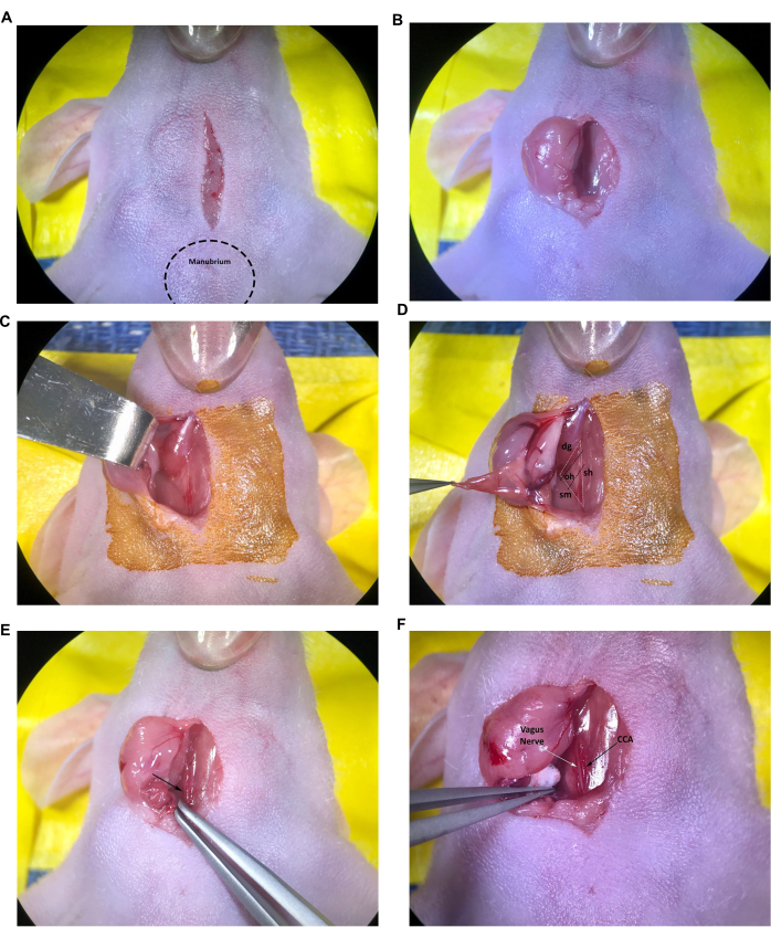
Figure 2: Incision and exposure of structures at the injection site. (A) Midline incision. (B,C) Retraction of the right salivary gland. (D) Muscular triangle formed by the trachea/sternohyoid muscle, sternocleidomastoid muscle, and the digastric muscle, omohyoid muscle is also visible. (E) Common carotid artery, indicated by the arrow. (F) Vagus nerve and common carotid artery, indicated by the arrows. Abbreviations: sh = trachea/sternohyoid muscle; sm = sternocleidomastoid muscle; dg = digastric muscle; oh = omohyoid muscle; CCA = Common carotid artery. Please click here to view a larger version of this figure.
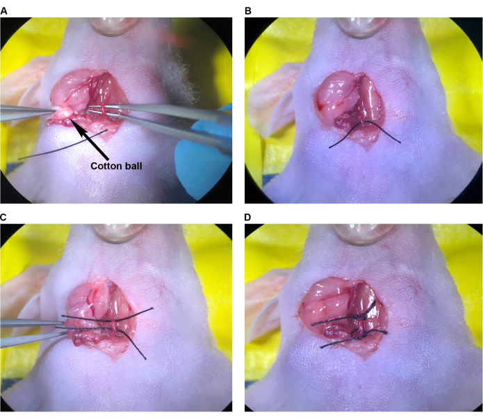
Figure 3: Preparing the CCA for injection. (A) Angled-tip forceps passed under the CCA. (B) The suture was pulled halfway under the CCA using angled-tip forceps. (C) The second suture was pulled halfway under the CCA. (D) Sutured tied in loose knows around the CCA. Abbreviation: CCA = Common carotid artery. Please click here to view a larger version of this figure.
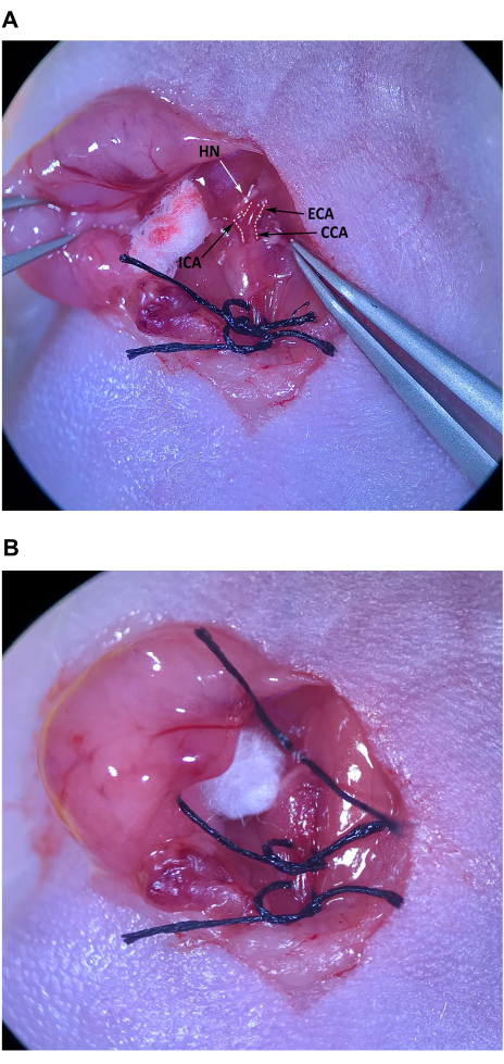
Figure 4: External carotid artery isolation and preparation. (A) CCA, external carotid artery, and internal carotid artery. (B) Suture pulled halfway through under the ECA. Abbreviations: CCA Common carotid artery; ECA = external carotid artery; ICA = internal carotid artery. Please click here to view a larger version of this figure.
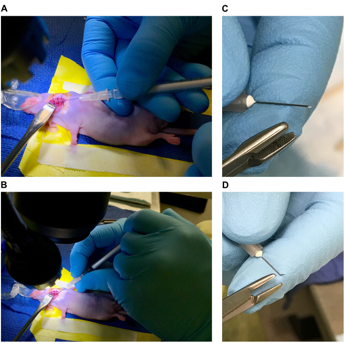
Figure 5: Needle and syringe preparation. (A) Injection with a straight needle with a syringe resting against the body of the mouse. (B) Injection with a bent needle, with a hand resting on the surgical table. (C,D) Preparing a bent needle. Please click here to view a larger version of this figure.
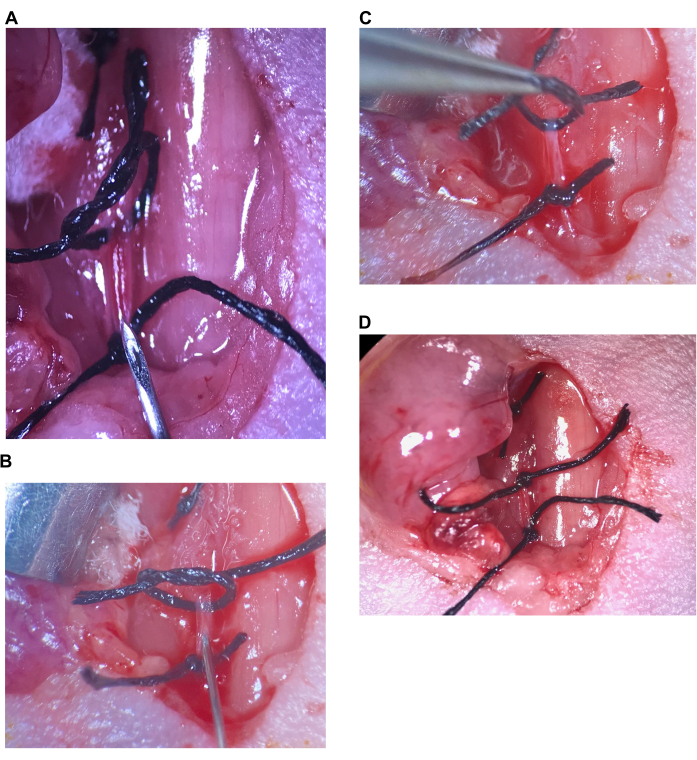
Figure 6: Intracarotid injection. (A) The upper suture is loose, the lower suture is tightened on the CCA, the needle is placed above the lower suture. (B) The needle is inserted just past the bevel, artery is sealed around the needle. (C) The upper suture is lifted to kink the artery upwards and prevent backflow. (D) The upper suture on the CCA is tightened. Abbreviation: CCA = Common carotid artery. Please click here to view a larger version of this figure.
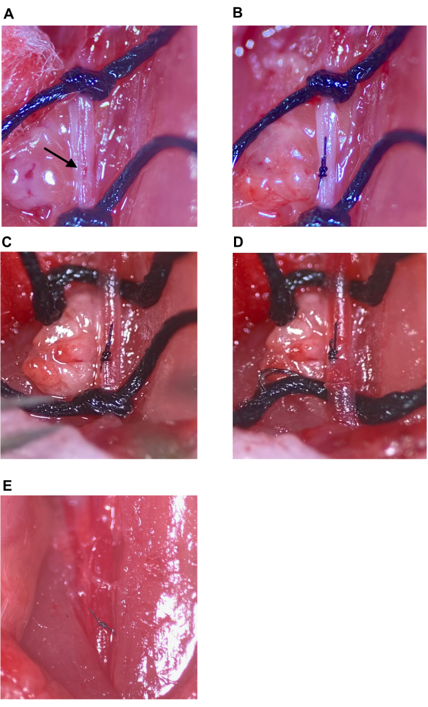
Figure 7: Injection site repair and restoring circulation. (A) The injection site indicated with an arrow. (B) The injection site closed with a surgeon's knot, a minimum of four throws. (C,D) Loosening of upper and lower sutures on the CCA after injection site repair; no bleeding seen after suture loosening. (E) Sutures are removed after the injection site is determined to be sufficiently repaired. Abbreviation: CCA = Common carotid artery. Please click here to view a larger version of this figure.

Figure 8: Intracarotid injection of GFP-BM-hMSCs and comparison of homing to intracranial glioma tumors after either CCA ligation or CCA repair with restored circulation. Brain tissue sections from tumor-bearing mice were stained with anti-GFP primary and Alexa Fluor 488 secondary antibodies to label GFP-BM-hMSCs (green). Nuclei were stained with Hoechst 33342 (blue). Representative low-power fields of labeled sections showing overall homing to tumor and high-power fields showing GFP-positive cell distribution after (A,C) CCA repair or (B,D) CCA ligation. (E) Overall homing of GFP-BM-hMSCs to tumors was evaluated by the total number of GFP-positive cells within the tumor boundary on two different slides and means were compared by t-test. No significant difference in overall homing was observed between the alternative procedures (P = 0.6858). (F) Dispersal of GFP-BM-hMSCs throughout the tumor was evaluated by counting GFP-positive cells across 10 high-powered fields within the tumor. Comparison of median values by the Wilcoxon Signed Rank test indicates no significant difference between individuals, regardless of procedure (P = 0.1914, 0.5000, 0.1641, 0.9512, 0.8828, 0.2207). Abbreviations: GFP = green fluorescent protein; GFP-BM-hMSCs = GFP-labeled bone marrow-derived human mesenchymal stem cells; CCA = Common carotid artery. Please click here to view a larger version of this figure.
Discussion
Intracarotid artery injections have been used increasingly in recent years to deliver therapeutics to brain tumors. Consequently, it is important to establish mouse models that mirror intracarotid artery injections in humans for research purposes. Previously, intracarotid artery injections in mice were performed with subsequent ligation of the artery, which limits the number of injections into the artery11,12. Additionally, occlusion of the carotid artery in mice can lead to cerebral ischemia in certain mouse strains that do not have a complete Circle of Willis13. We have developed a method to repair the injected carotid artery to overcome the limitations of prior methods. Repair of the injection site results in re-establishing blood flow to the injected artery, reducing the chance for cerebral ischemia, and facilitating subsequent injections into the same internal carotid artery.
Several steps, which are critical to success, require careful handling of surgical instruments or tissue, which include: insertion of the needle correctly into the lumen of the artery to avoid bleeding during intracarotid injection; careful dissection of connective tissue from the injection site prior to needle insertion; removal of all clumps and air bubbles in the syringe and needle prior to injection; and correct closing of the injection site to prevent closing of the lumen of the artery during the repair. To prevent bleeding after the needle is inserted, ensure that the needle is inserted into the artery past the bevel to form a seal around the needle shaft. To avoid a tear in the back wall of the artery, insert the needle at a shallow angle and subtly rock the syringe and needle back to keep the needle tip clear of the arterial back wall. If the injected solution leaks out during the injection, this suggests the needle was only inserted into the connective tissue surrounding the artery; careful dissection of the excess connective tissue from the injection site prior to injection will prevent this issue.
Regarding the choice of suture and closure technique, if the initial injection used a 33 G needle and made a clean insertion into the artery, one simple suture with 9-0 suture is sufficient to repair the artery. If a larger needle is used for injection (30 G etc) or any tearing occurs when inserting the needle (e.g., when the needle is off-center or the artery is moving because the mouse is breathing), this results in a slightly larger hole that needs to be repaired. Two simple sutures or a figure of eight is usually sufficient to repair this type of larger hole. The choice between these two techniques is based on the surgeon's preference in this situation. It is important to note that the repair technique has not been evaluated in situations where the injection site hole is significantly larger than in the situation mentioned above. If tearing at the injection site extends laterally (making a wider hole, greater than one-third of the circumference of the artery), repairing with this method may cause contraction of the artery and an increased risk of thrombosis.
If there is bleeding from the repaired injection site as the sutures are removed, it may be due to the stretching of the repaired site as normal circulation resumes; this may be rectified by gently covering the repaired injection site with sterile cotton and applying light pressure for 30 s. Alternatively, if there is bleeding from the repaired injection site with no visible blood flow and a proximal distended artery, it indicates that the suture needle passed through the back wall of the artery during repair. In this case, gently open the injection site edges during repair, pass the suture needle through the artery at a shallow angle, and visually confirm that the suture has not passed through the back wall before tying the suture knot.
With these measures in place, the method of injection site repair is precise and repeatable across cohorts of animals regardless of genetic background or age. In our experience, the success rate has been 100% with three different surgeons performing the procedure. With adequate experience and following the protocol provided carefully, we do not foresee any difficulty for other surgeons to perform this procedure. With practice, a skilled surgeon can complete the procedure in 15-20 min. If the experiment allows for it, the time per animal can also be reduced by leaving the upper and lower CCA sutures intact, forgoing repair of the injection site. However, as noted above, strain-specific differences in cerebral vascular anatomy have been documented and it is important to verify that the strain of the mouse used in the procedure can tolerate this prior to starting the experiment.
Since this is a surgical procedure, recovery of the mice must be taken into account. Stress tolerance and wound recovery are important considerations that will vary with different mouse strains. In addition, inflammation at the surgical site and scar tissue formation may increase recovery time after repeated surgeries. We have successfully performed multiple injections 7 days apart, but if more frequent injections are necessary, they should be evaluated carefully in the specific mouse strains to be used. Forceful handling and stress on the CCA (during isolation, tying and removing of sutures, and injection) can damage and weaken arterial walls leading to tearing during repeated injections. It is important to minimize the dissection of supporting connective tissue around the CCA and bifurcation and refrain from applying excessive tension to the artery.
Our results suggest that in this particular model, CCA ligation or CCA repair with restored circulation after injection do not differ in overall homing frequency or distribution of injected BM-hMSCs across intracranial tumors. While this may vary in different mouse strains, the use of injection site repair offers the advantage of returning blood flow to the injected artery, allowing for subsequent injections into the same artery, and importantly, resembling intracarotid artery injections in human patients. The choice of ligating versus repairing the injected artery is based on the type of experiment and the mouse model being used. If a second injection is needed, or if the mouse model does not have a complete Circle of Willis, injection site repair should be used. The ability to re-inject the CCA in mouse models can facilitate additional experimental manipulation. For example, to test multiple doses of a potential therapeutic given over time, repair of the injected artery is essential to perform subsequent injections. This method would also be useful in experiments involving the injection of combinations of therapeutic agents that need to be injected at different times. The increased flexibility in intracarotid injections afforded by repair of the injected artery improves the translational utility of mouse brain tumor models.
Disclosures
The authors have no relevant disclosures/conflicts of interest.
Acknowledgements
This study was supported by grants from the National Cancer Institute (R01CA115729, R01CA214749, and 1P50 CA127001) and by the generous philanthropic contributions to The University of Texas MD Anderson Cancer Center Moon Shots Program™, The Broach Foundation for Brain Cancer Research, The Elias Family Fund for Brain Tumor Research, The Priscilla Hiley Cancer Research Fund, The Bauman Family Curefest Brain Cancer Research Fund, Chuanwei Lu Fund, The Sweet Family Brain Cancer Research Fund, The Ira Schneider Memorial Cancer Research Foundation, The Jim & Pam Harris Fund, The Gene Pennebaker Fund for Brain Cancer Research, the Sorenson Fund for Brain Tumor Research, the Brian McCulloch Memorial Fund, the TLC Foundation from the Heart and the Mary Harris Pappas Endowed Fund for Glioblastoma Research, all to F.F.L.
Materials
| Name | Company | Catalog Number | Comments |
|---|---|---|---|
| 1 mL syringes (low dead space) | Air-tite Products Co. | A1 | |
| 26 G; 1/2" needle | Air-tite Products Co. | N2612 | |
| 33 G; 1/2" needle | JBP, Air-tite Products Co. | JBP3313B | |
| 3 cm Petri dish | Falcon, Fisher Scientific | 08-772A | |
| 3M durapore surgical tape | Fisher Scientific | 19-071-152 | |
| 6-0 suture thread | Fine Science Tools | 18020-60 | |
| 70% Ethanol | Fisher Scientific | 04-355-122 | |
| 9-0 microsurgical suture with needle | Fine Science Tools | 12052-09 | |
| Analgesic for major surgery | |||
| Artificial tears/ophthalmic ointment | Covetrus | 8897 | |
| Bead Sterilizer | Fisher Scientific | 14-955-341 | |
| Betadine/Chlorhexidine | McKesson, Fisher Scientific | NC1696484 | |
| Blunt hook retractor | Fine Science Tools | 17022-13 | |
| Dissecting microscope | Zeiss Microscopy, LLC | 491903-0010-000 | |
| Electric heating pad | Insource, Fisher Scientific | NC0667724 | |
| Extra narrow scissors | Fine Science Tools | 14088-10 | |
| Fine forceps - Dumont #5 forceps with micro-blunted tips | Fine Science Tools | 11253-20 | |
| Fine forceps - Dumont #5/45 angled tip forceps with micro-blunted tips | Fine Science Tools | 11253-25 | |
| Isoflurane vaporizer (or Ketamine/Xylazine cocktail) | Kent Scientific | VetFlo-1231 | |
| Light source | Laxco, Fisher Scientific | AMPSILED21 | |
| Mouse anesthesia nose cone | Braintree Scientific, Inc | XENO- M | |
| Needle driver | Fine Science Tools | 12002-12 | |
| Sterile cotton swabs | Texwipe, Fisher Scientific | 18-366-472 | |
| Sterile gauze pads | Covidien, Fisher Scientific | 22-037-907 | |
| Sterile saline (0.9%) | KD Medical, Fisher Scientific | 50-103-1363 | |
| Sterile surgical drapes | Fisher Scientific | 50-129-6666 | |
| Sterile surgical/downdraft table | |||
| Sterile suture pack (any suitable diameter for mouse wound closure) | Ethicon, Fisher Scientific | 50-209-2811 | |
| Surgical tools | |||
| Vinyl lab tape | Fisher Scientific | 15-901 |
References
- Loya, J., Zhang, C., Cox, E., Achrol, A. S., Kesari, S. Biological intratumoral therapy for the high-grade glioma part i: Intratumoral delivery and immunotoxins. CNS Oncol. 8 (3), (2019).
- Garfield, J., Dayan, A. D. Postoperative intracavitary chemotherapy of malignant gliomas. A preliminary study using methotrexate. J Neurosurg. 39 (3), 315-322 (1973).
- Kroin, J. S., Penn, R. D. Intracerebral chemotherapy: Chronic microinfusion of cisplatin. Neurosurgery. 10 (3), 349-354 (1982).
- Sendelbeck, S. L., Urquhart, J. Spatial distribution of dopamine, methotrexate and antipyrine during continuous intracerebral microperfusion. Brain Res. 328 (2), 251-258 (1985).
- Alter, R. A., et al. Long-term benefit of intra-arterial bevacizumab for recurrent glioblastoma. J Exp Ther Oncol. 12 (1), 67-71 (2017).
- Angelov, L., et al. Blood-brain barrier disruption and intra-arterial methotrexate-based therapy for newly diagnosed primary cns lymphoma: A multi-institutional experience. J Clin Oncol. 27 (21), 3503-3509 (2009).
- Chen, S. R., Chen, M. M., Ene, C., Lang, F. F., Kan, P. Perfusion-guided endovascular super-selective intra-arterial infusion for treatment of malignant brain tumors. J Neurointerv Surg. 14 (6), 533-538 (2022).
- Faltings, L., et al. Rechallenging recurrent glioblastoma with intra-arterial bevacizumab with blood brain-barrier disruption results in radiographic response. World Neurosurg. 131, 234-241 (2019).
- Srinivasan, V. M., et al. Advances in endovascular neuro-oncology: Endovascular selective intra-arterial (esia) infusion of targeted biologic therapy for brain tumors. J Neurointerv Surg. 12 (2), 197-203 (2020).
- Shinojima, N., et al. Tgf-β mediates homing of bone marrow-derived human mesenchymal stem cells to glioma stem cells. Cancer Res. 73 (7), 2333-2344 (2013).
- Liu, Z., et al. Improving orthotopic mouse models of patient-derived breast cancer brain metastases by a modified intracarotid injection method. Scientific Reports. 9 (1), 622(2019).
- Zhang, C., Lowery, F. J., Yu, D. Intracarotid cancer cell injection to produce mouse models of brain metastasis. Journal of visualized experiments : JoVE. (120), e55085(2017).
- Barone, F. C., Knudsen, D. J., Nelson, A. H., Feuerstein, G. Z., Willette, R. N. Mouse strain differences in susceptibility to cerebral ischemia are related to cerebral vascular anatomy. J Cereb Blood Flow Metab. 13 (4), 683-692 (1993).
- Benbenishty, A., et al. Maintaining unperturbed cerebral blood flow is key in the study of brain metastasis and its interactions with stress and inflammatory responses. Brain, behavior, and immunity. 62, 265-276 (2017).
- Doucette, T., et al. Mesenchymal stem cells display tumor-specific tropism in an rcas/ntv-a glioma model. Neoplasia. 13 (8), 716-725 (2011).
- Hata, N., et al. Platelet-derived growth factor bb mediates the tropism of human mesenchymal stem cells for malignant gliomas. Neurosurgery. 66 (1), 144-156 (2010).
- Nakamizo, A., et al. Human bone marrow-derived mesenchymal stem cells in the treatment of gliomas. Cancer Res. 65 (8), 3307-3318 (2005).
- Qiao, Y., et al. Magnetic resonance and photoacoustic imaging of brain tumor mediated by mesenchymal stem cell labeled with multifunctional nanoparticle introduced via carotid artery injection. Nanotechnology. 29 (16), 165101(2018).
- Yong, R. L., et al. Human bone marrow-derived mesenchymal stem cells for intravascular delivery of oncolytic adenovirus delta24-rgd to human gliomas. Cancer Res. 69 (23), 8932-8940 (2009).
Reprints and Permissions
