Campylobacter is the leading cause of bacterial foodborne gastroenteritis worldwide. Despite establishments enacting measures to reduce the prevalence throughout their facilities, contaminated products consistently reach consumers. The technique developed over the past twelve years addresses limitations of existing methods for isolating and detecting Campylobacter spp. from raw meat.
Method Article
Detection and Isolation of Campylobacter spp. from Raw Meat
In This Article
Summary
Abstract
This article presents a rapid yet robust protocol for isolating Campylobacter spp. from raw meats, specifically focusing on Campylobacter jejuni and Campylobacter coli. The protocol builds upon established methods, ensuring compatibility with the prevailing techniques employed by regulatory bodies such as the Food and Drug Administration (FDA) and the U.S. Department of Agriculture (USDA) in the USA, as well as the International Organization for Standardization (ISO) in Europe. Central to this protocol is collecting a rinsate, which is concentrated and resuspended in Bolton Broth media containing horse blood. This medium has been proven to facilitate the recovery of stressed Campylobacter cells and reduce the required enrichment duration by 50%. The enriched samples are then transferred onto nitrocellulose membranes on brucella plates. To improve the sensitivity and specificity of the method, 0.45 µm and 0.65 µm pore-size filter membranes were evaluated. Data revealed a 29-fold increase in cell recovery with the 0.65 µm pore-size filter compared to the 0.45 µm pore-size without impacting specificity. The highly motile characteristics of Campylobacter allow cells to actively move through the membrane filters towards the agar medium, which enables effective isolation of pure Campylobacter colonies. The protocol incorporates multiplex quantitative real-time polymerase chain reaction (mqPCR) assay to identify the isolates at the species level. This molecular technique offers a reliable and efficient means of species identification. Investigations conducted over the past twelve years involving retail meats have demonstrated the ability of this method to enhance recovery of Campylobacter from naturally contaminated meat samples compared to current reference methods. Furthermore, this protocol boasts reduced preparation and processing time. As a result, it presents a promising alternative for the efficient recovery of Campylobacter from meat. Moreover, this procedure can be seamlessly integrated with DNA-based methods, facilitating rapid screening of positive samples alongside comprehensive whole-genome sequencing analysis.
Introduction
Campylobacter spp. are the leading cause of bacterial foodborne gastroenteritis worldwide, with an estimated 800 million cases annually1. As a major zoonotic bacterium, Campylobacter naturally colonizes the gastrointestinal tracts of a wide range of animals, including wild birds, farm animals, and pets2. During slaughtering or food processing, Campylobacter spp. frequently contaminate carcasses or meat products3. Campylobacteriosis is usually associated with the consumption of undercooked poultry or cross-contamination of other foods by raw poultry juices2. It can cause serious complications, such as Guillain-Barré syndrome, reactive arthritis, and septicemia in immunocompromised individuals4. Detecting and isolating Campylobacter from food sources, especially poultry products, is essential for public health surveillance, outbreak investigation, and risk assessment.
Conventional culture-based methods are the traditional and standard methods for Campylobacter detection5,6. However, there are several limitations, including long incubation times (48 h or more), low sensitivity (up to 50%), and are not inclusive to all strains (some stressed Campylobacter cells may not grow well or at all in the media)7. Molecular methods, such as polymerase chain reaction (PCR), are more rapid and sensitive than culture-based methods, but they do not provide viable isolates for further characterization8,9.
Immunological methods are alternative and complementary methods for Campylobacter detection. These are rapid, simple, and versatile, but also have several limitations, including cross-reactivity (some antibodies may bind to non-Campylobacter bacteria or other substances that share similar antigens), low specificity (some antibodies may not bind to all Campylobacter strains or serotypes), and sample preparation requirements (immunological methods often require pre-treatment of the samples to remove interfering substances to enhance the binding of the antibodies)10.
Within the genus of Campylobacter, C. jejuni and C. coli cause most human Campylobacter infections (81% and 8.4%, respectively)11. Both are spiral-shaped, microaerophilic, and thermophilic bacteria containing a unipolar flagellum or bipolar flagella. Rotation of a flagellum at each pole is considered both the primary driving force for its characteristic corkscrew motility and crucial to its pathogenesis because it allows the bacterium to swim through the viscous mucosa of the host gastrointestinal tract. The motility of Campylobacter is controlled by its chemosensory system that allows the cells to move toward favorable environments12,13. Based on the cell morphology and physiological characteristics of Campylobacter, a few studies have utilized membrane filtration for the isolation of Campylobacter spp. from fecal and environmental samples14,15,16.
This study presents a rapid and robust protocol for the isolation and subsequent detection of C. jejuni and C. coli from raw meat, which overcomes the drawbacks of the existing methods and offers several advantages. Tentative colonies can be confirmed as Campylobacter spp. using a variety of methods, such as microscopy, biochemical tests (e.g., catalase and oxidase activity assays), or molecular methods6. The method identifies the isolates at the species level using a multiplex real-time PCR (mqPCR) assay that targets genes unique to C. jejuni and C. coli. This method is relatively inexpensive, rapid, and selective, which makes it suitable for use in a variety of settings, including food processing facilities, clinical laboratories, and research laboratories.
Protocol
All work associated with this protocol should be conducted within a biological safety cabinet (BSC) to maintain aseptic conditions and minimize the risk of sample contamination or operator exposure to microbial pathogens. When transferring samples outside the BSC, use sealed containers to prevent spillage in case of accidental drops, maintaining sample integrity. Preferably, disposable components should be used throughout the procedure to mitigate the possibility of cross-contamination. In cases where disposables are not feasible, ensure all equipment and materials are sterile prior to use. Proper waste management is crucial; all used disposable components should be discarded as biohazard waste. Autoclave materials before discarding to ensure proper sterilization and avoid containment of potentially hazardous materials. Adhering to these precautions not only safeguards sample integrity but also minimizes the risk of operator exposure to microbial pathogens. Figure 1 depicts the workflow of sample preparation, selective enrichment, filter-based isolation, and mqPCR differentiation of Campylobacter species. Supplemental File 1 depicts a more detailed workflow and images throughout the process.
1. Preparation of meat samples
- Acquiring meat samples
- Acquire various fresh meat packages, including chicken thighs, wings, drumsticks, and livers from local retailers.
- Transfer all samples to storage at 4 °C and process within 24 h after receipt.
NOTE: Storing the fresh samples at lower temperatures, such as below freezing, will affect the recovery.
- Processing meat samples
- Follow the ratio of components prescribed within the FSIS sampling guideline6,17.
- Cut 450 g chicken pieces from each package and place them in a stomacher bag (see Table of Materials).
NOTE: Stomacher bags are suggested because they have sufficient mechanical strength to ensure the downstream processes, and will not rupture or leak. - Prepare buffered peptone water (BPW).
- Dissolve 20 g of the powder (see Table of Materials) in 1 L of purified water. Autoclave the solution at 121 °C for 15 min. Dilute the solution in sterile water to a concentration of 0.1%.
- Add 200 mL 0.1% BPW to the stomacher bag containing the chicken.
- Manually massage/palpate the sample from the outside of the stomacher bag for 2 min.
- Collect all the chicken rinse from the filtered side of the bag using a motorized pipette controller (see Table of Materials).
- Dispense the chicken rinse into sterile centrifuge bottles. Centrifuge at 10,000 x g for 10 min at room temperature.
- Carefully collect the supernatant using a motorized pipette controller with a 25 mL disposable serological plastic pipette. Avoid disturbing the pellet.
- Repeat the process as necessary to ensure all of the supernatant is removed.
- Discard the collected supernatant.
2. Selective enrichment of Campylobacter from raw meat
- Preparation of Bolton Broth with supplements
- Dissolve 13.8 g of powder (see Table of Materials) in 500 mL of purified water. Sterilize the broth by autoclaving for 15 min at 121 °C.
- Add 25 mL laked horse blood (see Table of Materials) to the sterilized 500 mL Bolton Broth.
NOTE: The addition of horse blood acts as an oxygen quenching agent to aid in the recovery of injured Campylobacter cells from the food matrix. - Reconstitute 1 vial of antibiotic supplement (cefoperazone, cycloheximide, trimethoprim, and vancomycin, see Table of Materials) in 5 mL 50% ethanol.
- Add the reconstituted antibiotic supplement to the Bolton Broth.
- Enrichment procedure
- Resuspend the pellet in 50 mL Bolton Broth containing laked horse blood and antibiotics.
- Place samples (with loosened caps) inside a sealed container that maintains a gas mixture of 85% N2, 10% CO2, and 5% O2.
- Ensure the cap is loose, but the container is tightly sealed to produce the microaerophilic and thermophilic growth requirements of Campylobacter.
NOTE: Gas packs that maintain an atmosphere of 85% N2, 10% CO2, and 5% O2 can be used when environmental chambers are not available. - Incubate the samples at 42 °C for 24 h.
- Ensure the cap is loose, but the container is tightly sealed to produce the microaerophilic and thermophilic growth requirements of Campylobacter.
3. Isolation and purification of C. jejuni and C. coli from raw chicken
- Preparation of Brucella agar plates
- Dissolve 28 g of Brucella powder (see Table of Materials) in 1 L of purified water. Dissolve 15 g of agar in the Brucella solution.
- Sterilize the Brucella agar by autoclaving for 15 min at 121 °C. Cool the medium-agar mix in a 55 °C water bath.
- Pour 20 mL of Brucella agar into each 100 mm diameter Petri dish.
- Filter method and colony cultivation
- Evaluate the effect of moisture on Campylobacter passing through filters by drying Brucella agar plates with lids opened in a Biosafety cabinet for 0 h, 1 h, 2 h and 3 h.
- Prepare a no-filter control by directly spreading 80 µL of the sample on the Brucella agar plate.
- Place a cellulose acetate filter (0.45 µm or 0.65 pore-size, see Table of Materials) at the center of a Brucella agar plate.
- Pipette 4 drops/filter and 20 µL/drop of enriched sample onto the filter.
- Place the drops near the center of the filter to ensure the liquid that reaches the plate goes through the filter, not around the filter.
- Place the drops in a manner that ensures they will not spread and aggregate.
- Incubate drops at room temperature for 15 min and carefully remove the filters.
NOTE: This step permits sufficient time for the Campylobacter cells to traverse the membrane and reach the agar medium without excessive drying. - Incubate plates at 42 °C for approximately 24 h under the microaerobic conditions described earlier.
- Pick characteristic Campylobacter colonies with specific traits.
NOTE: Campylobacter colonies are typically round with smooth edges, glistening, and translucent yellowish or pinkish color6. - Streak colonies onto Brucella agar plates for purification. Repeat this step until plates with a single uniform colony morphology are obtained.
- Prepare samples for long-term storage.
- Prepare Bolton Broth as described earlier. Add one colony from the plate with uniform colony morphology.
- Grow overnight (24 h) under microaerophilic conditions described earlier. Add 900 µL of the overnight culture to a 2 mL cryovial containing 100 µL of DMSO.
- Rapidly cool in a dry ice-ethanol bath (approx. -72 °C) for 10 min. Transfer to a -80 °C freezer for long-term storage.
4. Identification of C. jejuni and C. coli species
- Perform species-level identification of C. jejuni and C. coli using a multiplex qPCR (mqPCR) assay previously developed18,19.
- Perform rapid cell lysis and genomic DNA extraction in a 96-well plate format.
NOTE: It is strongly recommended to consider using commercial kits (see Table of Materials) to ensure that the sample is sufficiently free of known inhibitors of PCR.- Disperse purified Campylobacter colonies into 100 µL of extraction solution.
- Lyse samples at 99 °C for 10 min followed by cooling at 20 °C for 2 min in a thermocycler.
- Centrifuge the plate at 8,000 x g for 10 min at room temperature. Remove 2 µL of aliquots of the supernatant for the mqPCR assay.
- Prepare a 20 µL reaction mixture consisting of 10 µL of 2x Master Mix, 2.0 µL of DNA sample, 104 copies of Internal Amplification Control (IAC) template, and 200 nM of each primer and probe (see Table of Materials).
NOTE: The primers and probes of hipO and cdtA are the exclusive target genes for C. jejuni and C. coli, respectively. The IAC consists of a 79-bp DNA segment of the human adenovirus and is included as a positive control to ensure consistent activity of DNA polymerase across all samples.
- Load all samples in triplicates in a 96-well optical plate covered with an optical film and place them in a Real-Time PCR system (see Table of Materials).
- Initiate a hot-start activation of the DNA polymerase at 95 °C for 10 min. Follow with 40 cycles of denaturation at 95 °C for 15 s and annealing/extension at 60 °C for 1 min.
- Perform rapid cell lysis and genomic DNA extraction in a 96-well plate format.
5. Enumerate cell suspensions
- Enumerate cell suspensions using a 6 x 6 drop plate procedure20. Refer to Supplemental File 1 for images depicting the 6 x 6 drop plate method.
- Air dry Brucella agar plates with the lid off in a laminar flow hood for 45 min.
- Add 200 µL of bacterial suspension to six rows (A-F) in the first column of a 96-well plate.
- Distribute 180 µL of Brucella medium across six rows of the remaining columns (2-12).
- Prepare ten-fold serial dilutions using 20 µL transfers.
- For instance, transfer 20 µL of sample from column one to column two. Repeat this process for a minimum of 6 columns.
- Reflux mix each suspension ten times using the pipette, changing the tips between each transfer.
- Use a multichannel pipette to deposit 7 µL drops from six rows of a column on the surface of a Brucella agar plate.
- Repeat to create a 6 x 6 array, ensuring rows are technical replicates across columns.
- Air dry the plates for 5 min, and then invert the plate.Incubate plates at 42 °C for 24 h.
- Count the number of colonies in each representative dilution.
Results
Effect of moisture in Brucella agar plates for passive filtration of Campylobacter
Campylobacter has a small genome and lacks several stress response genes commonly occurring in other bacteria, such as E. coli O157:H7 and Salmonella. Therefore, it is more sensitive to various environmental stresses and cannot tolerate dehydration or ambient oxygen levels. Conversely, an overly moist agar medium can flood the filter. This not only causes diffusion of the sample to outside the filter, but also increases exposure time to oxygen21.
To determine the appropriate conditions for filter-based isolation of Campylobacter, Brucella agar plates were dried with the lids removed for 0 h, 1 h, 2 h, and 3 h inside a biological safety cabinet and assessed for the efficiency of Campylobacter cells to traverse a 0.65 µm pore-size filter. Four 20 µL aliquots of C. jejuni S27 cultures at the concentrations of 1.53 x 10⁴ and 1.53 x 10⁵ CFU/mL were pipetted onto each filter membrane that had been placed on top of a Brucella plate. After 15 min of penetration, filters were removed, and plates were incubated overnight for cell growth.
Cells from 5 replicated plates were then counted and noted in Table 1. The results indicated that the agar plates dried for 2 h and 3 h performed similarly with nearly equal numbers of cells recovered from passive filtration. Noticeably, the plates dried under these conditions for 0 h and 1 h did not allow cells to fully traverse the membrane within the 15 min time period used.
Comparison of different pore-size filter membranes for isolating Campylobacter from chicken livers
Considering the Campylobacter cell sizes (0.5-5 µm in length and 0.2-0.9 µm in width) and a wide range of food particle sizes, cellulose acetate filters with 0.45 µm and 0.65 µm pore sizes were tested for the efficiency of Campylobacter passage when given a 15 min incubation time. Food samples consisting of 450 g of chicken livers spiked with 153 CFU of C. jejuni and then enriched overnight were used for the experiment. As a no-filter control, direct plating of the enrichment sample was included in parallel. The results (Figure 2) from 5 replicate plates consistently showed that the 0.65 µm pore size filter allowed more cells to traverse than the 0.45 µm pore-size filters, resulting in increases of ~29-fold more cells obtained. The 0.45 µm pore size filter retained too many cells on the upper side of the filter, resulting in a significantly lower recovery of Campylobacter from food compared to the 0.65 µm pore size filter. As expected, there was a lawn of different background organisms growing on the no-filter control plates.
Application of passive filtration in Campylobacter isolation from retail chicken
Because of the unusual motility of Campylobacter cells, the passive filtration technique was selected for the isolation of C. jejuni and C. coli from retail meat products, which are typically contaminated with numerous background organisms. Between the years 2014-2023, a total of 79 raw meat packages, including different parts of chicken meat, chicken livers, beef livers, and calf livers, were collected from various local supermarkets. From each package, 450 g was sampled for the isolation of Campylobacter spp. By combining selective enrichment of Campylobacter in blood-containing Bolton Broth and passive filtration of the cells through a 0.65 µm pore size cellulose acetate filter directly onto a Brucella agar plate, 49 Campylobacter strains have been successfully isolated from 79 meat samples (Table 2). Figure 3 represents the result of isolating a new Campylobacter strain from chicken livers. The method has been repeatedly proven to be sensitive, specific, and cost-effective.
Identification and differentiation of C. jejuni and C. coli isolates
To verify the genus and differentiate the species of Campylobacter isolates obtained from raw meat, a multiplex qPCR assay amplifying the specific gene targets (hipO and cdtA) for C. jejuni and C. coli, and an internal amplification control (IAC) was employed. The IAC was included as a false-negative indicator in the concurrent amplification of multiple genes. The assay was implemented in a 96-well format with rapid cell lysis and DNA extraction using a commercially available reagent (see Table of Materials). Table 2 summarizes the result of species identification of the C. jejuni and C. coli strains. As additional verification, whole-genome sequencing results confirmed the species of all the isolates (data not shown).

Figure 1: A workflow diagram for the isolation and identification of Campylobacter species from retailed meat. Please click here to view a larger version of this figure.
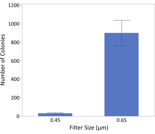
Figure 2: The impact of filter size on recovery of Campylobacter. The results indicate that the 0.65 µm filter can recover an average of 900 ± 138 colonies, while the 0.45 µm filter recovers 31 ± 7 colonies. The results were generated from N = 20 (5 plates with 4 drops/plate) and a Student's t-test indicates the means are statistically different (p < 0.0001). Please click here to view a larger version of this figure.

Figure 3: Isolation of Campylobacter by passive filtration of enriched poultry samples. (A) Depicts the four 20 µL drops of enriched sample deposited on the nitrocellulose membrane filter. The image was collected during the 15 min of passive filtration. (B) Depicts the plate after the nitrocellulose filter was removed. The four spots indicate where the enriched sample traversed the membrane. (C) Depicts the plate following 24 h incubation. Please click here to view a larger version of this figure.
Table 1: The effect of the drying time of Brucella agar plates on the passive filtration of C. jejuni. Please click here to download this Table.
Table 2: C. jejuni and C. coli strainsisolated from raw meat. During the period of time ranging from 2008 to 2023, 36 C. jejuni and 13 C. coli strains were isolated from 79 meat packages across 24 unique retail products. Please click here to download this Table.
Supplemental File 1: Images throughout the isolation and enumeration processes. Please click here to download this File.
Discussion
Significance of the protocol
C. jejuni and C. coli were the two major species of Campylobacter found to be prevalent in poultry22 and animal livers23,24. In this study, the meat samples of chicken parts (legs, wings, and thighs), chicken livers, and beef livers were randomly collected during different time periods, and from different retail stores and manufacturers for the isolation of Campylobacter spp. Of the 49 total Campylobacter strains isolated, 36 were identified as C. jejuni and 13 were C. coli, with no other Campylobacter species found, which is consistent with other reports25.
The assay is based on the spiral-shaped cell morphology and characteristic corkscrew-like motility of Campylobacter spp. A simple, yet effective, passive filtration technique26,27 that exploited its spiral-shaped cell morphology (long, slender, 0.2-0.9 by 0.5-5 µm) and strong corkscrew motility was used to separate Campylobacter from a mixture of background organisms. The high motility of Campylobacter allowed the cells to traverse the membrane filters and move towards favorable conditions found within the agar medium, while other background microorganisms from the meat products were unable to pass through. This method is relatively inexpensive, rapid, and selective, which makes it suitable for use in a variety of settings, including food processing facilities, clinical laboratories, and research laboratories.
A pioneering article often cited states that the 0.45 µm filter worked so well that 0.65 µm was not evaluated28. Results from this present study indicate the 0.65 µm pore size filter performed significantly better than the 0.45 µm pore size, resulting in a 29-fold increase in the number of cells recovered from the enrichment. This is important because the filters selected do not display reduced selectivity as previously reported29. Further, as it is known that filtering will significantly reduce the amount of Campylobacter recovered compared to direct plating30, therefore, increasing the size of the pore improves recovery of the microorganism, which is consistent with previously reported findings21. This is significant because all the cells that traversed the filters formed uniform Campylobacter colonies, indicating that both filters were sufficient at preventing other microflora and food particles from passing through. Additionally, the FSIS flowchart7 notes the potential for extended result production due to re-streaking isolates on Campy-Cefex plates containing antibiotics. Contrastingly, the protocol described in this manuscript, which combines the use of filtration and selective enrichment with cefoperazone, cycloheximide, trimethoprim, and vancomycin, has not necessitated re-streaking.
The current method employed is consistent with current FSIS Sampling and Verification programs17. As the level of Campylobacter contamination can be low (153 CFU/450 g chicken), the rinse is centrifuged to concentrate the sample by a factor of four, which increases the sensitivity of the assay. After concentrating the rinsate by a factor of 4x, samples are enriched for 48 h and screened with the Molecular Detection System (MDS) to replicate the method employed by FSIS laboratories (data not shown). Notably, the method described has yet to fail to identify positive strains within 24 h that were detected by the Molecular Detection System using 48 h of enrichment (data not shown). Lastly, an additional benefit of this protocol is that it can provide information related to the bacterial species and identify if the Campylobacter is C.coli, C. jejuni, or C. lari, while the MDS adopted in MLG 41.07 can only provide a binary positive/negative response for Campylobacter.
Critical steps
The protocol for Campylobacter isolation and identification necessitates precision during centrifugation, filtration, and molecular analysis. Accurate dilutions, proper incubation conditions, and meticulous adherence to qPCR assay conditions are pivotal for reliable species identification.
As a microaerophilic bacterium, Campylobacter is very fragile and sensitive to various environmental stresses and requires unique fastidious conditions for growth31,32,33. In food samples typically undergoing lengthy periods of transportation and storage, many Campylobacter cells are perhaps in a dormancy or sublethal/lethal injured state34,35. Thus, it is important to recover the stressed cells from their food matrices and grow them to a higher concentration. In the first step of the procedure, we used Bolton Broth supplemented with laked horse blood and antibiotics for selective enrichment of Campylobacter from food. The add-in blood served as an oxygen quenching agent to overcome the adverse effects of free oxygen radicals36. The antibiotics were used to inhibit the growth of background microflora37.
To minimize the exposure time of Campylobacter to ambient atmospheric oxygen, a 15 min incubation period was selected to allow for the cells to traverse the filter. Also, the moisture of the Brucella agar plate under the filter played an important role in the rate of passage. Specifically, the results from testing agar plates dried for 0 h, 1 h, 2 h and 3 h suggested that a high moisture content in the filter prevented cells from passing through. Equally critical is the precise placement of filters and drops on the plates and filters, both influencing the success of isolating cells.
Potential pitfalls and limitations
While presenting a structured approach for isolating and identifying Campylobacter species from raw chicken samples, several limitations of this protocol deserve attention. External contamination, insufficiently dried plates, clogging of filters impeding microbial movement, entrapment of the microorganisms within the pellet, incomplete sealing of the atmospheric chamber, and drops spreading beyond filter boundaries are among the primary pitfalls.
Inadequate separation of the microorganisms from the food surfaces or their confinement within the bulk of the sample may hinder their isolation using this method. Additionally, relying on microbial motility for traversal through passive filters presents a notable limitation; it is possible that the filter membranes retained some less motile Campylobacter strains, as it has been shown filters can reduce the capture efficiency of microbial pathogens in food38. Further limitations encompass the batch nature of centrifugation and filtration processes, susceptibility to filter clogging, and inefficiency in dispersing the pellet formed, which will impact the accuracy of microbial loads. These limitations collectively emphasize the need for caution and supplementary methodologies in ensuring comprehensive analysis, especially when dealing with varied sample types or seeking high-throughput capabilities.
Suggestions for troubleshooting
To preempt potential issues, initially ensure that all materials adhere to the necessary quality standards and have not expired. Troubleshoot clogged filters by potentially employing an additional filtration to remove any large contaminates that may restrict the passage of the Campylobacter through the nitrocellulose membrane. If contamination is observed, verify that the drops were not placed too close to the edge of the filter and permitted liquid to reach the agar by going around the filter as opposed to through the pores. If there is insufficient growth following enrichment, verify the seals of the atmospheric containers are tight and not leaking.
Potential refinement and expansion
Exploring alternative filter materials may enhance microbial traversal and enable this protocol to be expanded for use in isolating other motile microorganisms from heterogeneous mixtures such as food. Identifying controls to retain less motile Campylobacter variants without negatively impacting the specificity is advisable. Additionally, while the multiplexed qPCR assay utilized in this study was demonstrated to have the capabilities to detect C.lari18 other Campylobacter species of interest can be included within this assay.
In summary, through evaluating different parameters and settings, the appropriate conditions for filter-based isolation and species-level identification of C. jejuni and C. coli from food were established. The method has been demonstrated to be sensitive, specific, robust, and cost-effective. By applying it to real food samples, the protocol was able to isolate 36 C. jejuni and 13 C. coli strains from 79 meat packages.
The protocol is aligned with FSIS Directive 10,250.117, which outlines the procedure for raw chicken part sampling, and MLG 41.076 for isolation and identification of Campylobacter. The data suggests that concentrating the sample by 4x and enriching it for 24 h, coupled with filtration and plating, yields isolated, confirmed colonies within 48 h as opposed to 96 h. The protocol is compatible with DNA-based methods such as genome sequencing to provide a comprehensive characterization of Campylobacter strains, including their antimicrobial resistance profiles, virulence predictions, and phylogenetic relationships. The protocol represents a promising alternative for the efficient recovery and isolation of Campylobacter spp. from raw poultry, which can facilitate epidemiological studies and public health interventions.
Disclosures
All the authors declare that there is no conflict of interest.
Acknowledgements
This research was supported by the U.S. Department of Agriculture, Agricultural Research Service (USDA-ARS), National Program 108, Current Research Information System numbers 8072-42000-093 and 8072-42000-094-000-D. Mention of trade names or commercial products in this article is solely for the purpose of providing specific information and does not imply recommendation or endorsement by the U. S. Department of Agriculture. USDA is an equal opportunity provider and employer.
Materials
| Name | Company | Catalog Number | Comments |
|---|---|---|---|
| Agar - Solidifying Agent (Difco) | Becton, Dickinson and Company (BD) | 281230 | |
| Analytical Balance | Mettler Toledo | JL602-G/L | Equipment |
| Analytical Balance | Mettler Toledo | AB54-S | Equipment |
| Antibiotic supplements cefoperazone, cycloheximide, trimethoprim, vancomycin | Oxoid Ltd. | SR0183E | |
| Atmosphere Control Gas Pak (Campy, 85% N2, 10% CO2, and 5% O2) | Becton, Dickinson and Company (BD) | 260680 | |
| Atmosphere Control Vessel, GasPak EZ CampyPak container system | Becton, Dickinson and Company (BD) | 260678 | |
| Autoclave - Amsco Lab250, Laboratory Steam Sterilizer | Steris plc | LV-250 | Equipment |
| Biological Safety Cabinet, Type A2, Purifier Logic+ | Labconco Corporation | 302411101 | Equipment |
| Bolton broth | Oxoid Ltd. | CM0983 | |
| Brucella broth (Difco) | Becton, Dickinson and Company (BD) | 211088 | |
| Buffered Peptone Water | Bio-Rad Laboratories Inc. | 3564684 | |
| Centrifuge Microcentrifuge 5424 | Eppendorf | 5424 | Equipment |
| Centrifuge, Avanti J-25 | Beckman Coulter, Inc. | Equipment | |
| Chicken thighs, wings, drumsticks, livers | Local retailers | ||
| DNA Extraction - PreMan Ultra Sample Preparation Reagent | Thermo Fisher Scientific Inc. | 4318930 | |
| FAM probe for hipO (Sequence 5'-TTGCAACCTCACTAGCAAAATCC ACAGCT-3') | Integrated DNA Technologies | ||
| Filter - 0.45 µm sterile cellulose acetate filter | Merck-Millipore LTD | DAWP04700 | |
| Filter - 0.65 µm sterile cellulose acetate filter | Merck-Millipore LTD | HAWG04700 | |
| forward primer for cdtA (Sequence 5'-TGTCAAACAAAAAACACCAAGCT T-3' ') | Integrated DNA Technologies | ||
| forward primer for hipO (Sequence 5'-TCCAAAATCCTCACTTGCCATT-3') | Integrated DNA Technologies | ||
| forward primer for IAC (Sequence 5'-GGCGCGCCTAACACATCT-3') | Integrated DNA Technologies | ||
| HEX probe for cdtA (Sequence 5'-AAAATTTCCCGCCATACCACTTG TCCC-3') | Integrated DNA Technologies | ||
| Incubator - Inova 4230 refrigerated incubator shaker | New Brunswick Scientific | 4230 | Equipment |
| Inoculating Loop - Combi Loop 10µL and 1µL | Fisher Scientific International, Inc | 22-363-602 | |
| Internal Amplification Control (IAC) DNA fragment (Sequence 5'-TGGAAGCAATGCCAAATGTGTAT GTGGTGGCATTGTCTTCTCCC GTTGTAACTATCCACTGAGATG TGTTAGGCGCGCC-3') | Integrated DNA Technologies | ||
| Laked horse blood | Remel Inc. | R54072 | |
| Manual pipette Pipet-Lite LTS Pipette L-1000XLS+ | Mettler Toledo | 17014382 | Equipment |
| Manual pipette Pipet-Lite LTS Pipette L-100XLS+ | Mettler Toledo | 17014384 | Equipment |
| Manual pipette Pipet-Lite LTS Pipette L-10XLS+ | Mettler Toledo | 17014388 | Equipment |
| Manual pipette Pipet-Lite LTS Pipette L-200XLS+ | Mettler Toledo | 17014391 | Equipment |
| Manual pipette Pipet-Lite LTS Pipette L-20XLS+ | Mettler Toledo | 17014392 | Equipment |
| Manual pipette Pipet-Lite Multi Pipette L8-200XLS+ | Mettler Toledo | 17013805 | Equipment |
| Manual pipette Pipet-Lite Multi Pipette L8-20XLS+ | Mettler Toledo | 17013803 | Equipment |
| Media Storage Bottle -PYREX 1 L Square Glass Bottle, with GL45 Screw Cap | Corning Inc. | 1396-1L | Equipment |
| Media Storage Bottle -PYREX 2 L Round Wide Mouth Bottle, with GLS80 Screw Cap | Corning Inc. | 1397-2L | Equipment |
| Microtiter plate, 96 well plate, flat bottom, polystyrene, 0.34 cm2, sterile, 108/cs | MilliporeSigma | Z707902 | |
| Mixer - Vortex Genie 2 | Scientific Industries Inc. | SI-0236 | Equipment |
| Motorized pipette controller, PIPETBOY2 | INTEGRA Biosciences Corp. | Equipment | |
| PCR Mastermix 2× TaqMan Gene Expression | Thermo Fisher Scientific Inc. | 4369542 | |
| Petri Dish Rotator - bioWORLD Inoculation Turntable | Fisher Scientific International, Inc | 3489E20 | Equipment |
| Petri Dishes with Clear Lid (100 mm x 15mm) | Fisher Scientific International, Inc | FB0875713 | |
| Pipette Tips GP LTS 1000µL S 768A/8 | Mettler Toledo | 30389273 | |
| Pipette Tips GP LTS 20µL 960A/10 | Mettler Toledo | 30389270 | |
| Pipette Tips GP LTS 200µL F 960A/10 | Mettler Toledo | 30389276 | |
| Reagent Reservoir, 25 mL sterile reservoir used with multichannel pipettors | Thermo Fisher Scientific Inc. | 8093-11 | |
| Realtime PCR - 7500 Real-Time PCR system | (Applied Biosystems, Foster City, CA) | Equipment | |
| reverse primer for cdtA (Sequence 5'-CCTTTGACGGCATTATCTCCTT-3') | Integrated DNA Technologies | ||
| reverse primer for hipO (Sequence 5'-TGCACCAGTGACTATGAATAACG A-3') | Integrated DNA Technologies | ||
| reverse primer for IAC (Sequence 5'-TGGAAGCAATGCCAAATGTGTA -3') | Integrated DNA Technologies | ||
| Serological Pipettes, Nunc Serological Pipettes (10 mL) | Thermo Fisher Scientific Inc. | 170356N | |
| Serological Pipettes, Nunc Serological Pipettes (2 mL) | Thermo Fisher Scientific Inc. | 170372N | |
| Serological Pipettes, Nunc Serological Pipettes (25 mL) | Thermo Fisher Scientific Inc. | 170357N | |
| Serological Pipettes, Nunc Serological Pipettes (50 mL) | Thermo Fisher Scientific Inc. | 170376N | |
| Spreader - Fisherbrand L-Shaped Cell Spreaders | Fisher Scientific International, Inc | 14-665-230 | |
| Stomacher bag, Nasco Whirl-Pak Write-On Homogenizer Blender Filter Bags | Thermo Fisher Scientific Inc. | 01-812 | |
| TAMRA probe for IAC (Sequence 5'-TTACAACGGGAGAAGACAATGCC ACCA-3') | Integrated DNA Technologies | ||
| Thermocycler (GeneAmp PCR system 9700) | Applied Biosystems | Equipment | |
| Water Filtration - Elga Veolia Purelab Flex | Elga LabWater | PF2XXXXM1-US | Equipment |
References
- Rushton, S. P., et al. Climate, human or environment: Individual-based modelling of Campylobacter seasonality and strategies to reduce disease burden. J Transl Med. 17 (1), 34(2019).
- Facciolà, A., et al. Campylobacter: From microbiology to prevention. J Prev Med Hyg. 58 (2), E79-E92 (2017).
- Perez-Arnedo, I., Gonzalez-Fandos, E. Prevalence of Campylobacter spp. In poultry in three spanish farms, a slaughterhouse and a further processing plant. Foods. 8 (3), 111(2019).
- Nachamkin, I., Allos, B. M., Ho, T. Campylobacter species and guillain-barré syndrome. Clin Microbiol Rev. 11 (3), 555-567 (1998).
- Department of Food Administration. Bacteriological analytical manual. Department of Food Administration. , (1992).
- USDA Office of Public Health Science. Isolating and identifying Campylobacter jejuni/coli/lari from poultry rinsate, sponge and raw product samples. Microbiology Laboratory Guidebook. , (2022).
- Singh, H., Rathore, R. S., Singh, S., Cheema, P. S. Comparative analysis of cultural isolation and PCR based assay for detection of Campylobacter jejuni in food and faecal samples. Braz J Microbiol. 42 (1), 181-186 (2011).
- Kralik, P., Ricchi, M. A basic guide to real time pcr in microbial diagnostics: Definitions, parameters, and everything. Front Microbiol. 8, (2017).
- Linton, D., Lawson, A. J., Owen, R. J., Stanley, J. PCR detection, identification to species level, and fingerprinting of Campylobacter jejuni and Campylobacter coli direct from diarrheic samples. J Clin Microbiol. 35 (10), 2568-2572 (1997).
- Kabiraz, M. P., Majumdar, P. R., Mahmud, M. M. C., Bhowmik, S., Ali, A. Conventional and advanced detection techniques of foodborne pathogens: A comprehensive review. Heliyon. 9 (4), e15482(2023).
- Kaakoush, N. O., Castaño-Rodríguez, N., Mitchell, H. M., Man, S. M. Global epidemiology of Campylobacter infection. Clin Microbiol Rev. 28 (3), 687-720 (2015).
- Gilbreath, J. J., Cody, W. L., Merrell, D. S., Hendrixson, D. R. Change is good: Variations in common biological mechanisms in the epsilonproteobacterial genera Campylobacter and Helicobacter. Microbiol Mol Biol Rev. 75 (1), 84-132 (2011).
- Lertsethtakarn, P., Ottemann, K. M., Hendrixson, D. R. Motility and chemotaxis in campylobacter and helicobacter. Annu Rev Microbiol. 65, 389-410 (2011).
- Jokinen, C. C., et al. An enhanced technique combining pre-enrichment and passive filtration increases the isolation efficiency of Campylobacter jejuni and Campylobacter coli from water and animal fecal samples. J Microbiol Methods. 91 (3), 506-513 (2012).
- Engberg, J., On, S. L., Harrington, C. S., Gerner-Smidt, P. Prevalence of Campylobacter, Arcobacter, Helicobacter, and Sutterella spp. In human fecal samples as estimated by a reevaluation of isolation methods for Campylobacters. J Clin Microbiol. 38 (1), 286-291 (2000).
- Lastovica, A. J., Le Roux, E. Efficient isolation of campylobacteria from stools. J Clin Microbiol. 38 (7), 2798-2799 (2000).
- USDA. Salmonella and Campylobacter verification program for raw poultry products. FSIS Directive 10250.1. 10000 Series: Laboratory Services. USDA. , (2021).
- He, Y., et al. Simultaneous detection and differentiation of Campylobacter jejuni, c. Coli, and c. Lari in chickens using a multiplex real-time PCR assay. Food Anal Methods. 3, 321-329 (2010).
- Suo, B., He, Y., Tu, S. -I., Shi, X. A multiplex real-time polymerase chain reaction for simultaneous detection of salmonella spp., escherichia coli o157, and listeria monocytogenes in meat products. Foodborne Pathog Dis. 7 (6), 619-628 (2010).
- Chen, C. Y., Nace, G. W., Irwin, P. L. A 6 x 6 drop plate method for simultaneous colony counting and MPN enumeration of Campylobacter jejuni, listeria monocytogenes, and escherichia coli. J Microbiol Methods. 55 (2), 475-479 (2003).
- Speegle, L., Miller, M. E., Backert, S., Oyarzabal, O. A. Use of cellulose filters to isolate Campylobacter spp. From naturally contaminated retail broiler meat. J Food Prot. 72 (12), 2592-2596 (2009).
- Guirin, G. F., Brusa, V., Adriani, C. D., Leotta, G. A. Prevalence of Campylobacter jejuni and Campylobacter coli from broilers at conventional and kosher abattoirs and retail stores. Rev Argent Microbiol. 52 (3), 217-220 (2020).
- Noormohamed, A., Fakhr, M. K. A higher prevalence rate of Campylobacter in retail beef livers compared to other beef and pork meat cuts. Int J Environ Res Public Health. 10 (5), 2058-2068 (2013).
- Walker, L. J., et al. Prevalence of Campylobacter coli and Campylobacter jejuni in retail chicken, beef, lamb, and pork products in three Australian states. J Food Prot. 82 (12), 2126-2134 (2019).
- Zhao, C., et al. Prevalence of Campylobacter spp., Escherichia coli, and Salmonella serovars in retail chicken, turkey, pork, and beef from the greater Washington, D.C., area. Appl Environ Microbiol. 67 (12), 5431-5436 (2001).
- Honsheng, H., Manuel Mariano, G. Campylobacter. Guillermo, T. -I., Saeed, E. -A. , IntechOpen. Rijeka. (2022).
- He, Y., et al. Rapid identification and classification of Campylobacter spp. using laser optical scattering technology. Food Microbiol. 47, 28-35 (2015).
- Steele, T. W., Mcdermott, S. N. The use of membrane filters applied directly to the surface of agar plates for the isolation of Campylobacter jejuni from feces. Pathology. 16 (3), 263-265 (1984).
- Bourke, B., Chan, V. L., Sherman, P. Campylobacter upsaliensis: Waiting in the wings. Clin Microbiol Rev. 11 (3), 440-449 (1998).
- Berrang, M. E., Meinersmann, R. J., Cox, N. A. Passage of Campylobacter jejuni and Campylobacter coli subtypes through 0.45- and 0.65-micrometer-pore-size nitrocellulose filters. J Food Prot. 80 (12), 2029-2032 (2017).
- Hill, G. N., et al. Optimizing enrichment of Campylobacter on poultry. J App Poult Res. 26 (3), 307-315 (2017).
- Kim, J. C., Oh, E., Kim, J., Jeon, B. Regulation of oxidative stress resistance in Campylobacter jejuni, a microaerophilic foodborne pathogen. Front Microbiol. 6, 751(2015).
- Nennig, M., et al. Metaphenotypes associated with recurrent genomic lineages of Campylobacter jejuni responsible for human infections in luxembourg. Front Microbiol. 13, (2022).
- Moore, J. E. Bacterial dormancy in Campylobacter: Abstract theory or cause for concern. Int J Food Sci Technol. 36 (6), 593-600 (2001).
- Saha, S., Saha, S., Sanyal, S. Recovery of injured Campylobacter jejuni cells after animal passage. Appl Environ Microbiol. 57 (11), 3388-3389 (1991).
- Baylis, C. L., Macphee, S., Martin, K. W., Humphrey, T. J., Betts, R. P. Comparison of three enrichment media for the isolation of Campylobacter spp. From foods. J Appl Microbiol. 89 (5), 884-891 (2000).
- Line, J. E. Development of a selective differential agar for isolation and enumeration of Campylobacter spp. J Food Prot. 64 (11), 1711-1715 (2001).
- Armstrong, C. M., et al. Impacts of clarification techniques on sample constituents and pathogen retention. Foods. 8 (12), 636(2019).
Reprints and Permissions
