This article describes a protocol for the collection and detection of miR-15a from tears as a new diagnostic tool for diabetic retinopathy.
Method Article
Tear-Derived Exosomal miR-15a as New Diagnostic Tool for Diabetic Retinopathy
Opens in a new tab
In This Article
Summary
Loading...
$$\rightleftharpoonup{xx}$$
$$\longleftharp{xx}$$,
$$\longrightharp{xx}$$,
Abstract
Loading...
$$\rightleftharpoonup{xx}$$
$$\longleftharp{xx}$$,
$$\longrightharp{xx}$$,
Diabetic retinopathy (DR) is a major complication of diabetes mellitus, affecting a significant percentage of the diabetic population. Early diagnosis and intervention are critical for preventing irreversible vision loss. Current diagnostic methods, although effective, have limitations. miRNA, a small non-coding RNA that inhibits the stability and/or translation of an mRNA by binding to the 3'-untranslated regions (3'-UTRs) of target mRNAs, has been increasingly recognized as being significantly associated with diabetes and can be obtained from tears. This study investigated the potential of tear fluid-derived exosomal miR-15a as a novel diagnostic marker for DR. Tear Samples were obtained from 135 diabetic patients (36 with diabetic retinopathy [DR] and 50 without DR) and 49 healthy controls. Exosomes were isolated, and the expression levels of exosomal miR-15a were measured using droplet digital PCR (ddPCR). The results showed that exosomal miR-15a expression was altered in diabetic and DR patients compared to healthy individuals. These findings indicate that exosomal miR-15a derived from tear fluid may play a role in the molecular mechanisms underlying diabetes and its complications. Furthermore, it holds potential as a non-invasive and reliable diagnostic biomarker for diabetic retinopathy, offering high sensitivity and specificity.
Introduction
Loading...
$$\rightleftharpoonup{xx}$$
$$\longleftharp{xx}$$,
$$\longrightharp{xx}$$,
Diabetic retinopathy (DR) is one of the most prominent microvascular complications of diabetes, threatening the vision of millions around the globe. The global prevalence of DR from population data of IDP Atlas 2019 was estimated at 22.7% out of the whole diabetic population worldwide, with the number of new cases being continuously on the rise, particularly in developed and developing countries1. Clinically, DR is characterized by early-stage DR or non-proliferative diabetic retinopathy (NPDR), late-stage DR or proliferative diabetic retinopathy (PDR), and the presence of macular edema. Increased vascular permeability and capillary blockage are two common pathological changes seen in NPDR, and abnormal neovascularization of the iris, macula, optic disc, or retina are observed in the case of PDR2,3. The progression into PDR, which could potentially result in vitreous hemorrhage and retinal detachment, may cause significant vision impairment3. In all cases, pathological changes can be detected through funduscopy or fundus photography examination by trained ophthalmologists4. Using the standard fundus photography method, diabetic retinopathy can be detected with 78%-96% sensitivity and 86%-97% specificity5. However, this method only detects once damage has occurred to the retina. There is an unmet need for early detection of DR before the onset of clinically visible retinal signs, such as blot retinal hemorrhages, microaneurysms, exudates, and new vessel proliferation.
The advancement of molecular tools led to the discovery of exosomes as a potential diagnostic tool for various diseases, including cancer and cardiovascular diseases6,7. Exosomes are nanosized particles bound by a lipid bilayer membrane secreted by most of the cells into their extracellular vesicle (EV) space. These exosomes can be transported by biofluids such as blood, tears, saliva, and urine, making them readily accessible for non-invasive or minimally invasive diagnostic procedures8. They carry various origin cell markers which include proteins, lipids, and nucleic acids. Of particular interest are small non-coding RNAs carried within EVs. Although these RNAs are not encoded for proteins, they play pivotal roles in gene regulation and have been implicated in many physiological and pathological processes, including in the pancreatic β-cells, which are highly important in the development of diabetes9,10.
Specifically, miR-15a has been shown to have a strong association with the development of diabetes, mainly through the regulation of insulin production and pancreatic beta-cell functions11. Our group has recently demonstrated that miR-15a is altered in the plasma of diabetic retinopathy. In addition, the miR-15a was also shown to be secreted by the pancreatic beta-cells, and it travels through the bloodstream to the retina12. Tear fluid is considered an easily accessible fluid, readily obtainable from the eye in a non-invasively and cost-effective manner13. It contains a mixture of various proteins, lipids, electrolytes, and other molecules, which reflects the physiological state of the ocular surface and surrounding tissues, including the retina14. A higher number of miRNAs have been detected in tear fluid than in serum and aqueous humor solution15,16. The tear composition changes in response to various systemic conditions, including diabetes17. In fact, tear fluid analysis has been shown to have a promising potential for monitoring and diagnosis of other non-eyes-related diseases such as multiple sclerosis and breast cancer18,19. With the direct link of diabetic retinopathy to the eye, it would be more relevant to look for potential diagnostic properties of tear fluid in this disease. Levels of IL-1RA, IL-8, IL-6, and TNF-α in the tear fluids of patients with diabetic retinopathy have been observed to undergo changes, which also vary according to the severity of the disease20,21. Compared to the traditional funduscopy examination, tear fluid analysis for diabetic retinopathy offers potential advantages in terms of accessibility, non-invasiveness, and specificity and allows early detection before the onset of the disease22.
Hence, the main goal of this study is to investigate the potential of tear fluid-derived exosomal miR-15a as a novel diagnostic marker for diabetic retinopathy through miRNA isolation and quantification from tear fluids of the diabetic retinopathy population. With this, there would be a new detection method for diabetic retinopathy through tear fluid screening, which is detectable even before the onset of retinal complications, allowing early preventative or therapeutic measures.
Access restricted. Please log in or start a trial to view this content.
Protocol
Loading...
$$\rightleftharpoonup{xx}$$
$$\longleftharp{xx}$$,
$$\longrightharp{xx}$$,
The study was conducted with approval from the Universiti Malaya Medical Centre Medical Research Ethical Committee (Reference number: 20165-2446). Written consent was obtained from all human subjects prior to recruitment. A total of 135 subjects were included from among patients attending the ophthalmology clinic at the Universiti Malaya Medical Centre (UMMC), Kuala Lumpur. Inclusion criteria included patients who received services under UMMC and were aged between 20-80 years. Patients undergoing active insulin treatment, with a history of laser or anti-VEGF treatment in the last 3 months, or on regular anti-platelets or blood thinners were excluded from the study. Subjects were then divided into three groups: (1) healthy control (Control), (2) diabetic without retinopathy (DM No DR), and (3) diabetic with retinopathy (DR). A summary of the protocol is provided in Figure 1. Details of the reagents and equipment used in this study are listed in the Table of Materials.
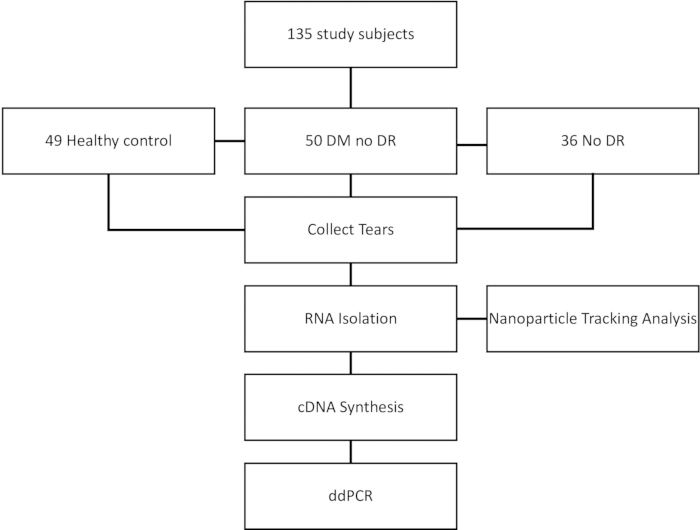
Figure 1: An overview of the protocol. Please click here to view a larger version of this figure.
1. Preparation of materials
- Lay out a sterile basic dressing set containing two forceps, six cotton balls, four gauze pads, a tray, and a bio-hazard bag.
- Pour 5 mL of 0.9% w/v sodium chloride irrigation solution into a tray.
- Open a pack of eyelid cleansing wipes and place it on the sterile field.
- Prepare a tube containing 250 µL of preservative solution.
- Fold a Schirmer strip at the wedge level while still in its packaging before placing the sterile strip onto the sterile field.
- Put on surgical gloves in an aseptic manner.
- Using a pair of sterile forceps, moisten the cotton balls with 0.9% w/v sterile sodium chloride irrigation solution.
2. Preparation of human subjects
- Allow the subject to sit comfortably and ask to close his/her eyes.
- Wipe the periorbital skin four times using cotton balls soaked in an irrigation solution.
- Wipe the periorbital skin with an eyelid cleansing wipe.
3. Collection of human tear fluid
- Collect tear fluids using the Schirmers strip placed at the temporal inferior fornix.
- When placing the partially folded strip onto the right eye, request the subject to gaze to the left. Pull down the lower lid to expose the inferior fornix.
- Using a pair of sterile forceps, place a Schirmer strip on the outer two-thirds of the lower conjunctival fornix. Avoid letting the strip from touching the skin to prevent contamination of the sample with sweat or other materials on the skin.
- Place a gauze pad underneath the strip to prevent skin contact.
- Allow tears to be absorbed into the strip until the 20 mm mark or a maximum of 5 min, whichever comes first.
- If the 20 mm level mark is not reached, document the level of tears based on the markings to allow standardization of tear fluid volume.
- Using a pair of sterile forceps, carefully remove the Schirmer strip and place the strip into a tube containing 1x phosphate buffer saline (PBS) solution.
- Place the tube into a cooler box for transportation to the laboratory.
- Clean the periorbital surface by wiping away any residual solution on the skin.
4. Pre-processing of tear samples
- Vortex the tube containing the Schirmer strip soaked in PBS for 5 min at room temperature.
- Agitate the tube on a rocker for 5 min at room temperature.
- Centrifuge the tube at 2000 x g for 15 min at 4 °C.
- Transfer the solution into a new tube, label and keep it at -80 °C until downstream processing.
- Discard the remaining strip.
5. Exosome isolation
NOTE: RNA isolation is performed using a commercially available kit following the manufacturer protocol (see Table of Materials).
- Thaw the tear solution on ice for 10 min.
- Equilibrate the spin column for 15 min at room temperature before use.
- Remove the lower outlet of the column and place it onto the waste collection plate provided.
- Remove the top sealing mat and allow the storage buffer to pass through the column through gravitational force.
- Add 250 µL of PBS and allow it to pass through the column.
- Repeat step 5.5.
- Apply a 110 µL sample to the top of each column and allow the sample to enter the column.
- Transfer the column onto a provided sample collection plate.
- Add 100 µL of PBS and allow it to pass through the column into the sample collection plate.
- Transfer the column onto the waste collection plate.
- Add 4x 200 µL PBS to the column and allow it to enter the column to remove the free proteins fraction from the first loading.
- Add 110 µL of sample into the column. Allow to enter the column.
- Place the column onto a sample collection plate. Add 100 µL of PBS and allow it to pass through the column.
- Briefly centrifuge the sample collection plate at 100 x g for 30 s. The isolated exosomes are now ready for RNA isolation.
6. RNA isolation
NOTE: RNA isolation is performed using a commercially available kit following the manufacturer protocol (see Table of Materials).
- Subject 200 µL of the isolated exosomes to RNA isolation.
- Add 60 µL of lysis buffer to the isolated exosomes. Vortex for 5 s.
- Incubate at room temperature for 3 min.
- Add 20 µL of inhibitor precipitation buffer, vortex for 20 s, and incubate at room temperature for 3 min.
- Centrifuge at 12,000 x g for 3 min at room temperature.
- Transfer the clear, colorless supernatant to a new tube.
- Add 1 volume of isopropanol.
- Vortex for 5 s and transfer the solution to the mini spin column provided in the kit.
- Centrifuge at room temperature for 15 s at 8000 x g. Discard the flow through.
- Wash the column with 700 µL of RWT wash buffer.
- Centrifuge for 15 s at 8000 x g. Discard the flowthrough.
- Wash the column with 500 µL of RPE wash buffer.
- Centrifuge for 15 s at 8000 x g. Discard the flowthrough.
- Add 500 µL of 80% ethanol to the sample mini spin column.
- Centrifuge for 2 min at 8000 x g. Discard the flowthrough and collection tube.
- Place the mini spin column into a new 2 mL collection tube.
- Centrifuge the column and tube at 18,000 x g for 5 min. Discard the flow through and the collection tube.
- Place the mini spin column into a new 1.5 mL tube and add 15 µL of RNase-free water at the center of the spin column membrane.
- Incubate for 3 min at room temperature.
- Centrifuge at 18,000 x g for 1 min to elute the RNA. RNA is ready for downstream processing.
- Perform Nanoparticle tracking analysis for quality control and verification of the RNA yield.
7. cDNA synthesis
- Perform cDNA synthesis using commercially available kits following the manufacturer protocols (see Table of Materials).
- Prepare the master mix according to Table 1 in a 1.5 mL microcentrifuge tube.
- Mix the solution by pipetting up and down.
- Pipette 5.1 µL of the master mix into 0.1 µL PCR tubes.
- Add 9.9 µL of sample to the allocated tube containing the master mix.
- Place the PCR tubes into a thermocycler and run according to Table 2.
NOTE: The cDNA is ready for further analysis.
| Component | Volume/ reaction |
| 10x RT Buffer | 1.5 µL |
| 100M dNTP mix | 0.15 µL |
| RNAse Inhibitor (200U/µL ) | 0.19 µL |
| Multiscribe RT Enzyme (50U/µl ) | 1 µl |
| 20x RT Primer | 0.75 µL |
| RNAse Free Water | 1.51 µL |
| Sample | 9.9 µL |
| Total Volume | 15 µL |
Table 1: Preparation of the PCR master mix used for cDNA synthesis from the isolated RNA.
| Step | Temperature | Duration |
| Reverse Transcription | 16 °C | 30 min |
| 42 °C | 30 min | |
| Stop | 85 °C | 5 min |
| Hold | 4 °C |
Table 2: Thermocycling conditions for the cDNA synthesis.
8. Digital droplet PCR
NOTE: Digital droplet PCR (ddPCR) is performed using commercially available kits following the manufacturer protocols (see Table of Materials).
- Prepare the master mix for ddPCR in a 1.5 mL microcentrifuge tube according to Table 3.
- Pipette 19.8 µL of master mix into 0.1 µL PCR tubes. Add 2.2 µL of samples and mix well by pipetting the mixture up and down for 10 times.
- Transfer 20 µL of the sample mixture into the sample lane of the ddPCR cartridge.
- Add 50 µL of oil to the oil lane of the cartridge .
- Attach a gasket onto the cartridge using the hook on the plate.
- Place the cartridge into the droplet generation machine. A click sound indicates a proper insertion of the cartridge into the droplet generation machine.
- Carefully transfer all of the generated droplets into wells of a 96-well plate. Add at least one well of no template control well as a negative control.
- Seal the plate using a pierceable foil in a plate sealer machine (see Table of Materials).
- Place the plate into a thermocycler and run according to Table 4.
- Transfer the plate into the droplet reader machine to count for positive and negative droplets.
- Quantify the copy number/mL using the copy number/20 µL value generated from the run.
| Component | Volume/ reaction |
| 2x ddPCR Supermix for Probes (No dUTP) | 10 µL |
| 20x Primers/ probe | 1 µL |
| Sample | 7 µL |
| RNAse Free Water | 2 µL |
| Total Volume | 20 µL |
Table 3: Preparation of the PCR master mix used for ddPCR.
| Step | Temperature | Duration | Ramp | Cycle |
| Enzyme Activation | 95 °C | 10 min | ~2 °C/ s for each steps | 1 |
| Denaturation | 94 °C | 30 s | 40 | |
| Annealing | 60 °C | 1 min | 40 | |
| Enzyme deactivation | 98 °C | 10 min | 1 | |
| Hold | 4 °C | N/A | N/A |
Table 4: Thermocycling conditions for the ddPCR.
Access restricted. Please log in or start a trial to view this content.
Results
Loading...
$$\rightleftharpoonup{xx}$$
$$\longleftharp{xx}$$,
$$\longrightharp{xx}$$,
Standard curve
A standard curve was created to determine the exact volume of tears collected from each patient. Basically, different volumes of preservative ranges between 3 µL to 25 µL were exposed to Schirmer strips. And the distance of the wet area on the Schirmer was observed. The procedure was repeated three times for each procedure, and the average reading was calculated. A standard curve of the volume of tears collected vs the distance of wet areas of Schirmer was generated (
Access restricted. Please log in or start a trial to view this content.
Discussion
Loading...
$$\rightleftharpoonup{xx}$$
$$\longleftharp{xx}$$,
$$\longrightharp{xx}$$,
Funduscopy examination, optical coherence tomography, and fluorescein angiography are the currently available detection techniques for diabetic retinopathy detection. The three techniques are often used together to provide a comprehensive assessment of various retinal and choroidal diseases. Funduscopy is a clinical examination routinely performed during diabetic retinopathy assessment. This method is carried out by trained ophthalmologists and is the gold standard for diagnosing diabetic retinopathy. This method enables...
Access restricted. Please log in or start a trial to view this content.
Disclosures
Loading...
$$\rightleftharpoonup{xx}$$
$$\longleftharp{xx}$$,
$$\longrightharp{xx}$$,
The authors declare that they have no competing interests.
Acknowledgements
Loading...
$$\rightleftharpoonup{xx}$$
$$\longleftharp{xx}$$,
$$\longrightharp{xx}$$,
This research was supported by Alcon Research Institute (IF011-2020) and FOM UMSC Care Postdoctoral Research Grant 2021.
Access restricted. Please log in or start a trial to view this content.
Materials
| Name | Company | Catalog Number | Comments |
|---|---|---|---|
| 0.9% w/v Sodium Chloride | Ain Medicare Sdn Bhd | GB16165967618 | |
| 1x Phosphate Buffer Saline | Thermo Fisher Scientific | 10010-023 | |
| Applied Biosystems TaqMan MicroRNA Reverse Transcription Kit | Applied Biosystems | 4366596 | |
| ddPCR Supermix for Probes (No dUTP) | BioRad Laboratories | 1863024 | |
| ddPCR 96-Well Plates | BioRad Laboratories | 12001925 | |
| DG8 Cartridges | BioRad Laboratories | 1864008 | |
| DG8 Gaskets | BioRad Laboratories | 1863009 | |
| Droplet Generation Oil for Probes | BioRad Laboratories | 1863005 | |
| Exospin-96 Exosome Isolation Kit | Cell Guidance Systems | EX07-96 | |
| miRNeasy Serum/Plasma Advanced Kit | Qiagen | 217204 | |
| PX1 PCR Plate Sealer | BioRad Laboratories | 1814000 | |
| QX100 droplet digital PCR System | BioRad Laboratories | QX100 | |
| QXDx Droplet Generator | BioRad Laboratories | 12001049 | |
| Schirmer Strip | Optitech | SCH-100 | |
| T100 Thermal Cycler | BioRad Laboratories | 1861096 | |
| Taqman MicroRNA Assay MTOMED 20 (Assay ID: 000389 ) | Applied Biosystems | 4440887 | Target Sequence: UAGCAGCACAUAAUGGUUUGUG |
References
Loading...
$$\rightleftharpoonup{xx}$$
$$\longleftharp{xx}$$,
$$\longrightharp{xx}$$,
- Teo, Z. L., et al. Global prevalence of diabetic retinopathy and projection of burden through 2045: Systematic review and meta-analysis. Ophthalmology. 128 (11), 1580-1591 (2021).
- Sayin, N., Kara, N., Pekel, G. Ocular complications of diabetes mellitus. World J Diabetes. 6 (1), 92-108 (2015).
- Wang, W., Lo, A. C. Y. Diabetic retinopathy: Pathophysiology and treatments. Int J Mol Sci. 19 (6), 1816(2018).
- Yang, Z., Tan, T. E., Shao, Y., Wong, T. Y., Li, X. Classification of diabetic retinopathy: Past, present and future. Front Endocrinol (Lausanne). 13, 1079217(2022).
- Goh, J. K. H., et al. Retinal imaging techniques for diabetic retinopathy screening. J Diabetes Sci Technol. 10 (2), 282-294 (2016).
- Parizadeh, S. M., et al. Circulating exosomes as potential biomarkers in cardiovascular disease. Curr Pharm Des. 24 (37), 4436-4444 (2018).
- Nair, S., Tang, K. D., Kenny, L., Punyadeera, C. Salivary exosomes as potential biomarkers in cancer. Oral Oncol. 84, 31-40 (2018).
- Dellar, E. R., Hill, C., Melling, G. E., Carter, D. R. F., Baena-Lopez, L. A. Unpacking extracellular vesicles: RNA cargo loading and function. J Extracell Biol. 1 (5), e40(2022).
- Fluitt, M. B., Kumari, N., Nunlee-Bland, G., Nekhai, S., Gambhir, K. K. miRNA-15a, miRNA-15b, and miRNA-499 are reduced in erythrocytes of pre-diabetic African-American adults. Jacobs J Diabetes Endocrinol. 2 (1), 014(2016).
- Belgardt, B. F., et al. The microRNA-200 family regulates pancreatic beta cell survival in type 2 diabetes. Nat Med. 21 (6), 619-627 (2015).
- Sun, L. L., et al. MicroRNA-15a positively regulates insulin synthesis by inhibiting uncoupling protein-2 expression. Diabetes Res Clin Pract. 91 (1), 94-100 (2011).
- Kamalden, T. A., et al. Exosomal microRNA-15a transfer from the pancreas augments diabetic complications by inducing oxidative stress. Antioxid Redox Signal. 27 (13), 913-930 (2017).
- Posa, A., et al. Schirmer strip vs. capillary tube method: Non-invasive methods of obtaining proteins from tear fluid. Ann Anat. 195 (2), 137-142 (2013).
- Ohashi, Y., Dogru, M., Tsubota, K. Laboratory findings in tear fluid analysis. Clin Chim Acta. 369 (1), 17-28 (2006).
- Nakagawa, A., Nakajima, T., Azuma, M. Tear miRNA expression analysis reveals miR-203 as a potential regulator of corneal epithelial cells. BMC Ophthalmol. 21 (1), 377(2021).
- Chan, H. W., et al. A pilot study on microRNA profile in tear fluid to predict response to anti-VEGF treatments for diabetic macular edema. J Clin Med. 9 (9), 2920(2020).
- Barmada, A., Shippy, S. A. Tear analysis as the next routine body fluid test. Eye (Lond). 34 (10), 1731-1733 (2020).
- Tomečková, V., et al. Experimental analysis of tear fluid and its processing for the diagnosis of multiple sclerosis. Sensors (Basel). 23 (11), 5251(2023).
- Daily, A., et al. Development and validation of a short-term breast health measure as a supplement to screening mammography. Biomark Res. 10 (1), 76(2022).
- Quevedo-Martínez, J. U., et al. Pro-inflammatory cytokine profile is present in the serum of Mexican patients with different stages of diabetic retinopathy secondary to type 2 diabetes. BMJ Open Ophthalmol. 6 (1), e000717(2021).
- Sorkhabi, R., Ahoor, M. H., Ghorbani Haghjo, A., Tabei, E., Taheri, N. Assessment of tear inflammatory cytokines concentration in patients with diabetes with varying severity of involvement. Exp Eye Res. 224, 109233(2022).
- Adigal, S. S., et al. Human tear fluid analysis for clinical applications: Progress and prospects. Expert Rev Mol Diagn. 21 (8), 767-787 (2021).
- Mackay, D. D., Garza, P. S., Bruce, B. B., Newman, N. J., Biousse, V. The demise of direct ophthalmoscopy: A modern clinical challenge. Neurol Clin Pract. 5 (2), 150-157 (2015).
- Saeedi, P., et al. Global and regional diabetes prevalence estimates for 2019 and projections for 2030 and 2045: Results from the International Diabetes Federation Diabetes Atlas, 9th edition. Diabetes Res Clin Pract. 157, 107843(2019).
- Kong, L., et al. Significance of serum microRNAs in pre-diabetes and newly diagnosed type 2 diabetes: A clinical study. Acta Diabetol. 48, 61-69 (2011).
- Baldeón, R. L., et al. Decreased serum level of miR-146a as sign of chronic inflammation in type 2 diabetic patients. PLoS One. 9 (12), e115209(2014).
- Liang, Y. Z., et al. Identification of neuroendocrine stress response-related circulating microRNAs as biomarkers for type 2 diabetes mellitus and insulin resistance. Front Endocrinol (Lausanne). 9, 132(2018).
- Wan, S., et al. Increased serum miR-7 is a promising biomarker for type 2 diabetes mellitus and its microvascular complications. Diabetes Res Clin Pract. 130, 171-179 (2017).
- Santovito, D., et al. Plasma microRNA signature associated with retinopathy in patients with type 2 diabetes. Sci Rep. 11 (1), 4136(2021).
- Smit-McBride, Z., et al. Unique molecular signatures of microRNAs in ocular fluids and plasma in diabetic retinopathy. PLoS One. 15 (7), e0235541(2020).
- Sun, Y., Liu, W., Zuo, C. Regulatory role of miRNA-23a in diabetic retinopathy. Exp Ther Med. 22 (6), 1-9 (2021).
- Wong, W. K. M., et al. MicroRNA profiling from tears as a potential non-invasive method for early detection of diabetic retinopathy. Methods Mol Biol. 2678, 117-134 (2023).
- Amorim, M., et al. Putative biomarkers in tears for diabetic retinopathy diagnosis. Front Med (Lausanne). 9, 873483(2022).
- Arroyo, C. A. -D., et al. Diurnal variation on tear stability and correlation with tear cytokine concentration. Cont Lens Anterior Eye. 45 (6), 101705(2022).
- Patel, S., Mittal, R., Kumar, N., Galor, A. The environment and dry eye: Manifestations, mechanisms, and more. Front Toxicol. 5, 1173683(2023).
- Benito, M. J., et al. Intra- and inter-day variation of cytokines and chemokines in tears of healthy subjects. Exp Eye Res. 120, 43-49 (2014).
- Bachhuber, F., Huss, A., Senel, M., Tumani, H. Diagnostic biomarkers in tear fluid: From sampling to preanalytical processing. Sci Rep. 11 (1), 10064(2021).
- Shoji, J., Aso, H., Inada, N. Clinical usefulness of simultaneous measurement of the tear levels of CCL17, CCL24, and IL-16 for the biomarkers of allergic conjunctival disorders. Curr Eye Res. 42 (5), 677-684 (2017).
- Gijs, M., et al. Pre-analytical sample handling effects on tear fluid protein levels. Sci Rep. 13 (1), 1317(2023).
- Vandermeid, K. R., Su, S. P., Krenzer, K. L., Ward, K. W., Zhang, J. Z. A method to extract cytokines and matrix metalloproteinases from Schirmer strips and analyze using Luminex. Mol Vis. 17, 1056-1063 (2011).
- Benlloch-Navarro, S., et al. Lipid peroxidation is increased in tears from the elderly. Exp Eye Res. 115, 199-205 (2013).
- Li, K., Chen, Z., Duan, F., Liang, J., Wu, K. Quantification of tear proteins by SDS-PAGE with an internal standard protein: A new method with special reference to small volume tears. Graefes Arch Clin Exp Ophthalmol. 248 (6), 853-862 (2010).
- Grigor'eva, A. E., et al. Exosomes in tears of healthy individuals: Isolation, identification, and characterization. Biochem (Moscow) Suppl B Biomed Chem. 10 (2), 165-172 (2016).
- Hu, L., et al. Discovering the secret of diseases by incorporated tear exosomes analysis via rapid-isolation system: iTEARS. ACS Nano. 16 (8), 11720-11732 (2022).
- Ter-Ovanesyan, D., et al. Framework for rapid comparison of extracellular vesicle isolation methods. eLife. 10, e70725(2021).
- Guo, J., et al. Establishment of a simplified dichotomic size-exclusion chromatography for isolating extracellular vesicles toward clinical applications. J Extracell Vesicles. 10 (11), e12145(2021).
- Aqrawi, L. A., et al. Proteomic and histopathological characterisation of sicca subjects and primary Sjögren's syndrome patients reveals promising tear, saliva and extracellular vesicle disease biomarkers. Arthritis Res Ther. 21 (1), 181(2019).
- Aqrawi, L. A., et al. Identification of potential saliva and tear biomarkers in primary Sjögren's syndrome, utilizing the extraction of extracellular vesicles and proteomics analysis. Arthritis Res Ther. 19 (1), 14(2017).
- McKay, T. B., Hutcheon, A. E. K., Zieske, J. D., Ciolino, J. B. Extracellular vesicles secreted by corneal epithelial cells promote myofibroblast differentiation. Cells. 9 (5), 1080(2020).
- Atienzar-Aroca, S., et al. Oxidative stress in retinal pigment epithelium cells increases exosome secretion and promotes angiogenesis in endothelial cells. J Cell Mol Med. 20 (8), 1457-1466 (2016).
- Liu, J., et al. Roles of exosomes in ocular diseases. Int J Nanomedicine. 15, 10519-10538 (2020).
- Kenny, A., et al. Proteins and microRNAs are differentially expressed in tear fluid from patients with Alzheimer's disease. Sci Rep. 9 (1), 15437(2019).
- Wijesinghe, P., et al. MicroRNAs in tear fluids predict underlying molecular changes associated with Alzheimer's disease. Life Sci Alliance. 6 (6), e202201757(2023).
- Urbizu, A., Arnaldo, L., Beyer, K. Obtaining miRNA from saliva-Comparison of sampling and purification methods. Int J Mol Sci. 24 (3), 2386(2023).
- Cirillo, P. D. R., Margiotti, K., Mesoraca, A., Giorlandino, C. Quantification of circulating microRNAs by droplet digital PCR for cancer detection. BMC Res Notes. 13 (1), 351(2020).
- Sangalli, E., et al. Circulating microRNA-15a associates with retinal damage in patients with early stage type 2 diabetes. Front Endocrinol (Lausanne). 11, 254(2020).
Access restricted. Please log in or start a trial to view this content.
Reprints and Permissions
Request permission to reuse the text or figures of this JoVE article
Request Permission