The protocol provided here describes the detailed steps to perform drug efficacy studies in a bioluminescent model of Trypanosoma cruzi infection, with a focus on data acquisition, analysis, and interpretation. Troubleshooting and quality control procedures to minimize technical issues are also provided.
Method Article
Demystifying In Vivo Bioluminescence Imaging of a Chagas Disease Mouse Model for Drug Efficacy Studies
In This Article
Summary
Abstract
To control and decrease the public health impact of human protozoan diseases such as Chagas disease, leishmaniasis, and human African trypanosomiasis, expediting the development of new drugs and vaccines is necessary. However, this process is filled with difficulties such as highly complex parasite biology and disease pathogenesis and, as typical for neglected tropical diseases, comparatively limited funding for research and development. Thus, in vitro and in vivo study models that can sufficiently reproduce infection and disease key features while providing rational use of resources are essential for progressing research for these conditions. One example is the in vivo bioluminescence imaging (BLI) mouse model for Chagas disease, which provides highly sensitive detection of long wavelength light generated by Trypanosoma cruzi parasites expressing luciferase. Despite this technique becoming the standard approach for drug efficacy in vivo studies, research groups might still struggle to implement it due to a lack of proper practical training on equipment handling and application of quality control procedures, even when suitable BLI equipment is readily available. Considering this scenario, this protocol aims to guide from planning experiments to data acquisition and analysis, with details that facilitate the implementation of protocols in research groups with little or no experience with BLI, either for Chagas disease or for other infectious disease mouse models.
Introduction
Chagas disease is endemic in Latin America and affects approximately seven million people worldwide1. Annually, more than 50,000 deaths and economic losses of around 7 billion dollars result from the disabling nature of this disease2. Chagas disease is caused by the protozoan Trypanosoma cruzi, a heteroxenic hemoflagellate parasite capable of infecting mammals (wild and domestic) and triatomine vectors (Hemiptera, Reduviidae)3 in the Americas, where the vectorial transmission is established. Other important infection routes include blood transfusion, organ transplantation, oral (by the ingestion of food contaminated with an infected triatomine)4, and congenital transmission. Non-vectorial transmission routes have contributed to the spread of Chagas disease to non-endemic areas3,5.
Chagas disease manifests in two clinical phases. The acute phase is, in most cases, asymptomatic. Symptomatic infections are usually associated with non-specific signs such as fever, fatigue, myalgia, lymphadenopathy, splenomegaly, and hepatomegaly. The acute phase is also often associated with patent parasitemia and systemic circulation of parasites. Death may occur in up to 10% of the diagnosed cases, especially in those of oral infection6. The chronic phase is often characterized by a long period of absence of any symptoms. With time, approximately one-third of the patients infected decades earlier exhibit cardiac manifestations, usually accompanied by fibrosis and myocardial inflammation, and/or gastrointestinal disorders mostly related to the development of megaesophagus and/or megacolon syndromes3,5,6.
The etiological treatment of Chagas disease consists of only two drugs: benznidazole and nifurtimox. These antiparasitic agents have been available for over 50 years and have considerable toxicity and limited efficacy5,7,8. Consequently, there is a pressing need to develop new, safe, and more efficacious treatments for Chagas disease patients.
More sophisticated and accurate techniques make it possible now to obtain answers to old questions that allow advances in the search for new treatments for Chagas disease. In this sense, the scientific community greatly benefits from genetically modified parasites for in vivo studies on the course of infection and evaluation of drug efficacy9,10,11,12. A longitudinal assay based on the bioluminescence imaging (BLI) system allows efficacy evaluation during and after the treatment regimen, leading to the identification of compounds with trypanocidal activity10,13. The BLI method provides a direct measurement of parasite load, both in circulation or in tissues and organs, through the quantitation of light produced by the genetically modified T. cruzi CL Brener Luc::Neon lineage11, which constitutively expresses the red-shifted firefly luciferase12.
Nonetheless, nearly 10 years after the establishment of the Chagas disease BLI animal model and drug efficacy studies, only a few research groups dominate this technique. This fact is due to not only the reduced access to proper imaging equipment but also the lack of training and availability of structured, detailed protocols. This method presents several advantages over other approaches, which rely on the assessment of parasitemia by microscopy, serology, or organ/tissue infection evaluation by qPCR for parasite DNA detection, as it allows improved mice well-being and reduction of animal use by the possibility of generation of more robust and integrated data in vivo. Furthermore, this method is, arguably, more sensitive, as it enables ready detection of parasite foci in visceral organs after drug treatment10,12. Therefore, this protocol aims to guide research groups on Parasitology and other infectious diseases to establish this methodology in their laboratories by detailing the technical procedures. Here, we share the experience obtained through implementing of the Chagas disease BLI model in Brazil, the first of its kind in Latin America, as part of the drug discovery efforts coordinated by the Drugs for Neglected Diseases initiative (DNDi).
Protocol
All procedures described in this protocol were submitted, approved, and conducted according to the guidelines14 preconized by the Animal Ethics Committee of Instituto de Ciências Biomédicas at Universidade de São Paulo: protocol CEUA ICB/USP no 5787250522.
1. Solutions
NOTE: Consider the preconized administrated volume of 10 mL/kg (200 µL for a mouse 20 g weight)15,16. For instance, prepare a 15 mg/mL working solution to reach 150 mg/kg of animal dosing.
- Hydroxypropylmethylcellulose suspension vehicle (HPMC-SV)
NOTE: The reagents required are 0.5% (w/v) Hydroxypropylmethylcellulose (HPMC), 0.4% (v/v) Tween 80, and 0.5% (v/v) Benzyl alcohol.- For 200 mL of vehicle solution, weigh 1 g of HPMC and dissolve in 64 mL of hot ultrapure water. Stir for 2 min.
- Add 120 mL of ice-cold ultrapure water and stir for 1 h. Add 1 mL of benzyl alcohol.
- Then add 0.8 mL of Tween 80 using the reverse pipetting technique and keep stirring until the solution becomes transparent.
- Adjust the solution for the final volume of 200 mL. Store the HPMC-SV under refrigeration (4 °C) for a maximum of 3 months.
- Benznidazole
- Calculate the necessary amount of compound solution, as follows:
volume of compound solution = (number of doses in each day x number of days x number of mice to be treated x administered volume in each mouse) + 30% extra. - Weigh the necessary amount of benznidazole (BZ), considering the desired dose. For curative treatment, an oral dose of 100 mg/kg once daily for 10 days achieves a 100% cure in the CD chronic model17.
- Considering the desired final volume, calculate the proper amount to reach a concentration of 5% (v/v) DMSO and completely dissolve BZ in neat DMSO.
- Add the proper volume of the vehicle to a glass tube to reach a concentration of 95% (v/v) HPMC-SV. Transfer the BZ dissolved in DMSO to the glass tube containing HPMC-SV. Vigorously homogenize the suspension and store it at 4 °C.
E.g.: Final volume of the BZ formulation = 10 mL (step 1.2.1)
Amount of BZ = 100 mg (step 1.2.2)
5% DMSO = 0.5 mL (step 1.2.3) and 95% HPMC-SV = 9.5 mL (step 1.2.4) - Before each drug administration, the formulations are placed in an ultrasonic water bath for 5 min at 37 °C and vigorously homogenized before the animal dosing.
- Calculate the necessary amount of compound solution, as follows:
- Cyclophosphamide
- Dissolve the necessary amount of cyclophosphamide in ultrapure water to achieve a 12.5 mg/mL solution.
- Sterilize the solution by filtration and store at 4 °C.
- Giemsa stain
- Dissolve 0.6 mg of Giemsa powder reagent in 50 mL of methanol. Add 25 mL of glycerol and mix. Remove precipitates using filter paper and a funnel. Store the stock solution at room temperature (RT), protected from light.
- Prepare a working solution by diluting 10 mL of Giemsa solution in 90 mL of Mix-Phosphate Buffer (20.5 M Na2HPO4, 65.4 M KH2PO4 at pH 7.2).
2. Trypanosoma cruzi culture
- Working in a biological safety cabinet (BSC), cultivate 4 x 10⁶ epimastigotes/mL of T. cruzi CL Brener Luc::Neon11 in LIT media (68.44 mM NaCl, 5.36 mM KCl, 112.7 mM Na2HPO4, 5 g/L tryptone, 5 g/L liver infusion broth, 0.03 M hemin, 4.16 mM glucose), supplemented with 10% (v/v) Fetal Bovine Serum (FBS), 100 µg/mL penicillin, 100 U/mL streptomycin and 150 µg/mL hygromycin as selective drug at 28 °C. The parasites typically achieve a stationary phase in 3-4 days.
- Induce metacyclogenesis to obtain infectious parasites by incubating 3 x 10⁸ epimastigotes in 10 mL of Grace's Insect medium with 10% (v/v) FBS in 15 mL conical tubes at 28 ˚C for 7-10 days.
- Evaluate the differentiation rate by Giemsa-staining smears. For that, spread 20 µL of the differentiated parasites in a glass slide and let dry for 10 min.
- In a fume hood, proceed with the sample fixation by covering the glass slide with neat methanol and let dry completely (1-2 min). Cover the glass slide with Giemsa working solution for 20 min.
- Gently wash the glass slide with distilled water. In a microscope, count the percentage of metacyclic trypomastigotes. If a differentiation rate of at least 10 percent is achieved, proceed to the next step.
- Centrifuge the parasites (120 x g for 10 min) and resuspend the pellet with 10 mL of DPBS. Centrifuge once more and resuspend the parasites in 5 mL of DMEM supplemented with 10% (v/v) of FBS, 100 µg/mL penicillin, and 100 U/mL streptomycin.
- In a 25 cm2 culture flask, perform the infection of 1.66 x 10⁵ LLC-MK2 cells (kidney epithelial cells from Macaca mulatta) at 37 °C in 5% CO2, in a humidified incubator18.
- Tissue-culture trypomastigotes (TCTs) are released in the medium after 8-9 days. Infect fresh LLC-MK2 cells monolayers with TCTs at a multiplicity of infection (MOI) of 1:40.
3. Analysis of Trypanosoma cruzi population homogeneity by flow cytometer
- Collect TCTs of CL Brener Luc::Neon and CL Brener wild-type strains, centrifuge at 120 x g for 10 min, and resuspend the TCTs in DPBS. Adjust the parasite density to achieve 1 x 10⁶/mL.
- Proceed with population analysis by flow cytometer based on mNeonGreen fluorescence (ex. 506/em. 517 nm)19, acquiring at least 20,000 events for each sample.
NOTE: The fluorescent population percentage is obtained by comparing the wild type against the transfected parasite. To proceed with the mice infection, a minimum of 95% of the fluorescent parasite population of the CL Brener Luc::Neon lineage should be achieved (Supplementary Figure 1).
4. Mice experimental infection
NOTE: To increase the amount of parasites, bloodstream trypomastigotes (BT) are frequently obtained from immunodeficient mice10,13. Here, chemical immunosuppression of BALB/C mice is used to obtain BT. For that, mild immunosuppression is achieved with four intraperitoneal injections of cyclophosphamide (CTX) at 62.5 mg/kg with 96 h intervals, which is performed concomitantly to T. cruzi infection.
- Administer 62.5 mg/kg of sterile cyclophosphamide by intraperitoneal injection (i.p.) 1 day before the T. cruzi infection.
- Infect each mouse with 1 x 10⁴ TCTs in 0.2 mL of DPBS through the intraperitoneal route. Attention to the high risk of incidents when handling pathogens and needles. Perform the infection in a BSC, wearing gloves and a face shield during the entire procedure.
- Monitor the parasitemia daily by direct observation of the BT in the blood (Pizzi-Brener method)20. At the parasitemia peak, around 13-17 days post-infection (dpi), conduct the blood collection. For that, anesthetize mice with 100 mg/kg ketamine and 10 mg/kg xylazine.
- When mice are completely anesthetized21, proceed with the cardiac puncture using a 1 mL syringe attached to a 24 G X¾ needle containing 50 µL of 3.8% (w/v) sodium citrate. Unplug the needle and gently place the blood in a centrifuge tube.
- In a BSC, perform a dilution of an aliquot of blood in ammonium-chloride-potassium (ACK) lysis buffer and count the parasites in the Neubauer chamber. Repeat this procedure at least twice, as small blood clots influence parasite counting.
NOTE: To prevent counting errors, adjust the dilution factor to avoid counts lower than 30 and higher than 300 trypomastigotes in the Neubauer chamber. - Adjust the parasite density to 5 x 10³ BT/mL by mixing the necessary amount of pure blood in DPBS.
- Infect non-immunosuppressed BALB/c mice with 1 x 10³ BT in 0.2 mL of DPBS per mouse (5 x 10³ BT/mL) by intraperitoneal route using a 26 G needle (narrower needles lead to parasite lysis)22. After injecting the volume intraperitoneally, hold the needle inside the mouse for 5 s to avoid backflow.
- Place the infected mice temporarily in a box with a paper towel to identify potential leaking or bleeding and then return mice to their cages.
5. In vivo imaging
NOTE: Glossary of the terms used here are as follows:
Imaging session: Bioluminescence acquisition performed in all groups of a certain experiment on a given day. Figure 1A shows an overview of the procedure.
Imaging round: Procedure performed in subgroups of three mice, from D-luciferin injection to anesthesia recovery. It is recommended that each experimental group contain 6 mice, due to the mice´s intrinsic variability of parasite load and other factors that influence BLI quantification described below.
Imaging acquisition: Photoshooting performed by the imaging equipment to quantify bioluminescence, which results in an image overlay of a photograph and the quantified bioluminescence shown as a pseudocolor scale.
Imaging procedure: All steps discussed in this session of the protocol.
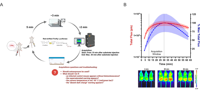
Figure 1: Acquiring BLI data. (A) Diagram of acquisition workflow applied for infectious diseases studies, using the Chagas disease bioluminescent mouse model as an example. Mice infected with genetically modified T. cruzi are analyzed by in vivo imaging on the established time points. On each imaging round, groups of up to three mice are injected with 150 mg/kg of enzyme-substrate (D-luciferin). After 5 min, anesthesia is administered at 2.5% (v/v) isoflurane in oxygen. When completely motionless, mice are placed in an imaging system, and acquisition is started following the defined settings. After imaging, mice recover from anesthesia and are returned to cages. Frequent questions and issues that the researchers might have and face during acquisition are highlighted in red. This scheme has been modified from Lewis et al. (2014)12 (created with BioRender.com: UD26KWEVS2). (B) In vivo kinetic of D-luciferin/red-shifted firefly luciferase (PpyRE9h). Mice at the T. cruzi parasitemia peak (n = 3) were anesthetized and injected with 150 mg/kg D-luciferin. Images were acquired for 1 h (exposure time: 2 min; binning: 4). Top: ventral total flux (p/s) quantification (left Y-axis, in red) and as percentage of highest mean measurement (right Y-axis, in blue). Data are shown as mean (curves) and standard deviation (shaded area). Bottom: Acquired images of the first (4 min), highest BLI signal (30 min), and last (62 min) time points. Please click here to view a larger version of this figure.
- Check the green bar in the Acquisition window to verify if the charge-coupled device (CCD) camera is cold (-90 °C). Click on the bar (green or red) to visualize the temperature.
NOTE: According to the imaging equipment manual, the imaging equipment and software should be turned on all the time to keep the CCD cold. - Verify if the auto background adjustment was performed. To do so, click on Acquisition > Background > View available Dark Charge. A list with several setting combinations will appear.
NOTE: This procedure reduces luminescence noise. If the chosen configuration results in a value higher than 1000, based on the formula "exposure time x (binning²)", then dark charge should not be ignored. Depending on the software configurations, binning can be shown as a number (binning factor) or a range from ‘small’ to ‘large’. The correspondence between the range and the binning factor can be found in the device manual. - If the Activity Window (white area at the bottom of the software) is not shown, enable it on Menu View > Activity Window.
- Set the imaging area by clicking on Acquisition Window > Field of View. Option C (13.2 cm) acquires 3 mice, and Option D (22 cm) acquires 5 mice.
- Select the autosave option by clicking on Acquisition > Auto-Save and select or create a new folder for each time point.
- Lay a matte black paper on the imaging area and place the partitioning slides perpendicularly between the inlets. If the manufacturer did not provide the slides, use pieces (3 cm x 15 cm) of matte black paper.
- Adjust the internal area of the imaging chamber to accommodate the glass nose cones to the anesthesia inlets. Use small pieces of black electrical tape to attach the anesthesia system to the platform (outside the image area).
NOTE: In some versions of imaging devices, a laser defines the imaging area. If this tool is unavailable, acquire images through the software to check the partitioning slides within the imaging area. - Set the isoflurane vaporizer to 2.5% (v/v) in oxygen for the imaging and induction chambers. Check by the pressure gauge if the gas is flowing through the system21.
NOTE: If the imaging device does not have an inhalation anesthesia system attached to it, other anesthesia options are possible, such as injectable xylazine and ketamine23. In this case, mice recovery is slow, and the risk of death is high24. - Fill syringes (31 G needles) with D-luciferin (a comprehensive protocol to prepare the D-luciferin is provided by different suppliers)25,26, protect them from direct light, and prepare a few plastic containers or extra cages by covering the interior with a paper towel.
- Keep a clock/watch nearby to easily track time and a lab notebook to register mice groups, luciferin injection time, and any other information during the procedure.
- Weigh all mice and register their weight. Mark each mouse with a pen marker or tattoo on the tail. Keep mice identification well displayed during the entire experiment. This step can also be performed on the day prior to the acquisition.
NOTE: The mark should not occupy a large body area because the ink may interfere with the bioluminescent signal. Permanent markers of different colors can also help to differentiate the groups. - To start the Imaging session, register a background luminescent image (settings: Exposure Time: 5 min; Binning: 'large' or '16', f/stop: 1) of the imaging chamber without mice. This procedure helps to identify auto luminescent light sources or undesired issues on the equipment.
NOTE: Because BLI is based on enzymatic reaction, time is an essential feature of the experiment. Check all the items listed above and equipment settings before starting animal handling. - Inject 150 mg/kg (i.p.) of D-luciferin in the first 3 mice without reaching internal organs. Place the mice in the container box to assess whether D-luciferin leaks (bright yellow solution). Take notes if this happens and register the mice group and D-luciferin injection time.
- Wait 5 min then transfer the mice to the anesthesia induction chamber. Turn on the system. It takes around 3 min for mice to be fully anesthetized (absence of responses like shaking legs, tail, etc.)21.
- Open the imaging device's door, turn on the anesthetic flow to the imaging chamber, and, at the same time, turn off the induction chamber.
- Place each mouse in the nose cones position: from mouse 1-3, from left to right side. Gently accommodate the mice with ventral side up inside the imaging area shown by the laser. During the accommodation, verify the mice's ID and position. Do not force the entrance of mice partially anesthetized in nose cones.
- Place the partitioning slides between mice and close the imaging equipment's door.
- Adjust the acquisition settings on the software Acquisition window, as follows:
- For non-infected mice, Exposure Time = 5 min / Binning = 'large' or '16'
(most mathematically sensitive set of configurations to set the threshold values). - For infected mice in the acute model: Exposure Time = 2 min/ Binning = 'medium' or '4'.
- For infected mice in the chronic model: Exposure Time = 5 min/ Binning = 'large' or '16'.
- For non-infected mice, Exposure Time = 5 min / Binning = 'large' or '16'
- Verify if the D-luciferin injection time was performed at least 10 min before starting the acquisition. If not, wait the necessary period to obtain the images during the peak of the luciferase signal (Figure 1B); otherwise, click on Acquire to begin the imaging acquisition.
- The Image Labels window will appear after the imaging starts. Fill the boxes with the appropriate information about each group, including luciferin injection time, experiment, mice identification, time point, and any other desired information that can help in keeping record. This information will be included in the measurements table file.
- Verify whether the warning sign Saturated Image is shown at the end of the imaging acquisition. Saturated images are not acceptable. If this happens, decrease the binning and acquire a new image. Take notes to consider in the analysis.
- Once the image is acquired, open the imaging machine door and turn the mice to the dorsal view (back side up). Perform the acquisition again and label it appropriately.
- At the end of this imaging acquisition, turn off the anesthetic flow. Gently remove the mice from the imaging chamber and place them in a container.
- Record the mice's body weight while observing the anesthesia recovery. Record any abnormal behavior or body abnormalities found during the procedure. Once the mice return to moving, place them in their cages.
NOTE: Verify the BLI intensity for each acquired image. After the acquisition ends, the recorded bioluminescent image and the photograph will be displayed on the software with an automatic scale that should not be considered. Set the scale defined in data analysis (described below) on Tool Palette window.
6. Data analysis
NOTE: The above protocol is based on commercial in vivo imaging software. However, a software license-free version can perform the most basic analysis. Software details can be found in the Table of Materials.
- Open files.
- Open the folder that contains the acquired data from a certain imaging time point by selecting Menu File > Browser > Select the folder (Figure 2A - Step 1).
NOTE: A new window will open in a table format with all acquired data saved in the chosen file. It will display the Click Number sign, which is the image ID automatically given by the software (data and hour numbers), the information provided by the researcher in the Image Labels window during the acquisition, and acquisition settings. All these data help sort the images that can be used in the analysis, given that saturated images must not be used. - Select multiple rows related to the chosen images, including the non-infected controls. Write down the image ID in the lab notebook along with the information registered during the imaging session.
- Assemble the selected images by clicking on Load as Group. This procedure creates a new sequence image. Therefore, it is possible to adjust features for all images simultaneously. Save the new sequence image in a new folder different from the one used in step 6.1 to facilitate the reanalysis of raw data if needed.
- In the sequence image, identify the binning used for each image by clicking on Options > Display > Select Binning Factor (Figure 2A - Step 2). The acquired binning factor will be displayed at the top of each image in the sequence.
- Correct all binning factors to the same number. To do so, double-click on the image to be adjusted. On the Tool Palette window > Image Adjust section, choose the appropriate binning factor (Figure 2A - Step 3) - see step 5.18 for binning factor choice. Perform this procedure in all divergent images.
NOTE: This procedure directly influences BLI quantification (Table 1) and is further addressed in the Discussion section.
- Open the folder that contains the acquired data from a certain imaging time point by selecting Menu File > Browser > Select the folder (Figure 2A - Step 1).
- Set scale.
- Distinguish valid bioluminescent signal (above 600 counts according to the device manual) based on the non-infected control. For that, double-click on the image of the non-infected mice. Then, in the upper-left corner of the new window, select the option Counts in the Units field. Then, adjust the Color Scale for the minimum value of 600. The resulting image will be the baseline to set the minimum scale in radiance.
- With the sequence window active, select the option Radiance in the Units field. Adjust the Color Scale and Color Table in the Tool Palette. For that, disable the box Individual in the Color Scale Limits area.
- Mark the boxes Logarithmic Scale and Manual (Figure 2A - Step 4). Set the minimum scale numbers according to the non-infected control (as observed in step 6.8) and the area with the highest signal as the maximum (as shown in Figure 2A by the arrows).
NOTE: In vivo 2D light mesurament is a relative quantification. Different scale values can be found due to differences in each device’s version and calibration. Therefore, the values demonstrated in the representative results could not fit other experiments performed on other devices and yet be equally valid according to the internal controls (non-infected and infected not treated). - After setting the same scale for all images, double-click on each image, maximize the window, and export the image view in an image format (.jpg, .tiff, etc.) by using the Export Graphics button, naming the files by treatment_miceID_time point. (Figure 2A - Step 5). Perform this procedure for all images.
- Perform measurements.
- Choose any image from the sequence and double-click on it. On the Tool Palette window > ROI tools section, click on the Square button (Figure 2B- Step 1) and draw a rectangle covering the whole mouse.
- Click on the border of the created ROI and copy and paste the same ROI for each mouse (resulting in three ROIs in the image). Save the ROIs by clicking save on the ROI Tools Section. Apply the saved ROIs for all images by selecting the box Apply to Sequence, then click on Load. The saved ROIs will be used for all mice in the entire experiment.
- Adjust the ROI position in each mouse to fit the animal better in the measurement area. When all mice are labeled, click on the Measure ROIs button (Figure 2B- Step 2). The ROI Measurements Table will appear. Select Measurement Types: Radiance; Image Attributes: All Possible Values; ROI Dimensions: cm. Then, click on Select All button, followed by Copy button.
- Paste the data directly into a table analysis software (spreadsheet). Alternatively, export a file in .csv or .txt format.
- Work on the data.
- In table analysis software, organize the data by groups (treatment, control non-infected, non-treated, etc.).
- Parasite load is shown as whole-body bioluminescence. Therefore, arrange the data to match the same mice's ventral and dorsal Total Flux values and sum them.
- Calculate the mean and standard deviation of the summed values considering each group (e.g.: Supplementary Table 1). Plot the data into graphing and statistics software.
- Prepare a panel with bioluminescent images in an image or slide presentation software. Place each mouse in a column and each time point in the row to build a drug efficacy matrix to facilitate visualization and interpretation of the data.
NOTE: There is an option to build the images directly into imaging software without performing step 6.2.4 (View > Image Layout Window). However, the software interface and tools limit data organization.

Figure 2: Data analysis steps from setting image scales to quantifying luminescence. (A) Living Image Software view for image data processing. Step 1: Upload the acquired data as a sequence using the Browser tool. Step 2: Identify the binning used for each acquisition (will appear on top of the individual images). Step 3: Set all images of infected mice to the same binning factor and apply binning factor 16 for the non-infected mice. Step 4: Set the color scale manually based on non-infected mice (purple arrow), which should be seen on the screen as non-bioluminescent, and infected and non-treated mice at the highest bioluminescent foci (red arrow), which should be seen as red. Step 5: To obtain an image free of any written information, double-click on each image and export the image using the Export Graphics button. (B) View of Regions of Interest (ROI) Tools. Step 1: Draw an ROI covering entirely a mouse and copy and paste the same ROI for each animal. Save the adjusted ROIs and apply them to all images in the experiment. Step 2: Click on the Measure ROIs button to generate the table to export as .csv or .txt. Please click here to view a larger version of this figure.
7. Ex vivo procedure
- Inject 150 mg/kg D-luciferin i.p. in the mouse (one per ex vivo round) and wait for 3 min. Anesthetize the mice using 100 mg/kg ketamine and 10 mg/kg xylazine i.p.
- In a biological safety cabinet, when the mouse is completely unresponsive to pinching of the paws, cut the mouse skin to expose the peritoneum (without opening the peritoneum cavity) and the tissue of the left-side axillary. Proceed with the exsanguination by cutting the axillary artery and vessel.
- Collect the blood using a disposable Pasteur Pipette. Then perfuse 10 mL of 0.3 mg/mL of D-luciferin in DPBS solution through the heart´s left ventricle.
- Lay out the selected organs on pre-established spots on a Petri dish and soak them in 0.3 mg/mL d-luciferin solution. This procedure must be performed within 30 min maximum.
- Acquire the luminescent image of the excised organs (heart, lungs, thymus, liver, quadriceps muscle, mesentery, spleen, kidney, adrenal gland, visceral fat, esophagus, stomach, small intestine, cecum, colon, brain, subcutaneous fat, uterus, gonadal fat, bladder, and peritoneum membrane) using 5 min of exposure time and binning 16, depending on signal intensity (saturated images are not acceptable)22.
NOTE: The same procedure must be performed in a non-infected mouse prior to the infected ones to set the threshold.
Results
Using an adequate mouse model based on transgenic parasites that constitutively express luciferase, it is possible to reproduce key human T. cruzi infection features causing minimum harm to the host, allowing real-time tracking of the parasites by whole-body BLI of the host in a longitudinal (lifelong) manner, for up to several months12.
Figure 3 shows a pilot experiment demonstrating the CL Brener Luc::Neon infection time-course from day 1 post-infection until 150 dpi in BALB/c mice. In the first 20 dpi, bioluminescence-inferred parasite burden increases, typical of parasitemia peak, followed by a sharp decrease in parasite load, due to mice immune control. Thus, the acute phase of the infection is considered the first 50 dpi. The chronic phase is defined when infection reaches a constant bioluminescent rate, from 100-150 dpi.
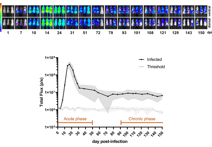
Figure 3: Understanding the model. Kinetics of Trypanosoma cruzi CL Brener Luc::Neon infection in BALB/c mice (n = 11). Top panel: ventral and dorsal bioluminescent images from different time points for 150 days post-infection (dpi) with 1 x 10³ bloodstream trypomastigotes by intraperitoneal injection. Heatmap scale (in Log10) of bioluminescent signal in radiance (p/s/cm2/sr). Color code: Purple = 5 x 10³; red = 1 x 10⁷. Bottom panel: bioluminescent signal quantification in Total Flux (p/s) of whole-body mice. Data are expressed as means (lines) and standard deviations (shaded regions). Periods defined for acute and chronic phases of infection in this mouse model are highlighted in orange. Luminescence mean values of non-infected mice (n = 3) are shown as the threshold (light gray line). Please click here to view a larger version of this figure.
The therapeutic outcomes can also be predicted since the main phases of the infection are well-reproduced in a mouse model. Thus, BLI is now applied to translational science through proof-of-concept studies that support decision-making about compound efficacy potential and progression in the development pipeline10,27. In the acute mouse model, the oral treatment should start when the infection reaches the parasitemia peak (around 14-21 dpi), characterized by a high number of trypomastigotes in the bloodstream (approximately 1 x 10⁵ trypomastigotes/mL - data not shown) and systemic infection. In the chronic phase, mice treatment starts at 100 dpi, when the parasite load is comparatively much lower, with stable subpatent parasitemia.
To demonstrate the application of the protocol described above in the evaluation of antiparasitic agents, we compared the treatment efficacy of benznidazole (BZ), the standard drug used for treatment of Chagas disease, and posaconazole (Posa), a sterol 14α-demethylase (CYP51) inhibitor that failed in clinical trials for Chagas disease, and that has also been shown to be inefficacious as antiparasitic agent against T. cruzi in vitro and in vivo, including in the BLI model described in this protocol13,18,28,29,30.
For the acute mouse model, cohorts of 6 mice per group were treated orally by gavage31 for 20 days with Posa at 20 mg/kg or BZ at 100 mg/kg, once daily. Mice were also treated with BZ for 5 days at 100 mg/kg once daily to evaluate the effect of short treatments. After compound elimination (10 days after the treatment ended), the BLI-negative mice were immunosuppressed, a condition that promotes infection relapse when a parasitological cure is not achieved (Figure 4A). Posa treatment resulted in a 99.49% ± 0.27% (mean ± standard deviation) reduction in bioluminescence-inferred parasite burden at the end of the treatment and remained at similar levels of non-infected mice during the drug compound elimination period (except for mouse #3, with a transient signal at 40 dpi, and mouse #2, that demonstrated a weak BLI spot at 44 dpi). Compared to the non-treated group, BZ treatment for 20 days reduced 100% ± 0.01% the bioluminescence-inferred parasite burden at the end of the treatment. In contrast, the short treatment with BZ for 5 days led to a 99.98% ± 0.03% reduction at 19 dpi (similar to the threshold level). However, in this instance, the BLI reduction was transient and in the subsequent acquisitions, all mice presented infection reactivation (Figure 4B).

Figure 4: Proof-of-concept experiment design and results obtained with bioluminescence imaging in Chagas disease acute model. (A) Schematic chart of timeline process in acute Chagas disease mouse model for preclinical studies for assessment of compound efficacy. Black dots: imaging time points. Medicine flask and orange bar: treatment start and end, respectively. Syringe icon: cyclophosphamide injections. dpi: day post-infection. Color code: gray = non-treated infection; orange = treatment period; blue = compound elimination phase; yellow = immunosuppression period. (B) Time-course of treatment of Trypanosoma cruzi. Left panel: ventral bioluminescence images of BALB/c mice (i) non-treated or treated for 20 days with (ii) posaconazole (Posa) at 20 mg/kg, (iii) benznidazole (BZ) at 100 mg/kg treated for 20 days and (iv) BZ at 100 mg/kg treated for 5 days with (n = 6/group). All treatments were orally administered by gavage (10 mL/kg) once a day. Log10 scale heatmap indicates the bioluminescent signal intensity from low level (blue) to high (red). Right graph: whole-body quantification of bioluminescence imaging data. Data are expressed as means (lines) and standard deviations (shaded regions). Key: Medicine flask: treatment start (14 dpi); orange bar: treatment end (18 dpi/33 dpi); syringe icon cyclophosphamide injections (from day 44-53); orange dot: mouse analyzed by ex vivo procedure; § data excluded due to D-luciferin leakage in urine. (C) Ex vivo procedure for T. cruzi foci detection in organs and tissues. Plate distribution key is presented at the bottom of the figure (created with BioRender.com: ZY26LG8AOF). Same radiance scale as shown in vivo panel. Please click here to view a larger version of this figure.
In this model, antiparasitic treatment may reduce parasite load below the BLI detection limit of 1000 parasites. Thus, mice appear as BLI negative10. To ensure that a parasitological cure has been achieved, it is necessary to promote immunosuppression of mice using cyclophosphamide treatment29,32. Mice that still have an undetectable parasite load after drug treatment will become BLI-positive after immunosuppression. This effect is shown more extensively by Posa treatment in the acute model, which promotes parasite relapse. In this experiment, the ex vivo procedure was performed on mice with the lowest bioluminescent signal to evaluate parasite tissue tropism (Figure 4C). Non-treated mice shows strong bioluminescent signals, especially in the gastrointestinal tract (TGI) and associated tissues such as visceral fat and mesentery. In addition, the skin was a site associated with parasite persistence. Mice treated with Posa presented lower-intensity bioluminescent spots that were more restricted to the colon, mesentery, and visceral fat. On the other hand, in mice treated with a curative treatment regimen, as BZ 100 mg/kg for 20 days, no bioluminescent signal is detected even under immunosuppression. Therefore, considering that after promoting conditions for bioluminescent signal rising above the threshold level of 1000 parasites10, and increasing sensitivity by exposing the visceral organs, it is considered that BZ 100 mg/kg for 20 days provides sterile cure. The assessment of sterile cure was previously validated by different techniques10,13,17,33,34.
A similar study design was applied to the chronic model (Figure 5A), in which mice (n = 6/group) started to be treated at day 100 post-infection. At this point, Posa 20 mg/kg, BZ at 100 mg/kg, or 10 mg/kg once daily were administered orally for 20 days. After the drug elimination period, the BLI-negative mice were immunosuppressed. In addition, to assess sterile cure, mice that were still BLI-negative at the end of the immunosuppression phase were subjected to ex vivo analysis.
In chronic infection, treatment with Posa at 20 mg/kg led to a decrease of 95.03% ± 6.18% in bioluminescence-inferred parasite burden at the end of the treatment and remained at similar levels of non-infected mice during the drug elimination period. After the immunosuppression, the majority of mice showed a variable level of BLI signal and distribution (Figure 5B). Ex vivo procedure revealed that one whole-body BLI-negative mice treated with Posa had a bioluminescent spot in the colon detectable via ex vivo examination (Figure 5C).
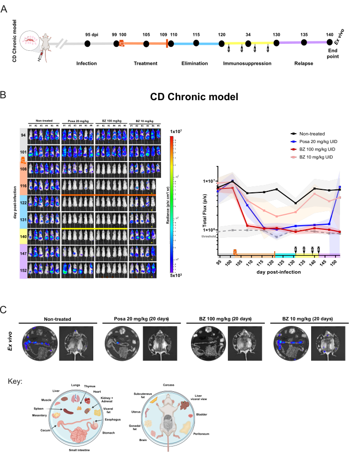
Figure 5: Drug assessment experiment design and results achieved in the chronic model of Trypanosoma cruzi infection by bioluminescence imaging. (A) Schematic chart of experiment design of chronic Chagas disease mouse model. Black dots: imaging time points. Medicine flask and orange bar: treatment start and end, respectively. Syringe icon: cyclophosphamide injections. Dpi: day post-infection. Color code: Gray = non-treated infection; orange = treatment period; blue = compound elimination phase; yellow = immunosuppression; lilac = relapse. (B) Longitudinal evaluation of treatment efficacy in the Chagas disease chronic model. Left panel: ventral BLI of BALB/c mice (i) non-treated or treated for 20 days with (ii) posaconazole (Posa) at 20 mg/kg; (iii) benznidazole (BZ) at 100 mg/kg, and (iv) BZ at 10 mg/kg (n = 6/group). All treatments were orally administered by gavage once a day. Right graph: sum of ventral and dorsal total flux raw data. Key: medicine flask: treatment start (102 dpi); orange bar: treatment end (121 dpi); yellow bar and syringe icon: cyclophosphamide injections (from day 131-142). Orange dot: mouse analyzed by ex vivo procedure. ! mouse died during anesthesia.  Mouse displays abdominal abnormalities. Heatmap Scale (Log10) indicates the bioluminescence intensity in radiance unit from low level (3 × 10⁵ as blue) to high (1 × 10⁷ as red). (C) Ex vivo analysis of T. cruzi tropism in tissue. Bioluminescence detection of excised organs from immunosuppressed in vivo BLI-negative mice after treatment with Posa and BZ at 100 mg/kg or mice with the lowest signal in non-treated and BZ at 10 mg/kg groups. Plate distribution key is shown at the bottom of the figure (created with BioRender.com: ZY26LG8AOF). Radiance scale is equal to in vivo panel. Please click here to view a larger version of this figure.
Treatment with BZ at 100 mg/kg reduced the bioluminescence to threshold levels (93.87% ± 2.14%) at 122 dpi and forward until the end of the experiment. Considering ex vivo analysis, BZ achieved a cure rate of 100% (6/6 mice), as no mice presented a return of bioluminescent signal even when immunosuppressed and internal organs were examined. That means a lack of relapse. Nonetheless, the BZ treatment regimen with a lower concentration resulted in a slight reduction of bioluminescence (85.95% ± 18.43%) but no cure, as mice continued to show detectable bioluminescent signal foci at all subsequent time points. Thus, this model allows quantitative differentiation between inefficacious treatments (posaconazole and suboptimal treatment with benznidazole) and efficacious treatments (optimal dosing and treatment length with benznidazole).
Supplementary Figure 1: Preinfection analysis of T. cruzi population expression of luciferase by measurement of the reporter gene mNeonGreen fluorescence. Flow cytometry of mNeonGreen fluorescent protein constitutively expressed by the CL Brener Luc::Neon parasite. Normalized histogram of parasite number (Y-axis) by mNeonGreen fluorescence intensity (X-axis). Internal legend demonstrates the analyzed strain and form, fluorescence median, and percentage of fluorescence population. Gate lines are defined by of CL Brener wild type (WT) strain as non-fluorescent control. Please click here to download this File.
Supplementary Table 1: Example of analysis table in BLI measurement. Raw data obtained from bioluminescence imaging quantification of day 19 post-infection in the acute model used in the representative results shown in Figure 4B. Groups description: Control as non-infected mice (n = 3/group); vehicle as non-treated infected mice; posaconazole (Posa) at 20 mg/kg for 20 days; benznidazole (BZ) at 100 mg/kg for 20 days; BZ at 100 mg/kg treated for 5 days (n = 6/group). Please click here to download this File.
Discussion
Bioluminescence imaging is a breakthrough method that allows the detection of a report gene using a visible and infrared spectrum of electromagnetic radiation. Therefore, there is no need for radiolabeled markers to trace your specimen35. BLI is suitable for rodent models and other small species. It is very useful for preclinical studies because it is safer and allows several image rounds, causing minimal animal discomfort. Besides, in vivo imaging is very flexible due to the possibility of combining bioluminescence, fluorescence, and other techniques such as positron emission tomography36.
Optical imaging is ruled by optical physical properties, like absorption and scattering. All tissues absorb and scatter light of distinguished wavelengths differently37. One critical step is selecting a reporter gene without taking into account the emitted wavelength of the light produced by the chemical reaction. While a reporter gene expression level may be high in vitro in bioluminescent assays, the same levels of expression may not be achieved when progressing to the in vivo setting. In this protocol, we used red-shifted Photinus pyralis luciferase (PpyRE9H)38 codon-optimized version for trypanosomatids9, which emits light at 617 nm, one of the most suitable for in vivo studies39. Wavelengths longer than 600 nm are less absorbed and scattered by body endogenous chromophores, especially hemoglobin and melanin. Thus, red lights can be transmitted through several centimeters of tissue, allowing the photons to reach the CCD camera even from within visceral tissues39,40.
One area of concern in imaging settings is the lack of a comprehensive understanding of their function and effects. Binning, a pre-processing technique, combines the information acquired by contiguous detectors into a larger pixel. This process enhances the signal-to-noise ratio, reducing background noise and improving sensitivity. However, it decreases spatial resolution accuracy, resulting in a pixelated image41,42. This trade-off is an important consideration in your imaging strategy.
Based on the Target Product Profile and Target Candidate Profile for Chagas disease34, the proof-of-concept study is focused on sensitivity to detect T. cruzi and help establish if a new drug candidate can achieve a sterile cure (represented by lack of relapse after several immunosuppression rounds). Therefore, we execute the BLI using the highest binning factor without supersaturating the image. When the image supersaturates, a new acquisition is performed using a lower binning factor. During the analysis, a mathematical correction is applied to the images that required different binning. This way, the final data should be presented using the same binning. Table 1 demonstrates the different values obtained when distinctive binning factors were applied in the same image and ROIs.
Table 1: Influence of binning settings on the BLI quantification. Quantification of three ROIs on the image of acute model (d13) and chronic model (d118), analyzed in different binning factors. Please click here to download this Table.
Due to the current scenario of Chagas disease in the clinic, drug discovery efforts aim to completely eliminate parasites (parasitological cure)27,34. Therefore, the in vivo preclinical protocol includes approaches that overcome the limitations of BLI technical sensibility. One of the approaches is treating the mice with cyclophosphamide to decrease the immune response that controls the parasite load. Another strategy is diminishing the tissue deepness and removing layers of muscle, skin, and fur that obstruct the light path to the camera. Through the ex vivo procedure, small bioluminescent spots can be detected, revealing parasite foci below the in vivo BLI threshold, as shown in Figure 5C in ex vivo result of mouse treated by Posa.
Designing a pilot experiment to evaluate the model itself and the infection dynamics is crucial to establishing an accurate experiment for antiparasitic drug efficacy assessment. Hence, the researcher will be able to define the proper BLI settings and infection time course in advance. In an exploratory experiment, one tool that can be helpful to define the acquisition settings is the 'Autoexposure'. With this tool, the researcher establishes the priority of three settings (Exposure time, Binning, and F/Stop) to acquire the best image possible. In particular, the researcher should ensure that images are acquired within the dynamic range of the CCD camera, without supersaturation or underexposure, which can be checked through the minimum and maximum limits of the scale and the autoexposure feature (Menu Edit > Preferences > Tab Acquisition > Tab Autoexposure). In this protocol, the camera aperture was set at the maximum value (F/Stop: 1), and different exposure times and binning factors were defined for acute and chronic models. These settings give time predictability to perform different image rounds simultaneously. Considering the reporter method is based on an enzymatic reaction, both the biodistribution of the substrate into the mouse and the luciferase kinetics influence the bioluminescent signal and, thus, infection quantification (Figure 1B). Consequently, acquiring images in different moments of enzymatic kinetics introduces data variability that cannot be accounted for or corrected and impacts the total flux (photons/second) or radiance (photons/second/cm2/steradian) calculation. Besides, T. cruzi infection displays dynamic spatial positioning in mice (different areas and tissues, deepness, and parasite load). Hence, establishing a value of counts to acquire could miss weaker signal sources (low number of parasites in a certain spot deeper in the tissue) if another stronger signal's source meets the defined auto-exposure criteria.
A tricky feature of Living Image software is displaying the acquired image in an automatic color scale. There is no option to pre-set the scale to exhibit automatically a brand-new acquired image according to the selected scale values (see protocol step 6.2). This situation forces the researcher to manually change the images one by one to the chosen max and min values. As a consequence, the non-experienced and not well-trained users do not have the proper readout during the acquisition session, and they could mislead the data or lose important information at that time point. For that, the pilot experiment is beneficial.
One of the most common questions about the proof-of-concept experiment design is how to choose the treatment duration and dose. For new chemical entities, these parameters are usually defined by the compound potency and selectivity in vitro, in combination with data generated by drug metabolism and pharmacokinetics (DMPK) and tolerability studies conducted prior to testing in vivo for efficacy. In summary, after identifying compounds that are able to selectively kill the parasite inside cells, the first ADME experiments (absorption, distribution, metabolism and excretion) are performed in vitro to estimate compounds' aqueous solubility, cell permeability and metabolic stability, among other parameters. If compounds show a good balance of in vitro properties (usually defined in target candidate profiles), then these candidates are progressed to in vivo pharmacokinetics (PK) studies in healthy mice, which outline the compound exposure in the blood (and possibly in tissues as well) and provide a general idea of the tolerability at different dose levels17,34,43. Ideally, the goal of the PK assessment in most infectious diseases is to determine the feasibility to reach free plasma concentrations (corrected for plasma protein binding) that are above the EC50/EC90 concentrations44 - the effective concentration that kills or at least inhibits the growth of 50% or 90 % of parasites, respectively - for a sufficiently long period of time. If enough exposure is achieved at a certain dose level, then this regimen can used during the efficacy studies using the Chagas BLI model. For drug repositioning studies, in vitro and in vivo PK data should be available. A good start for drug reprofiling is chemical databases such as PubChem45, which provide recognized data that can be converted to mice using allometric scaling46 to estimate safe and non-toxic treatment regimens to be tested. However, that is not always the case. PK studies are still an overlooked field in academic science, and few pharma companies publish their PK results. The drug discovery science community recommends including in vivo PK assessment along with in drug efficacy assays (pharmacodynamics)47. Therefore, preclinical imaging is compatible with compound measurements simultaneously, and this associated approach enhances data robustness.
In addition, the mice's handling, weight, and health conditions are monitored throughout the entire experiment. Signs of toxicity and side effects such as hunching, shaking, loss of balance, unwillingness to move, reluctance to feed or drink, prostration, or any other abnormalities present in the group or by individual mouse conditions should be registered and reported in preclinical studies. One of the goals of optical imaging is to ensure the animals' well-being. Thus, humane endpoints should be applied in mice with pain signs described in the 'Grimace scale48. Also, mice were weighed weekly during BLI acquisition and, more often, during drug dosing and CTX treatment. Following animal welfare regulations, mice that lose more than 20% of body weight must be immediately humanely euthanized.
The T. cruzi bioluminescent model is now the state-of-the-art experimental model for the discovering and developing of new treatments for Chagas disease. A model that replicates key features of T. cruzi infection and Chagas disease49, allowing for real-time monitoring of parasitemia and differentiation of compounds with varied efficacy profiles associated with known modes-of-action. BLI is a technique that enhances assertiveness in identifying infected tissues. It enables the precise selection of infected tissues to be used in a broad range of approaches, including all classic methods already applied in T. cruzi research50,51. Additionally, it allows researchers to explore cutting-edge technologies and develop new ones33. In addition, BLI provides improvement of animal well-being and more rational use according to 3Rs principles10,35, all at once.
Several research groups focus on neglected tropical diseases are placed in countries where in vivo imaging devices are unavailable. To overcome the current scenario, new international networks like Global BioImaging and their associated consortia promote actions to provide open access to imaging core facilities and improve staff and imaging scientists' training52,53. These initiatives, along with friendly user protocols like this one, can afford conditions democratizing high-end technologies for all researchers. The implementation of this method in the preclinical drug discovery offered a solid efficacy readout and predictive value of clinical outcome facilitating drug discovery for Chagas disease.
Disclosures
Authors have no conflicts of interest to declare.
Acknowledgements
The authors thank Amanda Franscisco, John Kelly, and Fanny Escudié for providing BLI training and support on drug efficacy assays, John Kelly and Simone Calderano for providing parasites, and Gabriel Padilla for support with animal studies. A.C.S received a CAPES PSDE Scholarship for training at the London School of Hygiene and Tropical Medicine (United Kingdom). The authors also would like to thank the Flow Cytometry and Imaging Research (FLUIR) Platform at the Core Facility for Scientific Research - University of Sao Paulo (CEFAP-USP) for technical support with the IVIS Spectrum equipment analysis, and the Laboratory of Genetics and Sanitary Control ICB-USP for the post-experimentation assays for quality control of mice as specific pathogen-free. This project was funded by DNDi. DNDi is grateful to its donors, public and private, who have provided funding for all DNDi activities since its inception in 2003. A full list of DNDi's donors can be found at https://dndi.org/about/donors/.
Materials
| Name | Company | Catalog Number | Comments |
|---|---|---|---|
| BD LSRFortessa™ X-20 Cell Analyzer | BD Biosciences | ||
| Weighing Balance (animal facility) | Available from several suppliers | ||
| IVIS Spectrum In Vivo Imaging System | Revvity (former PerkinElmer) | ||
| FlowJ Software v10.7.1 | BD Biosciences | ||
| Living Image Software for Spectrum v4.7.1 | Revvity (former PerkinElmer) | License Free Analysis Software called 'Aura Imaging' could be used for the most basic features provided by Spectral Instruments Imaging (Bruker company) (https://spectralinvivo.com/software/) | |
| Microsoft Office software | Microsoft | ||
| GraphPad Prism v8.4.0 | GraphPad Software Inc. | ||
| DMEM Low Glucose | Vitrocell | D0025 | |
| Sodium bicarbonate | Sigma-Aldrich | S5761-500G | |
| Foetal Bovine Serum (FBS) | Gibco | 16000-044 | |
| Penicillin-Streptomycin | Gibco | 15140-122 | |
| Trypsin 0.5% EDTA | Gibco | 25300-062 | |
| LIT medium | In house | ||
| Hygromycin B (50 mg/mL) | Gibco | 10687010 | |
| Grace′s Insect Medium | Sigma-Aldrich | G9771 | |
| HEPES | Sigma-Aldrich | 54457 | |
| IVISBrite d-luciferin potassium salt | Revvity (former PerkinElmer) | 122799 | Also could be used: VivoGlo Luciferin, in vivo grade (Promega/P1043); D-Luciferin, Monopotassium Salt (Thermo Scientific/88293) or PierceD-Luciferin, Monosodium* Salt (Thermo Scientific/88291); D-Luciferin, Potassium Salt (GoldBio/LUCK or eLUCK); D-Luciferin, Sodium* Salt (GoldBio/LUCNA or eLUCNA) *Sodium or potassium salt differences relies minimal chances on solubility, however do not affect in vivo performance. |
| DPBS | Gibco | 21600-044 | |
| Cyclophosphamide (CTX) | Sigma-Aldrich | C0768-5g | |
| Dimethyl sulfoxide (DMSO) | Sigma-Aldrich | D5879 | |
| (Hydroxypropyl)methyl cellulose (HPMC) | Sigma-Aldrich | 09963-25G | |
| Benzyl alcohol | Sigma-Aldrich | 402834 | |
| Tween 80 | Sigma-Aldrich | P1754-1L | |
| Benznidazole | ELEA | ||
| Posaconazole (Noxafil commercial formulation) | Schering-Phough | ||
| Giemsa | Available from several suppliers | ||
| gavage needle (stainless-steel straight) - 22GA | Aton | CA2003 | |
| 1 mL Syringe and 31G needle | Available from several suppliers | ||
| 1 mL Syringe and removable 26G needle | Available from several suppliers | ||
| 1 mL Syringe and removable 24G X¾ needle | Available from several suppliers | ||
| Sterile Syringe Filter 0.2 µm | Available from several suppliers | ||
| A4 Matte Black paper 120gr or thicker | Paper Color/ Canson (Available from several suppliers) | ||
| aluminum foil | Available from several suppliers | ||
| Neubauer chamber | Available from several suppliers |
References
- WHO fact sheet. Chagas disease (also known as American trypanosomiasis). WHO, World Health Organization. , Available from: https://www.who.int/news-room/fact-sheets/detail/chagas-disease-(american-trypanosomiasis) (2023).
- Lee, B. Y., Bacon, K. M., Bottazzi, M. E., Hotez, P. J. Global economic burden of Chagas disease: a computational simulation model. Lancet Infect Dis. 13 (4), 342-348 (2013).
- Bern, C. Chagas' disease. N Engl J Med. 373 (5), 456-466 (2015).
- Shikanai-Yasuda, M. A., Carvalho, N. B. Oral transmission of Chagas disease. Clin Infect Dis. 54 (6), 845-852 (2012).
- Lidani, K. C. F., et al. Chagas disease: From discovery to a worldwide health problem. Front Public Health. 7, 166(2019).
- Perez-Molina, J. A., Molina, I. Chagas disease. Lancet. 391 (10115), 82-94 (2018).
- Field, M. C., et al. Anti-trypanosomatid drug discovery: An ongoing challenge and a continuing need. Nat Rev Microbiol. 15 (7), 447(2017).
- Kratz, J. M. Drug discovery for chagas disease: A viewpoint. Acta Trop. 198, 105107(2019).
- McLatchie, A. P., et al. Highly sensitive in vivo imaging of Trypanosoma brucei expressing 'red-shifted' luciferase. PLoS Negl Trop Dis. 7 (11), e2571(2013).
- Lewis, M. D., Francisco, A. F., Taylor, M. C., Kelly, J. M. A new experimental model for assessing drug efficacy against Trypanosoma cruzi infection based on highly sensitive in vivo imaging. J Biomol Screen. 20 (1), 36-43 (2015).
- Costa, F. C., et al. Expanding the toolbox for Trypanosoma cruzi: A parasite line incorporating a bioluminescence-fluorescence dual reporter and streamlined CRISPR/Cas9 functionality for rapid in vivo localisation and phenotyping. PLoS Negl Trop Dis. 12 (4), e0006388(2018).
- Lewis, M. D., et al. Bioluminescence imaging of chronic Trypanosoma cruzi infections reveals tissue-specific parasite dynamics and heart disease in the absence of locally persistent infection. Cell Microbiol. 16 (9), 1285-1300 (2014).
- Francisco, A. F., et al. Limited ability of posaconazole to cure both acute and chronic Trypanosoma cruzi infections revealed by highly sensitive in vivo imaging. Antimicrob Agents Chemother. 59 (8), 4653-4661 (2015).
- du Sert, N. P., et al. The arrive guidelines 2.0: Updated guidelines for reporting animal research. PLoS Biol. 18 (7), e3000410(2020).
- Turner, P. V., Brabb, T., Pekow, C., Vasbinder, M. A. Administration of substances to laboratory animals: routes of administration and factors to consider. J Am Assoc Lab Anim Sci. 50 (5), 600-613 (2011).
- Turner, P. V., Pekow, C., Vasbinder, M. A., Brabb, T. Administration of substances to laboratory animals: equipment considerations, vehicle selection, and solute preparation. J Am Assoc Lab Anim Sci. 50 (5), 614-627 (2011).
- Francisco, A. F., et al. Nitroheterocyclic drugs cure experimental Trypanosoma cruzi infections more effectively in the chronic stage than in the acute stage. Sci Rep. 6, 35351(2016).
- Moraes, C. B., et al. Nitroheterocyclic compounds are more efficacious than CYP51 inhibitors against Trypanosoma cruzi: implications for Chagas disease drug discovery and development. Sci Rep. 4, 4703(2014).
- Shaner, N. C., et al. A bright monomeric green fluorescent protein derived from Branchiostoma lanceolatum. Nat Methods. 10 (5), 407-409 (2013).
- de Araújo-Jorge, T. C., de Castro, S. L. Chagas Disease: Manual for Animal Experimentation. , FIOCRUZ Publisher. Rio de Janeiro. (2000).
- JoVE. JoVE Science Education Database. Anesthesia Induction and Maintenance. JoVE. , (2023).
- Taylor, M. C., et al. Exploiting genetically modified dual-reporter strains to monitor experimental Trypanosoma cruzi infections and host-parasite interactions. Methods Mol Biol. 1955, 147-163 (2019).
- Keyaerts, M., et al. Inhibition of firefly luciferase by general anesthetics: effect on in vitro and in vivo bioluminescence imaging. PLoS One. 7 (1), e30061(2012).
- Jiron, J. M., et al. Comparison of isoflurane, ketamine-dexmedetomidine, and ketamine-xylazine for general anesthesia during oral Procedures in rice rats (Oryzomys palustris). J Am Assoc Lab Anim Sci. 58 (1), 40-49 (2019).
- GoldBio. GoldBio Luciferin In Vivo Handbook - a detailed method for Luciferin preparation and administration for model animals. GoldBio Protocol. , Available from: https://goldbio.com/documents/1068/Luciferin%20in%20vivo%20handbook.pdf (2013).
- Revity. Preparation of IVISbriteTM D-Luciferin for in vitro and in vivo bioluminescent assays. Revvity Standard Operate Procedure (Tech Notes). , Available from: https://resources.perkinelmer.com/lab-solutions/resources/docs/SOP_LuciferinPrep_InVitroInVivo_BLI-Assays.pdf (2023).
- Chatelain, E., Scandale, I. Animal models of Chagas disease and their translational value to drug development). Expert Opin Drug Discov. 15 (12), 1381-1402 (2020).
- Khare, S., et al. Antitrypanosomal treatment with benznidazole is superior to posaconazole regimens in mouse models of Chagas disease. Antimicrob Agents Chemother. 59 (10), 6385-6394 (2015).
- Bustamante, J. M., Craft, J. M., Crowe, B. D., Ketchie, S. A., Tarleton, R. L. New, combined, and reduced dosing treatment protocols cure Trypanosoma cruzi infection in mice. J Infect Dis. 209 (1), 150-162 (2014).
- Molina, I., et al. Randomized trial of posaconazole and benznidazole for chronic Chagas' disease. N Engl J Med. 370 (20), 1899-1908 (2014).
- JoVE. Science Education Database. Compound Administration II. JoVE. , (2023).
- Pukhalsky, A. L., Toptygina, A. P., Viktorov, V. V. Immunosuppressive action of cyclophosphamide in mice: Contribution of some factors to determination of strain differences. Int J Immunopharmacol. 15 (4), 509-514 (1993).
- Francisco, A. F., et al. Comparing in vivo bioluminescence imaging and the Multi-Cruzi immunoassay platform to develop improved Chagas disease diagnostic procedures and biomarkers for monitoring parasitological cure. PLoS Negl Trop Dis. 16 (10), e0010827(2022).
- Kratz, J. M., et al. The translational challenge in Chagas disease drug development. Mem Inst Oswaldo Cruz. 117, e200501(2022).
- Youn, H., Hong, K. -J. In vivo noninvasive small animal molecular imaging. Osong Public Health Res Perspect. 3 (1), 48-59 (2012).
- Refaat, A., et al. In vivo fluorescence imaging: success in preclinical imaging paves the way for clinical applications. J Nanobiotechnology. 20 (1), 450(2022).
- Pirovano, G., Roberts, S., Kossatz, S., Reiner, T. Optical imaging modalities: Principles and applications in preclinical research and clinical settings. J Nucl Med. 61 (10), 1419-1427 (2020).
- Branchini, B. R., Southworth, T. L., Khattak, N. F., Michelini, E., Roda, A. Red- and green-emitting firefly luciferase mutants for bioluminescent reporter applications. Anal Biochem. 345 (1), 140-148 (2005).
- Zhao, H., et al. Emission spectra of bioluminescent reporters and interaction with mammalian tissue determine the sensitivity of detection in vivo. J Biomed Opt. 10 (4), 041210(2005).
- O'Neill, K., Lyons, S. K., Gallagher, W. M., Curran, K. M., Byrne, A. T. Bioluminescent imaging: a critical tool in pre-clinical oncology research. J Pathol. 220 (3), 317-327 (2010).
- Mennel, L., et al. A photosensor employing data-driven binning for ultrafast image recognition. Sci Rep. 12 (1), 14441(2022).
- Yoo, Y., Im, J., Paik, J. Low-light image enhancement using adaptive digital pixel binning. Sensors. 15 (7), 14917-14931 (2015).
- Moraes, C. B., et al. Accelerating drug discovery efforts for trypanosomatidic infections using an integrated transnational academic drug discovery platform. SLAS Discov. 24 (3), 346-361 (2019).
- Sebaugh, J. L. Guidelines for accurate EC50/IC50 estimation. Pharm Stat. 10 (2), 128-134 (2011).
- Kim, S., et al. PubChem 2023 update. Nucleic Acids Res. 51, D1 D1373-D1380 (2023).
- Sharma, V., McNeill, J. H. To scale or not to scale: the principles of dose extrapolation. Br J Pharmacol. 157 (6), 907-921 (2009).
- Barrow, J. C., Lindsley, C. W. The importance of PK-PD. J Med Chem. 66 (7), 4273-4274 (2023).
- Langford, D. J., et al. Coding of facial expressions of pain in the laboratory mouse. Nat Methods. 7 (6), 447-449 (2010).
- Lewis, M. D., Kelly, J. M. Putting infection dynamics at the heart of Chagas disease. Trends Parasitol. 32 (11), 899-911 (2016).
- Brener, Z. Therapeutic activity and criterion of cure on mice experimentally infected with Trypanosoma cruzi. Rev Inst Med Trop Sao Paulo. 4, 389-396 (1962).
- Cummings, K. L., Tarleton, R. L. Rapid quantitation of Trypanosoma cruzi in host tissue by real-time PCR. Mol Biochem Parasitol. 129 (1), 53-59 (2003).
- Keppler, A., Grebnev, G., Reis, Y. Global BioImaging. , . (2024).
- Pfander, C., et al. Euro-BioImaging - Interdisciplinary research infrastructure bringing together communities and imaging facilities to support excellent research. iScience. 25 (2), 103800(2022).
Reprints and Permissions
