This protocol presents a comprehensive and efficient method for analyzing mandibular bone repair. We describe a reproducible technique for non-stabilized mandibular fracture in a mouse model, allowing analysis of the process of endochondral bone repair with minimal tissue damage and bone loss.
Method Article
Less-Invasive Technique for Non-stabilized Mandibular Fracture in Mouse Models
In This Article
Summary
Abstract
Non-stabilized fractures can be made at mandibular sites in mice, thus making it possible to analyze bone repair using an endochondral ossification mode. To most accurately reflect this process in vivo, it is necessary to have a standardized protocol to avoid excessive bone loss and soft tissue damage, particularly at the mandibular site, an anatomical site characterized by minimal access. To our knowledge, we describe for the first time a less-invasive protocol of non-stabilized mandibular fracture in mice. Adult mice are anesthetized with isoflurane and receive a preoperative dose of buprenorphine by subcutaneous injection. A submandibular approach is performed, with a skin incision made along the inferior border of the mandible. The masseter muscle is elevated in a subperiosteal plane along the mandibular ramus with a periosteal elevator. A vertical and complete fracture of the ramus is performed from the basilar border to the coronoid notch (between condylar and coronoid processes), using a piezoelectric bone surgery device under saline serum irrigation at the basilar edge and scissors to complete the fracture of the mandibular ramus. The skin approach is closed by silk sutures. This detailed procedure for non-stabilized mandibular fractures offers an efficient procedure allowing minimal bone loss and soft tissue damage. This technique ensures successful and consistent results to accurately reflect the process of endochondral bone repair in mouse models.
Introduction
Bone repair implies both membranous and endochondral ossification processes, depending on the fracture or osteotomy stabilization. Bone repair following stabilized bone osteotomies or fractures relies on membranous ossification, whereas non-stabilized fractures or poorly stabilized osteotomies consolidate according to a mode of endochondral ossification1. Membranous bone repair is characterized by bone formation by direct differentiation of mesenchymal cells into osteoblasts, whereas endochondral bone repair involves the formation of a transient cartilaginous template, which differentiates into osteoblasts to form the bone callus2.
Bone repair depends on multiple tissue and cellular interactions; it is also influenced by site-specific factors and by the force exerted on the callus3. Mandibular bone is a mobile bone supporting masticatory forces, mostly formed by cortical bone. The process of mandibular bone repair is influenced by the site and the direction of the fracture. It is well known that muscular and periosteum injuries may impact bone consolidation as well as important bone loss at fracture site4,5,6.
Analyzing endochondral mandibular bone repair implies standardized and reproducible protocols for valuable, non-biased comparisons. Regarding literature, only a few authors report protocols for non-stabilized mandibular fractures in mice models3,7, and mostly with an anterior direction at molar level8. By developing this technique, we aim to avoid muscular and periosteum injuries and limit bone loss at the fracture site to best analyze the process of endochondral bone repair without confounding factors. In addition, the purpose of developing a new technique for mandibular fracture in mice was to minimize post-operative pain and animal death frequent in such surgery in small animals. For the first time, we describe a less-invasive protocol for non-stabilized mandibular fracture at the ramus level in mice. This in vivo protocol avoids excessive muscular and periosteum damage or bone loss at the fracture site and helps reduce the level of postoperative pain and the risk of death of the animal.
Protocol
This protocol follows the animal care guidelines of Imagine Institute and has been validated by ethical committees and the French ministry (APAFIS 26995).
1. Animals
- Determine the number of animals per group. We recommend 12 animals per group. Determine the sex and age of mice. This protocol is adapted for 6-week-old C57BL/6J mice (male or female).
- Give animals, at least 1 week before the procedure, nutritional supplement gel for acclimation.
2. Surgical procedure
- Prepare the material required. Autoclave the instruments: scissors, needle driver, forceps, insert and handpiece of the piezoelectric bone surgery device, and elevator. Prepare sterile gloves, silk sutures (4/0), shaver, cotton gauze, 10% povidone-iodine solution, heating table, isoflurane, and the snout mask for isoflurane inhalation.
- Prepare buprenorphine at 0.1 mg/kg, diluted in sterile injectable physiological serum. Administer buprenorphine by subcutaneous injection with a 25G needle 30 min before surgery.
- Prepare the heating plate (37 °C), place foam support on it, and fix the snout mask with small needles or adhesive tape.
- Place the mouse in the isoflurane tank for induction, with isoflurane diffusion (4%). Test anesthesia depth by performing a toe pinch. In case of sufficient induction, a lack of pedal reflex will be observed.
- Place the anesthetized mouse on its back on the heating plate covered by a sterile field kept at 37 °C during the entire procedure. Position the mask on the snout for continuous isoflurane inhalation (maintenance at 2.5%). Fix the mouse extremities with adhesive tape.
- Apply eye lubricant bilaterally to prevent eye dryness induced by general anesthesia. Shave the submandibular hair, wear sterile gloves, and disinfect the skin with several swipes of 10% povidone-iodine solution and alcohol.
- Perform the submandibular cutaneous approach as described below.
- Perform a skin incision from the mandibular angle to the 1/3 posterior - 2/3 anterior junction of the horizontal branch of the mandible with micro scissors.
- Dissect the subcutaneous tissues to expose the masseter muscle.
- Disinsert the masseter muscle attaches with the use of a periosteal elevator from the inferior border of the mandible.
- Elevate the masseter muscle in a subperiosteal plane along the mandibular ramus with the elevator.
- Create a small osteotomy (3 mm length) at the inferior border of the mandible using the Piezoelectric bone surgery device under saline serum irrigation (10 mL/min maximum) at the inferior border of the mandible, anteriorly to the mandibular angle, in the posterior part of the concavity (Figure 1, Figure 2).
- Locate the posterior border of the mandible and the coronoid notch (with the micro scissors or the forceps) and complete the fracture with the use of small straight scissors inserted along the mandibular ramus to avoid additional masseter muscle aperture and injury. To allow correct orientation of the fracture, place the scissors perpendicularly to the inferior border of the mandible.
- Verify that the fracture is totally complete: total mobility between the two bone segments should be observed during soft mobilization of the 2 segments with forceps.
- Close the skin approach with silk sutures (4/0) with separated sutures. No suture is needed in the periosteum.
NOTE: Alternatively, the skin approach may be sutured with other non-adsorbable sutures (e.g., Polypropylene sutures). - Perform cephalic lateral X-rays to confirm the direction of the fracture (Figure 3).
- Place the mouse again in the isoflurane tank for short reinduction before performing the X-rays. Then, place the mouse in extension, on the lateral side, with the head slightly in hyperextension for X-ray analysis.
- In case of fracture too anteriorly placed at the level of a mandibular molar or when the fracture detached the mandibular angle (Figure 4), observe an abnormal increase in the volume of the bone callus. In these cases, exclude the animals from the study to avoid any bias in analysis.
- Check the mice until they regain consciousness before placing them back in the cage.
3. Post-operative care
- Prepare a mouse cage without kibbles and hard enrichment to avoid any masticatory forces.
- Isolate the operated mouse in a single cage for the 1st week until the skin has completely healed. At 1 week post-operatively, group operated female mice together in the same cage.
- Administer buprenorphine (0.1 mg/kg) by subcutaneous injections at 4 h, 24 h, 48 h, and 72 h after the procedure.
- Feed mice with soft diet gel during all post-operative periods from day 0 to the end of the consolidation period (i.e., 28 days) to avoid pain and secondary displacement of the fracture.
- Daily weigh the mice from day 0 to day 4 post fracture, in the morning, before buprenorphine injection, and then 3x per week until animal sacrifice (14 days post-fracture).
- Evaluate post-operative animal well-being to optimize post-operative animal care. Use the score scale in Table 1 to evaluate the postoperative state of the animal and adapt the follow-up. When the score is > 1, buprenorphine injection is given, and when the score is > 3 and/or in case of weight loss > 20% of preoperative weight despite food enrichment, euthanasia must be discussed (Table 1).
Results
As for in vivo surgical procedures, to optimally perform this procedure of non-stabilized mandibular fracture in a mouse model, specific learning and training are required.
This technique for non-stabilized mandibular fracture allows the study of the endochondral bone repair process. Following euthanasia, mandibular samples were collected in 4% paraformaldehyde (24 h). After full decalcification with EDTA pH 8, sections were embedded in paraffin. Sagittal serial sections were performed, deparaffined in deparaffinization solution, and then rehydrated prior to Sirus Red/Alcian Blue staining, using standard protocols. The histological images confirmed the formation of a soft callus with a cartilaginous template (Figure 5). Bone healing is completed at 4 weeks after the fracture, as shown by micro-CT analyses. We suggest studying the bone repair process at different key points from the onset of callus formation (e.g., every 7 days (day 7, 14, 21) or more depending on the aim of the study) to the end of bone healing (i.e., day 28), with histomorphological and micro-CT morphometric analyses.
Based on a retrospective analysis of this procedure performed on 58 wild-type mice (C57BL/6J background), the success rate of the protocol is 86.3% and an exclusion rate of 13.7%. Exclusion was due to premature spontaneous death in the early postoperative period (3%), to the decision of euthanasia for mice presenting a critical score (1%; score > 3 and/or in case of weight loss > 20%), or in case of abnormal position of the osteotomy, with abnormal split of the mandibular angle (5%), or when the fracture was at the molar level (n=1/58). Infection was observed in the latter case, with the abnormal splitting of the fracture at the molar level (n= 1/58; Table 2).
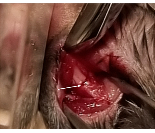
Figure 1: Image of the submandibular cutaneous approach of the mandibular ascending branch. The inferior border of the mandible is fractured with a piezotome insert, allowing minimal bone loss (arrow). Please click here to view a larger version of this figure.

Figure 2: Staining of mouse mandible. Lateral view of a 6-week-old wild type mouse mandible (stained with alizarin red / alcian blue staining): 1. Condyle, 2. Coronoid process, 3. Coronoid notch, 4. Angle process, 5. Posterior border of the ramus, 6. Basilar (or inferior) border of the mandible. Please click here to view a larger version of this figure.
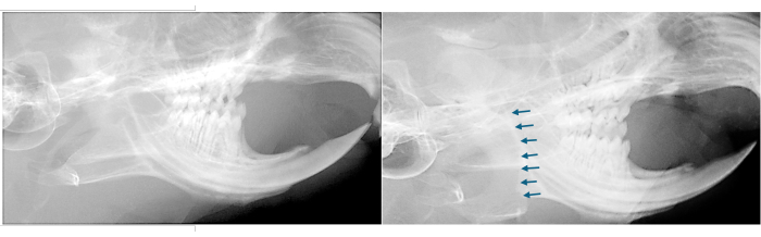
Figure 3: X-ray of the mouse skull. Lateral X-rays of the skull of a wild type 6-week-old mouse (sagittal view) showing the aspect of the mandible of an unfractured mouse (left) and the aspect of the mandible of a fractured mouse and the direction of the non-stabilized mandibular fracture (arrows), from the inferior border to the coronoid notch, placed anteriorly to the condyle (right). Please click here to view a larger version of this figure.

Figure 4. X-ray of the mouse skull under suboptimal conditions. Lateral X-rays of the skull of a wild-type 6-week-old mouse (sagittal view) showing abnormal fracture of the mandibular angle (arrows). Please click here to view a larger version of this figure.

Figure 5: Representative results. Results at day 14 post-fracture in a wild-type mouse (C57BL/6J). Representative 3D coronal views of a reconstructed µCT scan of the mandibular fracture site, showing the bone callus (white arrows). Representative images of the callus cartilage (Sirus Red/Alcian Blue staining) confirming the presence of an endochondral bone repair process. Abbreviations: B = Bone, C = Cartilage. Scale bar: 200 µm. Please click here to view a larger version of this figure.
| 0 | 1 | 2 | |
| Animal aspect | Normal | Toilet reduction | Ruffled hairs, arched back |
| Behavior | Normal | Hypomobility | Hypomobility, reduction of food intake, weight loss > 10 % |
| Clinical signs | Normal | Scar inflammation | Opened scar, infection |
Table 1. Post-operative well-being score scale.
| n = 58 mice | |
| Success rate | 86.3 % |
| Exclusion rate : | 13.7 % |
| Premature spontaneous death | 3% |
| Critical score < 3, and/or weight loss > 20 % | 1% |
| Abnormal fracture split to the mandibular angle | 5% |
| Abnormal fracture split, at molar level | 1.7 % |
Table 2. Success and exclusion rates of the protocol.
Discussion
The technique allows the realization of non-stabilized mandibular fractures in a mouse model within a minimal open approach in a short procedure time (around 10 min or less). This short-time procedure limits the risk of animal morbidity and death, especially in small animals such as mice. To obtain comparable results in the analysis of the endochondral bone repair process, particular care should be taken to perform the mandibular fractures in the same direction without abnormal split of the mandibular angle, as this undesirable effect can abnormally increase the volume of the bone callus and delay the consolidation period. This protocol for mandibular fracture does not impact mandibular incisor growth post-surgery. To improve the effectiveness of this protocol, we advise the experimenter to first exercise on euthanized animals of similar age and dissect immediately after fracturing the mandible to directly visualize the fracture direction if necessary.
Mesenchymal progenitors migrating from adjacent muscles to the callus contribute to bone repair6. It has been demonstrated that periosteum and muscle injury impact bone consolidation5,6. In addition, extensive muscle sections lead to an increased risk of bleeding and may lead to post-operative infection. Piezosurgery has been largely developed in the field of craniofacial bone surgery; it has been reported to have lower rates of soft tissue injury and postoperative pain when compared with conventional osteotomies7,9. To minimize masseter muscle and periosteum injury, we used piezosurgery to initiate the fracture at the inferior border of the mandible and completed the fracture with scissors at the upper level of the ramus to avoid extensive masseter muscle section. This implies controlling the position of the posterior border of the mandible and the coronoid notch before fracturing the upper part of the ramus with the scissors. We encountered no difficulty in completing the fracture with the use of small straight scissors after having initiated it with piezosurgery at the lower edge of the mandible. We experienced this procedure in adult mice with different genetic backgrounds (C57BL/6J, CD1 (results not shown)), with different bone densities and mandibular sizes (prognathism, micrognathia). However, we advise checking that the length of the scissors is suitable for fracturing the upper ramus only once to avoid any abnormal fracture splits.
The risk of bleeding is minimized in this procedure due to this minimal open approach. In the event of excessive bleeding that may occur during muscular elevation, a short compression of the surgical site with gauze stops the bleeding. A potential risk of operating site infection may occur in case of molar tooth fracture when mandibular fracture is too anteriorly placed; this adverse effect can be detected on post-operative lateral X-rays. In this case, the animal must be excluded from the study. Post-operative infection of the operating site may also be due to a hematoma; this may be prevented by performing separated cutaneous sutures, allowing spontaneous evacuation when abnormal bleeding occurs preoperatively. General anesthesia with isoflurane versus intraperitoneal injection avoids excessive mortality rates linked to the adverse traumatic effects of intraperitoneal injections. In our experience, we did not observe any animal death directly linked to general anesthesia with isoflurane.
Mice may present post-operative reduction of food intake. In case of insufficient food intake or insufficient weight gain, sifted kibble powder may be added to the gel. To improve postoperative food intake, we recommend giving nutritional supplement gel with kibbles at least 1 week before the procedure for accommodation. We recommend performing additional X-rays during the follow-up (every week, during the follow-up) and at the time of the sacrifice to evaluate potential fracture displacement. In non-pathological conditions, bone consolidation is complete 4 weeks after the fracture.
Several authors have described different protocols for non-stabilized fractures of the mandible in mice3,10,11. The instruments used to perform the osteotomy can be different, namely, drills, surgical tweezers, etc. However, the use of drills may lead to larger bone loss and soft tissue injuries. To the best of our knowledge, this procedure of non-stabilized mandibular fractures is the first to associate piezosurgery and scissors to complete the osteotomy. This technique has the advantage of preserving soft tissues while permitting a precise osteotomy with minimal bone loss.
As for all techniques, this method has some limitations. We preferred performing the fracture with the use of scissors after initiating it with a piezoelectric surgery insert to avoid any extended masseter muscle section and periosteum injury that may exacerbate a potential consolidation delay and/or formation of pseudarthrosis. However, this technique implies controlling the position of the coronoid notch without total visualization of the mandibular ramus to perform the fracture within the same direction. A learning curve is thus expected, depending on the surgical experience. In addition, the direction of the mandibular fracture on X-rays may be difficult to visualize depending on the X-ray device. We did not use in vivo micro-CT to confirm the fracture direction, as it could compromise mouse recovery because of the increased duration of general anesthesia.
Studying the endochondral bone repair process implies the realization of non-stabilized fractures, thus allowing a better understanding of the bone repair process. This step is necessary if we want to consider preclinical trials in mouse models with bone repair defects with the aim of developing new clinical applications for treating defective bone repair and pseudarthrosis in patients in the context of genetic diseases or in the case of post-traumatic or post-tumoral sequelae.
Disclosures
The authors declare no conflicts of interest.
Acknowledgements
The study is supported in part by the Philanthropy Department of Mutuelles AXA through the Head and Heart Chair and the ANR BonyBrain. We acknowledge all the members of the platform SFR Necker INSERM US24, LEAT Imagine, Paris, France, for their contribution to the realization of the procedure.
Materials
| Name | Company | Catalog Number | Comments |
|---|---|---|---|
| Buprenorphine : Buprecare 0.3 mg/kg | Animalcare | ||
| Elevator OBWEGESER width 6mm Length 17,5cm | Collin Medical | HA 5905 | |
| Forceps : Pad Plate MORIA 11 cm N°5 | MORIA | 9906 | |
| Heating table Bioseblab 55 cm x 33 cm | Bioseblab | 707 | |
| Needle HOLDER : Micro Halsey Needle Holder - Metal | LABODERM | 21,100 | |
| Neo Clear | Merck Millipore, Darmstadt, Germany | 109843 | |
| Ocry Gel Tube 10 g (eye lubricant) | tvm lab | 3.70045E+12 | |
| Piezotome 2 ASS FINAL | ACTEON | X57402 | |
| Piezotome insert Piezocision PZ3 | ACTEON | F87574 | |
| Scissors micro | MORIA | MC52 | |
| Silk sutures PERMA HAND SEIDE / MERSILK | ETHICON | REF 18501G | |
| Straight scissors | MORIA | 4877A | |
| Vetflurane (isoflurane) 250 ml | VIRBAC | VET066 (Centravet) |
References
- Thompson, Z., Miclau, T., Hu, D., Helms, J. A. A model for intramembranous ossification during fracture healing. J Ortho Res. 20 (5), 1091-1098 (2002).
- Bahney, C. S., et al. Cellular biology of fracture healing. J Ortho Res. 37 (1), 35-50 (2019).
- Yu, Y. Y., Lieu, S., Hu, D., Miclau, T., Colnot, C. Site specific effects of zoledronic acid during tibial and mandibular fracture repair. PLoS One. 7 (2), e31771(2012).
- Landry, P. S., Marino, A. A., Sadasivan, K. K., Albright, J. A. Effect of soft-tissue trauma on the early periosteal response of bone to injury. J Trauma. 48 (3), 479-483 (2000).
- Colnot, C. Skeletal cell fate decisions within periosteum and bone marrow during bone regeneration. J Bone Miner Res. 24 (2), 274-282 (2009).
- Julien, A., et al. Direct contribution of skeletal muscle mesenchymal progenitors to bone repair. Nat Commun. 12 (1), 2860(2021).
- McGuire, C., Boudreau, C., Prabhu, N., Hong, P., Bezuhly, M. Piezosurgery versus conventional cutting techniques in craniofacial surgery: A systematic review and meta-analysis. Plast Reconstr Surg. 149 (1), 183-195 (2022).
- Wong, S. A., et al. Chondrocyte-to-osteoblast transformation in mandibular fracture repair. J Orthop Res. 39 (8), 1622-1632 (2021).
- Vercellotti, T. Technological characteristics and clinical indications of piezoelectric bone surgery. Minerva Stomatol. 53, 207-214 (2004).
- Wong, S. A., et al. Chondrocyte-to-osteoblast transformation in mandibular fracture repair. J Orthop Res. 39 (8), 1622-1632 (2021).
- Tian, Y., et al. HIF-1α regulates osteoclast activation and mediates osteogenesis during mandibular bone repair via CT-1. Oral Dis. 28 (2), 428-441 (2022).
Reprints and Permissions
