The blood-brain barrier is a significant hurdle in the delivery of therapies for glioblastoma, a disease for which there is no cure. Here, we report an in vivo image-guided iron oxide therapeutic nano platform that can bypass this physiological barrier by virtue of size and accumulate in the tumor.
Method Article
Nanoparticle Delivery of an Oligonucleotide Payload in a Glioblastoma Multiforme Animal Model
Bryan D. Kim1,2 , Sujan K. Mondal1,3 , Elizabeth Kenyon1,3 , Ming Chen1,3 , Christiane L. Mallett3,4 , Ana C. deCarvalho5 , Zdravka Medarova6 , Anna Moore1,3
1Precision Health Program, Michigan State University, 2Department of Biomedical Engineering, College of Engineering, Michigan State University, 3Department of Radiology, College of Human Medicine, Michigan State University, 4Institute for Quantitative Health Science and Engineering, Michigan State University, 5Henry Ford Health, 6Transcode Therapeutics Inc.
Opens in a new tab
In This Article
Summary
Loading...
$$\rightleftharpoonup{xx}$$
$$\longleftharp{xx}$$,
$$\longrightharp{xx}$$,
Abstract
Loading...
$$\rightleftharpoonup{xx}$$
$$\longleftharp{xx}$$,
$$\longrightharp{xx}$$,
Glioblastoma multiforme (GBM) is the most common and aggressive form of primary brain malignancy for which there is no cure. The blood-brain barrier is a significant hurdle in the delivery of therapies to GBM. Reported here is an image-guided, iron oxide-based therapeutic delivery nano platform capable of bypassing this physiological barrier by virtue of size and accumulating in the tumor region, delivering its payload. This 25 nm nano platform consists of crosslinked dextran-coated iron oxide nanoparticles labeled with Cy5.5 fluorescent dye and containing antisense oligonucleotide as a payload. The magnetic iron oxide core enables tracking of the nanoparticles through in vivo magnetic resonance imaging, while Cy5.5 dye allows tracking by optical imaging. This report details the monitoring of the accumulation of this nanoparticle platform (termed MN-anti-miR10b) in orthotopically implanted glioblastoma tumors following intravenous injection. In addition, it provides insight into the in vivo delivery of RNA oligonucleotides, a problem that has hampered the translation of RNA therapeutics into the clinic.
Introduction
Loading...
$$\rightleftharpoonup{xx}$$
$$\longleftharp{xx}$$,
$$\longrightharp{xx}$$,
Glioblastoma multiforme (GBM) is the highest grade of astrocytoma for which there is virtually no cure. Approximately 15,000 people are diagnosed with glioblastoma annually, which has a dismal median survival of about 15 months and a 5-year survival rate of 5%1. In the past decades, there has been marginal improvement in prognosis despite multiple efforts to advance therapeutic options. The current standard of care for GBM includes maximal surgical resection, when feasible, followed by radiotherapy and chemotherapy2. Temozolomide (TMZ), the chemotherapy of choice, was the latest therapy for glioblastoma discovered to show notable clinical efficacy; however, at least 50% of GBM tumors show TMZ resistance3. In spite of this rigorous therapeutic regimen, there is still a significant clinical need for improved glioblastoma therapy.
The development of therapeutics for GBM and other brain-related diseases is significantly hampered by the selective nature of the blood-brain barrier (BBB). The BBB is a physiological barrier comprised of endothelial cells, pericytes, and astrocyte feet-ends, which creates the semi-permeable membrane between the circulatory system and the brain, restricting the free passage of molecules and cells into the brain4. While protective in normal physiology and critical for brain homeostasis, the BBB prevents many therapeutics from reaching the brain, complicating the treatment of GBM. Efforts to enhance the delivery of therapeutics to GBM have led to the development of nanoparticle-based delivery vehicles, focused ultrasound drug delivery enhancement, and receptor-mediated drug delivery5,6.
Nanoparticles have emerged as a promising medium for developing therapeutics for a myriad of diseases, including cancers. The application of nanoparticles for imaging and therapeutic purposes in GBM has been attempted using various nanoparticle constructs7,8. With the focus on delivering drugs to GBM in conjunction with in vivo imaging of the delivery, the proposed approach utilizes magnetic nanoparticles (MN) consisting of an iron oxide core and covered by dextran for stability. The magnetic properties of these nanoparticles afford for their detection by magnetic resonance (MR) imaging, while simple conjugation chemistry to the aminated dextran coating allows for conjugation of therapeutic moieties such as RNA molecules, additional targeting moieties, or imaging moieties (such as Cy5.5 near-infrared optical dye)9,10. In addition to the imaging capabilities, the nano platform is able to extend the half-life of RNA therapeutics by protecting the oligonucleotide from endogenous nucleases, improving therapeutic delivery. Here, the application of this nano platform for in vivo delivery of therapeutic oligonucleotides (termed MN-anti-miR10b) to GBM, monitored by in vivo imaging, is presented. Previously, the ability of this nano platform to accumulate was demonstrated in GBM cells in vitro, causing significant loss of viability of tumor cells11. Prior to performing therapeutic in vivo studies, it is necessary to demonstrate in vivo delivery of this nano platform to GBM tumors in animal models. To achieve this, orthotopic GBM animal models were produced, and intravenous administration of the construct was performed followed by in vivo imaging. Outlined here are the protocols of these studies showing accumulation in the tumor region confirmed by in vivo imaging and ex vivo microscopy.
Protocol
Loading...
$$\rightleftharpoonup{xx}$$
$$\longleftharp{xx}$$,
$$\longrightharp{xx}$$,
All procedures involving animal subjects have been approved by the Michigan State University Institutional Animal Care and Use Committee (IACUC). Female outbred athymic nude mice were purchased from Jackson Labs (strain #007850) at 7 weeks of age and allowed to acclimate for 1 week prior to implantation surgery. Mice were approximately 21-25 g at the time of implant. U251 cells expressing firefly luciferase were generated and provided by Dr. Ana deCarvalho12.
1. Cell culture and preparation for implantation
- Culture luciferase-labeled, human glioblastoma cells, U251 in Dulbecco's Modified Eagle Medium (DMEM) containing D-glucose, L-glutamine, and sodium pyruvate and supplemented with 10% fetal bovine serum and 1% penicillin/streptomycin at 37 °C with 5% CO2.
- Once the cells reach 70% confluency, trypsinize with 0.25% trypsin/EDTA for 1-2 min at 37 °C. Use 2-4 mL for a T-75 flask, or 4-6 mL for a T-175 flask.
- After cells have detached from the flask, quench the reaction with a 2:1 ratio of supplemented DMEM and mix gently by pipetting to dissociate cells into a single-cell suspension. Transfer the cells into a conical tube and pellet the cells by centrifugation at 300 x g for 5 min.
- Carefully aspirate the supernatant and resuspend the cell pellet in 1x phosphate-buffered saline (PBS). Count the cells with a hemocytometer using trypan blue staining to exclude dead cells and calculate the total number of live cells. Pellet the cells again, as described above.
- Resuspend the cell pellet to a concentration of 1 x 10⁸ cells/mL in PBS and store the cell suspension on ice for intracranial implantation.
2. Freehand orthotopic tumor implantation
NOTE: This protocol is adapted from Irtenkauf et al. (Dr. Ana deCarvalho's procedure; Henry Ford Health Hermelin Brain Tumor Center)12. All steps should be carried out in a biosafety cabinet to ensure safety for both subjects and researchers. The freehand implantation method allows for a faster procedure to achieve larger sample sizes while maintaining the quality of intracranial implantation. Alternatively, a stereotaxic device can be used to implant the tumor at the same coordinates described below.
- Weigh the nude, athymic mouse and administer an anesthetic solution of ketamine/xylazine (100 mg/kg/10mg/kg) by intraperitoneal injection with a 28G syringe. Place the mouse in a recovery cage on top of a heating pad to maintain body temperature. Confirm anesthetization by checking the pedal reflex.
- Apply ophthalmic eye ointment, administer ketoprofen (5 mg/kg) subcutaneously with a 28G syringe, and give identifying ear punches.
- Sterilize the surgical site at the top of the head with 70% ethanol followed by betadine scrub and repeat 3x. Then, make a 5-7 mm incision with small surgical scissors to the right of the midline of the head and retract the skin with fine forceps to expose the skull and suture lines of the skull.
- Scrub the skull with a cotton swab to remove the periosteum, allowing access to the bone. Apply a solution of 3% hydrogen peroxide using a new cotton swab to dry out the bone. Using a new cotton swab, wipe away excess hydrogen peroxide and any foam that may form. Repeat this step 3x until the bregma is visible on the skull and the bone is sufficiently dry for drilling. A sufficiently dry skull will have starkly whitened suture lines and reduced redness from the starting appearance.
- With respect to the bregma, measure -2.5 mm medial/lateral and -1 mm anterior/posterior with a ruler and mark the drill site with a marker. Measure again before drilling to ensure the mark is in the correct position.
- Take an electric micro-surgery drill (11,000 RPM) with a 0.5 mm drill burr and carefully make a hole in the skull where previously marked. Absorb any blood or fluid that may accumulate at the drill site with a fresh cotton swab.
- Prepare a 33G microliter syringe fitted with a needle sleeve to an exposed needle length of 3 mm. Draw up 5 µL of the previously prepared 1 x 10⁸ cell/mL cell suspension after briefly resuspending with a pipette and ensure there are no bubbles in the injection solution. Carefully wipe away any excess cell suspension left on the exterior of the needle and needle sleeve with a 70% isopropanol wipe.
- Insert the needle vertically into the drill site until the needle sleeve stops further insertion of the needle. Carefully retract the needle approximately 0.5 mm to create space for the injected cells to accumulate.
- Slowly inject the cell suspension over a period of 1 min. Once the full volume has been injected, hold the needle at depth for an additional minute to reduce the efflux of the injection solution. Then, retract the needle carefully.
- With a clean cotton swab, wipe away any liquid that accumulates at the injection site and then apply bone wax to seal the hole.
- Close the incision with a veterinary bond and allow it to dry. With a clean cotton swab, apply topical lidocaine as an analgesic.
- Monitor the mouse until fully recovered and ambulatory. Administer ketoprofen (5 mg/kg) subcutaneously for 2 days post-implantation for pain management.
3. Nanoparticle platform synthesis
- Synthesize and conjugate the nanoparticles with Cy5.5 and oligonucleotide (anti-miR10b) as previously described11.
4. MN-anti-miR10b administration
NOTE: In this study, the injections were done once a week for 6 weeks beginning 7 days after implantation, but the same protocol can be used for various injection frequencies.
- Weigh the mouse and calculate the volume of MN-anti-miR10b to administer to the mouse. Doses of MN-anti-miR10b are given at 20 mg Fe/kg in murine GBM models. For example, a 20 g mouse receives an 80 µL dosage of 5 mg Fe/mL MN-anti-miR10b. Control mice receive an equal volume of PBS consistent with this dosage scheme.
- Prepare a 28G syringe with the dose of MN-anti-miR10b and ensure there are no bubbles in the injection solution.
- Anesthetize the mouse under 2%-4% isoflurane for induction and 1.5%-2% for maintenance with a 1.5 L/min flow rate in an induction box. Move the mouse to a nose cone and continue to administer anesthesia. Check the depth of anesthesia and apply vet ointment.
- Sterilize the tail with a 70% isopropanol wipe and allow excess alcohol to dry.
- Rotate the tail to position the lateral caudal veins to the top of the tail. Then, aiming for the vein, insert the needle into the tail and establish a vacuum by pulling back the plunger.
- Insert the needle until blood enters the syringe, indicating successful entry into the vein.
- Slowly inject MN-anti-miR10b and retract the needle after complete delivery. Apply pressure to the injection site with gauze until the bleeding stops.
- Remove the mouse from the nose cone. Monitor the mouse until fully recovered and ambulatory.
NOTE: Bolus administration of nanoparticles may cause respiratory distress. Carefully monitor the mouse and immediately apply light chest compressions if there are signs of respiratory distress.
5. In vivo bioluminescence and fluorescence imaging
NOTE: In vivo bioluminescence and fluorescence imaging are conducted in the In Vivo Imaging System (IVIS) before and 24 h after the nanoparticle is injected.
- Prepare sterile-filtered D-luciferin at a concentration of 30 mg/mL in PBS without Mg2+ and Ca2+.
- Open the IVIS operation software and begin initialization to prepare the machine. While the system initializes, select a folder to save images by selecting Acquisition > Auto-Save To and define the folder for the files to be saved.
- At imaging time points, weigh the mouse and calculate the volume of D-luciferin to administer for a dosage of 150 mg/kg. Prepare a 28G syringe with the volume, protecting the solution from direct light. This study included imaging 2 days after implantation surgery to confirm seeding, and weekly imaging started on day 7 post-implant immediately prior to weekly treatments.
- Anesthetize the mouse under 2%-4% isoflurane for induction and 1.5%-2% for maintenance with a 1.5 L/min flow rate in an induction chamber. Once anesthetized, gently scruff the mouse to avoid further injuring the surgical site and inject the dose of D-luciferin intraperitoneally. Allow the mouse to recover and wait for 10 min before imaging to allow the bioluminescence signal to develop and stabilize.
- While the bioluminescence signal develops, prepare the IVIS scanner for bioluminescence imaging. Place down the black low fluorescence mat on the imaging stage and configure the nose cone array for the number of subjects to be imaged.
- In the software, click Imaging Wizard. Select Bioluminescence Imaging and then select Open Filter. On the next screen, select Imaging Subject and Field of View. The IVIS is now set for bioluminescence imaging.
- At 3 min before imaging, begin priming the induction box by allowing the isoflurane/oxygen mixture to fill the box. At 2 min before imaging, place mice in the induction box for anesthetization.
- Once anesthetized, move the mice from the induction box to the IVIS for imaging and lay each down in the prone position. Provide maintenance anesthesia using 2% isoflurane during imaging.
- Once mice have been placed in the IVIS scanner, click Acquire Sequence. Take an image using auto-exposure settings with a minimum signal threshold of 3,000 counts. Bioluminescence imaging visualizes the localization of luciferase-labeled U251 glioblastoma cells. Auto-exposure will capture an image with an exposure length calculated based on the brightest signal in view up to the user-defined maximum length of 5 min.
NOTE: A typical cage of mice requires a single image, as seeding is generally uniform. In cases with one or more bright mice, mice may be removed, and a repeat acquisition can be taken to better capture signals from dim mice. - After bioluminescence imaging, change the IVIS settings to prepare epi-fluorescence imaging and take fluorescence scans.
- In the Imaging Wizard, select Fluorescence > Filtered Pair with Epi-Illumination. In the next screen, select the probes to be imaged, such as Cy5.5. Select the Imaging Subject and Field of View. The IVIS scanner is now set for fluorescence imaging of the probes selected in the previous screen.
- Take an image using auto-exposure settings with a minimum signal threshold of 6,000 counts. Cy5.5 fluorescence imaging visualizes the localization of MN-anti-miR10b. Auto-exposure will capture an image with an exposure length calculated based on the brightest signal in view up to the user-defined maximum length of 5 min.
NOTE: Fluorescence imaging rarely takes longer than 1 min exposures, even in control PBS-injected mice. A single image is always sufficient as the signal deviation between treated mice is small. - Remove the mouse from the IVIS after imaging. Monitor until fully recovered from anesthesia and ambulatory.
6. In vivo magnetic resonance imaging
NOTE: MR imaging is performed before and 24 h after nanoparticle injection and can be performed in the same animals that undergoes optical and bioluminescence imaging.
- Anesthetize the subject mouse in an induction box under 2%-4% isoflurane for induction and 1.5%-2% for maintenance with a 1.5 L/min flow rate. Once anesthetized, place the mouse prone on the MRI bed on top of the respiratory monitoring balloon. Fit the nose cone snuggly to the snout to deliver anesthesia during imaging and apply ophthalmic eye ointment.
- Immobilize and position the head for scanning using a bite bar and ear bars.
- Install the lubricated rectal temperature probe and ensure respiration and temperature monitoring are functioning.Position the mouse brain coil over the head by fitting the pegs on the mouse bed into the holes on the coil and tape the coil into place to reduce movement during scanning.
- Place a small warm water circulating pad over the top of the mouse to maintain body temperature while anesthetized. Take care to allow some distance between the coil and the heating blanket to limit distortion of the image by water interference.
- Move the mouse and imaging bed into position for scanning.
- Tune and match the MRI coils by starting the Wobble setup step in the acquisition software. At the service end of the MRI, use the tune and match knobs on the coil to ensure that the trace is centered (tune) and as deep as possible (match).
- Acquire a three-plane localizer scan and adjust the imaging bed to position the brain at the isocenter of the magnet, if necessary.
- Acquire 2D T2 weighted scans to detect the injected agent in the tumor with the following parameters: repetition time (TR) = 2500 ms, echo time (TE) = 25 ms, the field of view (FOV) 20 x 20 mm, 18 coronal slices, 0.2 mm slice gap, 150 x 150 µm in-plane resolution, 0.5 mm slice thickness.
- Acquire a B0 map of the whole brain to calculate a localized shim using the Mapshim utility. Then, use 3D T2 weighted images to visualize the nanoparticles with TR = 30 ms, TE = 10 ms, FOV 20 x 15 x 12 mm, resolution 100 µm isotropic.
- Acquire T2* map for further nanoparticle imaging using the 2D T2 weighted image as a reference to position scan over the tumor. Use TR = 800 ms, TE = 3.5 ms. FOV 20 x 20 mm, 5 coronal slices, no slice gap. Resolution in plane 100 µm in plane. Acquire 10 positive echo images with 5 ms echo time spacing.
- Monitor respiration and body temperature throughout the imaging sequences and adjust isoflurane and water temperature if necessary.
- After imaging, remove the mouse from the MRI scanner and place it in a heated recovery cage. Monitor the mouse until it is fully recovered from anesthesia and ambulatory.
7. Ex vivo bioluminescence and fluorescence imaging
- Perform ex vivo bioluminescence and fluorescence imaging in the IVIS, similar to the in vivo imaging. Carry out the IVIS initialization and scan setting steps as described above.
- At the experimental endpoint, weigh the mouse and calculate the volume of D-luciferin to administer for a dosage of 150 mg/kg. Inject the dose of D-luciferin and conduct live imaging 10 min post-injection, as previously described.
- After final in vivo bioluminescence and fluorescence imaging, quickly euthanize the mouse by cervical dislocation while still under anesthesia, dissect the mouse, and collect the major organs, including the brain, on a Petri dish.
- Place the Petri dish with excised organs in the IVIS scanner and image the organs with both bioluminescence and fluorescence modalities using the same acquisition settings as the in vivo imaging.
- Flash freeze any organs necessary for further analysis in OCT by slowly lowering the cryomold with the specimen in OCT into a shallow pool of liquid nitrogen with long forceps until the OCT turns completely white. Do not allow liquid nitrogen to touch liquid OCT or lower the mold too quickly, as bubbling of the OCT will occur. Store frozen samples at -80 °C.
- Store the brain containing the tumor in OCT and flash freeze for cryosectioning. Store at -80 °C until needed for analysis.
8. Fluorescence microscopy
- Prepare the cryostat for cryosectioning. Set the chamber and specimen head temperatures between -15 °C and -20 °C.
- Take the flash-frozen brain in OCT out of -80 °C storage and place it inside the cryostat chamber. Allow the samples to warm to the chamber temperature (-20 °C) before sectioning.
- Using a single edged razor cut the brain coronally at the injection site. This is usually visible as a dimple on the surface of the brain. Mount the sample onto the specimen disc using a small amount of OCT to position the cut side of the brain to be sectioned. Apply ample pressure with the chilled weight inside the chamber to ensure stable mounting of the sample.
- Move the mounted sample and specimen disc onto the specimen head in preparation for sectioning. Trim the tissue to the desired depth by taking 20 µm sections, adjusting the angle of the sample to section the tissue as near to level with the tissue as possible.
- Once the desired tissue depth has been reached, adjust the cryostat to take 5 - 7 µm sections of the sample. Mount the sections onto glass slides pre-labeled with descriptors such as mouse ID, tissue sectioned, and slide number by quickly lowering the charged side of the microscope slide onto the section.
- Make fresh 4% paraformaldehyde (PFA). Submerge the slide with tissue in the PFA solution for 15 min to fix the tissue. Rinse the slide with DPBS 3x after fixation.
- To the now fixed tissue section on the glass slide, add a 40 µL drop of mounting medium containing 4',6-diamidino-2-phenylindole (DAPI), which stains cell nuclei. Carefully place a 24 mm x 50 mm glass coverslip on top of the mounting media and ensure that the entire tissue has been encased in the mounting media.
- Visualize the tissue using DAPI (emission (em) at 359 nm, excitation (ex) at 457 nm) and Cy5.5 (ex 683 nm, em 703 nm) fluorescence microscopy.
Results
Loading...
$$\rightleftharpoonup{xx}$$
$$\longleftharp{xx}$$,
$$\longrightharp{xx}$$,
MN-anti-miR10b was synthesized and characterized, as described previously11. Transmission electron microscopy of MN-anti-miR10b shows the morphology and polydispersity of the nano platform (Figure 1B). This nano platform has an average size of 25.12 ± 0.34 nm with a zeta potential of 13.18 ± 1.47 mV (Figure 1C,D). In these studies, nude athymic mice were orthotopically implanted with U251 human glioblastoma cells following the protocol described above. Systemic intravenous injections of MN-anti-miR10b were performed at 20 mg Fe/kg. Mice were euthanized, and tissues were collected 7 days after the final dosage of nanodrug. The Cy5.5 fluorescence and bioluminescence signals in MN-anti-miR10b injected mice showed clear co-localization (Figure 2A,B), indicating delivery of the nanoparticles to the tumor, while control mice showed no fluorescence signal. Ex vivo fluorescence imaging showed the localization of the nanoparticles in major clearance organs such as the liver and kidneys, which is expected for this type of nanoparticles (Figure 2C). In a separate study, mice were orthotopically implanted with GBM cells, as above, and subjected to in vivo MR imaging. T2* weighted images and T2* maps were obtained before administration of the nanoparticles and 24 h following a single dose of 20 mg Fe/kg. A characteristic decrease in T2 weighted MRI signal demonstrates the potential of MN-anti-miR10b as an MRI contrast agent to cross the blood-brain barrier and its accumulation in the tumor region (Figure 3). Following in vivo imaging, fluorescence microscopy was conducted to further verify the delivery of nanoparticles to the tumor. As described above, tissues were sectioned and prepared for microscopy with DAPI staining. Cy5.5 fluorescence signal was shown in the tumor region, confirming delivery (Figure 4).
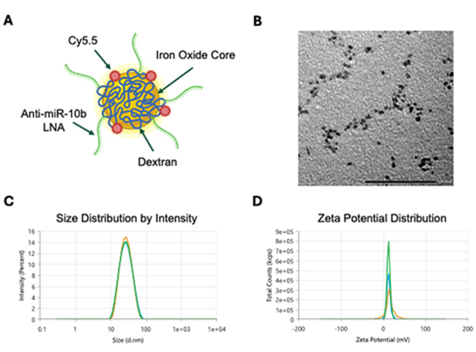
Figure 1. Characterization of MN-anti-miR10b. (A) A schematic structure of an MN-anti-miR10b. The nano platform consists of an iron oxide core covered with dextran, labeled with a Cy5.5 optical dye, and conjugated with therapeutic oligonucleotide (anti-miR10b). Created with BioRender.com. (B) Transmission electron microscopy of MN-anti-miR10b. (C) The hydrodynamic size of the construct is 25.12 ± 0.34 nm. (D) The zeta potential of MN-anti-miR10b is 13.18 ± 1.47 mV. Please click here to view a larger version of this figure.
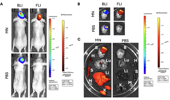
Figure 2. In vivo and ex vivo studies in GBM model. (A) In vivo bioluminescence imaging (left) and fluorescence imaging (right) show co-localization of the magnetic nanoparticle signal in the tumor region. (B) Ex vivo bioluminescence and fluorescence imaging of excised brains confirms co-localization of the signal. (C) Biodistribution of the organs shows accumulation to the major clearance organs. B: Brain; H: Heart; Lu: Lungs; Li: Liver; S: Spleen; K: Kidneys; M: Muscle; (n=6 for all groups). Please click here to view a larger version of this figure.
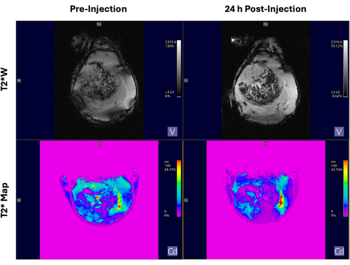
Figure 3. In vivo MRI in GBM model. T2* weighted (top row) and T2* maps (bottom row) images obtained before (left) and 24 h post-injection (right) of MN-anti-miR10b. The decrease in signal intensity in the post-injection image is indicative of iron oxide accumulation (n=3). Please click here to view a larger version of this figure.
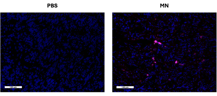
Figure 4. Ex vivo fluorescence microscopy of tumor tissues. Representative microscopic images show nanoparticle accumulation. Blue - DAPI, Magenta - Cy5.5. Scale Bar = 100 µm; (n=6 for all groups). Please click here to view a larger version of this figure.
Discussion
Loading...
$$\rightleftharpoonup{xx}$$
$$\longleftharp{xx}$$,
$$\longrightharp{xx}$$,
Several critical steps across the different methods of validating the accumulation of the nanoparticles across the BBB can be decisive for the success of the protocol. Beginning with the orthotopic implantation of GBM cells, it is important to ensure that the suture lines of the skull are visible after drying the bone; this aids in the accurate placement of the tumor cells. For drilling through the skull, it is best to apply light pressure to the drill site and begin drilling to make a shallow impression in the bone. Once the initial mark is made and in the correct location, it is easier to drill with minimal drill slippage. Immediately stop drilling after breaking through the skull to minimize damage to the brain. During the intravenous injection of nanoparticles and other agents, it is critical to ensure that there are no air bubbles in the injection solution to avoid potentially fatal venous air embolisms. Some indicators of successful entry into the tail vein following this protocol are blood being drawn into the syringe and the loss of color in the tail vein around the tip of the needle. For ex vivo bioluminescence and fluorescence imaging of excised tissues, it is best to work quickly as the bioluminescence signal begins to decay over time. Bioluminescence signal kinetics may vary between cell lines, so it is important to understand specific models.
Certain steps can be modified to aid in the detection of nanoparticle localization. During in vivo bioluminescence imaging, variations in tumor cell implantation and tumor growth rates can lead to differences in bioluminescence signal. In certain circumstances, the mouse with the highest BLI signal can shorten the exposure time of the scan, leading to diminished signal detected in other mice. In this instance, it is advisable to continue fluorescence scanning so that mice remain in the same position for BLI and FLI co-localization purposes. Then, reimage for bioluminescence after removing mice that showed signal in the initial BLI scan. This will result in the exposure time calculation being based on these lower signal mice and, ultimately, the detection of the tumor. While taking in vivo fluorescence images, nanoparticle localization in the body may be better ascertained by covering the tail with black paper, which may have a high fluorescence signal from residual nanoparticle presence from the tail vein injections. In a similar line of reasoning, during ex vivo fluorescence scanning of the organs, imaging the brain alone may better show the localization of the nanoparticle in the brain and tumor region as the accumulation of the nanoparticle in the reticuloendothelial system organs may obscure signal detection.
A limitation of this method for therapeutic RNA delivery is the systemic distribution of the nanoparticles in the body. As is the case with all other systemic injections, the therapeutic is removed from the bloodstream by the organs such as the liver, kidneys, and spleen, ultimately resulting in decreased delivery of the nanoparticle to the region of interest. To address this issue, MN-anti-miR10b has been synthesized with a crosslinked-dextran coating to increase the circulation time in the body13. Despite efforts to increase circulation time and accumulation by optimizing the nanoparticle structure, multiple injections of the nanoparticle may be required for significant accumulation in the tumor region, depending on the specific model and disease state.
The delivery of therapeutic molecules in GBM represents a significant challenge due to the highly selective BBB. Many studies have tested nanoparticle-based delivery vehicles, which ultimately are ineffective in the penetration of the BBB due to their size and charge5,6,7. The 25 nm nanoparticle construct, MN-anti-miR10b, described here, is able to pass this physiologic barrier and accumulate in the tumor region when systemically injected. This nanoparticle platform has the potential to mediate the delivery of RNA molecules to GBM and allows monitoring of the accumulation of the delivery vehicle using imaging. The biodistribution of iron oxide nanoparticles follows an expected pattern, where they accumulate in reticuloendothelial system organs for clearance. Similar biodistribution has been reported with analogous nanoparticles used for imaging or therapeutic purposes. Importantly, there were no indications of associated off-target toxicities in the major organs in these previous studies, even at a dosage of 30 mg Fe/kg14,15.
An advantage to this nanoparticle platform system is the in vivo imaging capabilities that allow for non-invasive tracking of its accumulation. The Cy5.5 near-infrared fluorescent dye conjugated to the dextran coat allows for rapid optical imaging to detect its presence. In addition to this modality, the superparamagnetic iron oxide nanoparticle core of MN-anti-miR10b makes the construct suitable as an MRI contrast agent and represents a developing area of theranostics16. The anatomical resolution of MRI in combination with the iron oxide nanoparticle contrast can precisely show where the nanoparticles accumulate in the brain. These two modalities together make pre-clinical in vivo tracking of the nano platform possible to monitor the delivery of the RNA or other payloads across the BBB.
Disclosures
Loading...
$$\rightleftharpoonup{xx}$$
$$\longleftharp{xx}$$,
$$\longrightharp{xx}$$,
Z.M. and A.M. are co-founders and shareholders of TransCode Therapeutics Inc. The remaining authors have no conflicts of interest to disclose.
Acknowledgements
Loading...
$$\rightleftharpoonup{xx}$$
$$\longleftharp{xx}$$,
$$\longrightharp{xx}$$,
The Funding for this study was provided in part by the grant from the Henry Ford Health Systems Michigan State University Health Sciences Alliance to A.M. and A.dC. We thank Dr. Danielle R. Ferguson for overseeing animal studies at Michigan State University and for approving this video.
Materials
| Name | Company | Catalog Number | Comments |
|---|---|---|---|
| Athymic nude "J:NU" mice | Jackson Laboratory | RRID:IMSR_JAX:007850 | Immunocompromised mouse model |
| 0.25% Trypsin | Gibco | 25200-056 | Cell culture reagent for U251 |
| 1.7 mL microcentrifuge tube | DOT Scientific | RN1700-GMT | For tissue collection |
| 10 µL, Neuros Syringe, Model 1701 RN, 33 gauge, Point Style 4 | Hamilton | 65460-06 | Syringe for intracranial implantation of tumor cells |
| 3M Vetbond | 3M | 1469SB | Tissue adhesive for surgical site closure |
| 4% Paraformaldehyde | Thermo Scientific | J199943-K2 | Tissue fixing solution |
| 70% isopropoyl alcohol wipe | Cardinal | MW-APL | Topical antiseptic wipe for tumor implantation and tail vein injection |
| Aperio Versa | Leica | For scanning of stained tissue section slides | |
| Betadine Surgical Scrub | Purdue | 6761815101 | Topical antiseptic for tumor implantation |
| BioSpec 70/30 | Bruker | Magnetic resonance imaging scanner | |
| Bone Wax | Medline | DYNJBW25 | Bone wax for sealing implantation site |
| Burrs for Micro drill | F.S.T. | 19007-05 | Drill burr used to make hole in skull for tumor implantation |
| DAPI Fluoromount-G | SouthernBiotech | 0100-20 | Tissue mounting media containing DAPI stain |
| Dulbecco’s Modified Eagle Medium (DMEM) | Gibco | 11995-065 | Cell culture media for U251 |
| Extra Fine Graefe Forceps | F.S.T. | 11150-10 | Sugical tool for tumor implantation |
| Fetal bovine serum | Corning | 35-010-CV | Cell culture media supplement for U251 |
| Fine Scissors - Sharp 10.5cm | F.S.T. | 14060-10 | Sugical tool for tumor implantation |
| Glydo (Lidocaine) | Sagent | 673-76 | Topical analgesic for surgical site |
| Ideal Micro Drill | CellPoint Scientific | 67-1200A | Drill used to make hole in skull for tumor implantation |
| Insulin syringe 1CC 29G X 1/2" | Becton, Dickinson | 324704 | Syringe for D-Luciferin injection and tail vein injection of nanoparticles |
| Isoflurane | Covetrus | 11695067772 | Anethesia |
| Isoflurane vaporizer | SOMNI Scientific | VS6002 | Anethesia apparatus |
| IVIS SpectrumCT In Vivo Imaging System | PerkinElmer/Revvity | 128201 | Bioluminescence and fluorescence imaging scanner |
| IVISbrite D-Luciferin Potassium Salt | PerkinElmer/Revvity | 122799-100MG | Substrate for bioluminescence imaging |
| Ketaset (Ketamine) | Zoetis | 10004027 | Anesthetic for tumor implantation surgery |
| Ketofen (Ketoprofen) | Zoetis | 10004031 | Analgesic for tumor implantation surgery |
| Leica CM1950 | Leica | CM1950 | For cryosectioning of OCT-embedded samples |
| PBS | Gibco | 14190-144 | Cell culture reagent and cell suspension solution for implantation of U251 |
| Penicillin-streptomycin | Gibco | 15140-122 | Antibiotic for cell culture media for U251 |
| Puralube vet ointment | MWI Veterinary | 27505 | Opthalmic eye ointment for protection during tumor implantation |
| Ruler | F.S.T. | 18000-30 | Used to measure drill site for implanation |
| Tissue-Tek Cryomold Intermediate 15 x 15 x 5 mm | Sakura | 4566 | Collection mold for collecting tissue samples |
| Tissue-Tek O.C.T. Compound | Sakura | 4583 | Freezing compound for collecting tissue samples |
| U-251 MG cell line human | Millapore Sigma | 9063001 | Human glioblastoma cell line |
| Xylazine Injectable Solution, 100 mg/ml | Covetrus | 1XYL006 | Paralytic for tumor implantation surgery |
References
Loading...
$$\rightleftharpoonup{xx}$$
$$\longleftharp{xx}$$,
$$\longrightharp{xx}$$,
- Tan, A. C., et al. Management of glioblastoma: State of the art and future directions. CA Cancer J Clin. 70 (4), 299-312 (2020).
- Cihoric, N., et al. Current status and perspectives of interventional clinical trials for glioblastoma - analysis of clinicaltrials.Gov. Radiat Oncol. 12 (1), 1(2017).
- Lee, S. Y. Temozolomide resistance in glioblastoma multiforme. Genes Dis. 3 (3), 198-210 (2016).
- Dong, X. Current strategies for brain drug delivery. Theranostics. 8 (6), 1481-1493 (2018).
- Teleanu, D. M., Chircov, C., Grumezescu, A. M., Volceanov, A., Teleanu, R. I. Blood-brain delivery methods using nanotechnology. Pharmaceutics. 10 (4), 268(2018).
- Ohta, S., et al. Investigating the optimum size of nanoparticles for their delivery into the brain assisted by focused ultrasound-induced blood-brain barrier opening. Sci Rep. 10 (1), 18220(2020).
- Teplyuk, N. M., et al. Therapeutic potential of targeting microrna-10b in established intracranial glioblastoma: First steps toward the clinic. EMBO Mol Med. 8 (3), 268-287 (2016).
- Dube, T., Kumar, N., Bishnoi, M., Panda, J. J. Dual blood-brain barrier-glioma targeting peptide-poly(levodopamine) hybrid nanoplatforms as potential near infrared phototheranostic agents in glioblastoma. Bioconjug Chem. 32 (9), 2014-2031 (2021).
- Yoo, B., et al. Design of nanodrugs for mirna targeting in tumor cells. J Biomed Nanotechnol. 10 (6), 1114-1122 (2014).
- Medarova, Z., Pham, W., Farrar, C., Petkova, V., Moore, A. In vivo imaging of sirna delivery and silencing in tumors. Nature Medicine. 13 (3), 372-377 (2007).
- Chen, M., et al. Co-administration of temozolomide (tmz) and the experimental therapeutic targeting mir-10b, profoundly affects the tumorigenic phenotype of human glioblastoma cells. Front Mol Biosci. 10, 1179343(2023).
- Irtenkauf, S. M., et al. Optimization of glioblastoma mouse orthotopic xenograft models for translational research. Compar Med. 67 (4), 300-300 (2017).
- Moore, A., Marecos, E., Bogdanov, A., Weissleder, R. Tumoral distribution of iron oxide nanoparticles in a rodent model. Radiology. 214 (2), 568-574 (2000).
- Moore, A., Medarova, Z., Potthast, A., Dai, G. In vivo targeting of underglycosylated muc-1 tumor antigen using a multimodal imaging probe. Cancer Res. 64 (5), 1821-1827 (2004).
- Yoo, B., et al. Therapy targeted to the metastatic niche is effective in a model of stage iv breast cancer. Sci Rep. 7, 45060(2017).
- Dadfar, S. M., et al. Iron oxide nanoparticles: Diagnostic, therapeutic and theranostic applications. Adv Drug Deliv Rev. 138, 302-325 (2019).
Reprints and Permissions
Request permission to reuse the text or figures of this JoVE article
Request Permission