This study presents an original and portable tPBM technology under electroencephalographic (EEG) control of deep or non-rapid eye movement (NREM) sleep in non-anesthetized male C57BL/6 mice of different ages to stimulate lymphatic clearance of Aβ from the brain into the peripheral lymphatic system (the deep cervical lymph nodes, dcLNs).
Method Article
Photobiomodulation Under Electroencephalographic Controls of Sleep for Stimulation of Lymphatic Removal of Toxins from Mouse Brain
Inna Blokina1 , Egor Iluykov2 , Dmitry Myagkov2 , Dmitry Tuktarov2 , Sergey Popov2 , Timofey Inozemzev2 , Ivan Fedosov2 , Alexander Shirokov1,3 , Andrey Terskov1 , Alexander Dmitrenko1 , Arina Evsyukova1 , Daria Zlatogorskaya1 , Viktoria Adushkina1 , Matvey Tuzhilkin1 , Maria Manzhaeva1 , Valeria Krupnova1 , Alexander Dubrovsky2 , Inna Elizarova1 , Maria Tzoy2 , Oxana Semyachkina-Glushkovskaya1
Opens in a new tab
In This Article
Summary
Loading...
$$\rightleftharpoonup{xx}$$
$$\longleftharp{xx}$$,
$$\longrightharp{xx}$$,
Abstract
Loading...
$$\rightleftharpoonup{xx}$$
$$\longleftharp{xx}$$,
$$\longrightharp{xx}$$,
The meningeal lymphatic vessels (MLVs) play an important role in the removal of toxins from the brain. The development of innovative technologies for the stimulation of MLV functions is a promising direction in the progress of the treatment of various brain diseases associated with MLV abnormalities, including Alzheimer's and Parkinson's diseases, brain tumors, traumatic brain injuries, and intracranial hemorrhages. Sleep is a natural state when the brain's drainage processes are most active. Therefore, stimulation of the brain's drainage and MLVs during sleep may have the most pronounced therapeutic effects. However, such commercial technologies do not currently exist.
This study presents a new portable technology of transcranial photobiomodulation (tPBM) under electroencephalographic (EEG) control of sleep designed to photo-stimulate removal of toxins (e.g., soluble amyloid beta (Aβ)) from the brain of aged BALB/c mice with the ability to compare the therapeutic effectiveness of different optical resources. The technology can be used in the natural condition of a home cage without anesthesia, maintaining the motor activity of mice. These data open up new prospects for developing non-invasive and clinically promising photo-technologies for the correction of age-related changes in the MLV functions and brain's drainage processes and for effectively cleansing brain tissues from metabolites and toxins. This technology is intended both for preclinical studies of the functions of the sleeping brain and for developing clinically relevant treatments for sleep-related brain diseases.
Introduction
Loading...
$$\rightleftharpoonup{xx}$$
$$\longleftharp{xx}$$,
$$\longrightharp{xx}$$,
Meningeal lymphatic vessels (MLVs) play an important role in the removal of toxins and metabolites from brain tissues1,2,3. Damage of MLVs in various brain diseases, including tumors, traumatic brain injuries, hemorrhages, and neurodegenerative processes, is accompanied by a decrease in the MLV functions leading to the progression of these pathologies1,2,3,4,5,6. Therefore, the development of methods for the stimulation of MLVs opens new horizons in the emergence of effective technologies for the treatment of brain diseases. Recently, non-invasive technology for effective transcranial photobiomodulation (tPBM) has been proposed to stimulate MLVs and remove toxins such as blood and Aβ from the brain5,7,8,9,10,11,12. It is interesting to note that deep sleep is a natural factor for the activation of lymphatic drainage processes in the brain13,14. Based on this fact, it is logical to assume that the tPBM of MLVs during sleep may have more effective therapeutic effects than during wakefulness9,11,12,15. However, there are currently no commercial technologies for tPBM during sleep16. In addition, animal experiments to study the therapeutic effects of tPBM are performed under anesthesia, which is required to accurately deliver light to the brain. However, anesthesia significantly affects the brain's drainage, which reduces the quality of research results17.
Aβ is a metabolic product of normal neural activity18. As it was established in cultured rat cortical neurons, Aβ is released from them at high rates into the extracellular space (2-4 molecules/neuron/s for Aβ)19. There is evidence that the dissolved form of Aβ, located in the extracellular and perivascular spaces, is most toxic to neurons and synapses20. The soluble Aβ is rapidly cleared from the human brain during 1-2.5 h21. MLVs are the tunnels for removal of the soluble Aβ from the brain1,7 that declines with age, leading to the accumulation of Aβ in the aged brain1,22. There is evidence that extracellular abnormalities of Aβ levels in the brain correlate with cognitive performance in aging and are associated with the development of Alzheimer's disease (AD)23,24. Therefore, aged and old rodents are considered alternatives to transgenic models for the study of amyloidosis, including AD25,26.
This study presents an original and portable tPBM technology under electroencephalographic (EEG) control of deep or non-rapid eye movement (NREM) sleep in non-anesthetized male C57BL/6 mice of different ages to stimulate lymphatic clearance of Aβ from the brain into the peripheral lymphatic system (the deep cervical lymph nodes, dcLNs).
Protocol
Loading...
$$\rightleftharpoonup{xx}$$
$$\longleftharp{xx}$$,
$$\longrightharp{xx}$$,
All procedures were performed in accordance with the "Guide for the Care and Use of Laboratory Animals", Directive 2010/63/EU on the Protection of Animals Used for Scientific Purposes, and the guidelines from the Ministry of Science and High Education of the Russian Federation (Nº 742 from 13.11.1984), which have been approved by the Bioethics Commission of the Saratov State University (Protocol No. 7, 22.09.2022).
1. Hardware assembly
- Cut a piece of foil textolite that is 1.5 mm thick to the dimensions of 1.5 mm x 2 mm. This part will be referred to as the light-emitting diode (LED) printed circuit board (PCB) (Figure 1C).
- Solder an LED to a PCB, as shown in Figure 1.
- Solder two stranded copper wires to the pins of an LED and then cover them with a sleeve.
- Print out the 3D model of the frame (Figure 1A); place an LED (Figure 1B) and magnets (Figure 1D) on the frame; place a washer (Figure 1E) on the mouse's head.
- To assemble the circuit (Figure 2), follow these steps.
- First, connect the resistor (Figure 2; R1) between an LED anode and 5 V port on the Arduino.
- Next, connect an LED cathode to the metal-oxide-semiconductor field-effect transistor (MOSFET) (Figure 2; Q1) drain. Then, connect the MOSFET source to the ground.
- Connect the pull-down resistor (Figure 2; R2) between the MOSFET gate and the ground. Finally, connect the MOSFET gate to pin 3 on the Arduino.
- Connect a liquid crystal display (LCD) keypad shield to the Arduino.
- Print out the 3D models of the case, the cover plate, and the buttons. Insert the Arduino board with LCD keypad shield, MOSFET, and LED connector into the case, as shown in Figure 3.
2. Software guide (Figure 4)
- Download the Arduino sketch (.ino file) and open it via the Arduino integrated development environment (IDE) (Supplementary Coding File 1).
- Select the correct communication (COM) port and flash the firmware.
- The interface includes two columns. Use the buttons Left and Right to navigate between these columns. The selection indicator is located to the left of the column.
- The left-hand column on the screen is the pulse width modulation (PWM) duty cycle selection field. Use the Up and Down buttons to adjust the duty cycle. Achieve the 10/20/30 Dj/cm2 dose of PBM with a corresponding 2%/4%/6% PWM duty cycle in a 17 min session.
- The right-hand column on the screen is the RUN/Off field. To select this column, press the Right button on the keypad and then press Select. When the process is running, the activity indicator will blink, and the text on the RUN button will change to OFF.
NOTE. Mice are prepared for the experiments over a period of 10 days, including implantation of EEG electrodes, implantation of a chronic catheter into the right lateral ventricle for injection of fluorescent Aβ, and placement of a plate for PBM.
3. Implantation of an EEG recording system (Figure 5)
- Weigh the mouse and anesthetize it with a mixture of Ketamine and Xylazine (100 mg/kg; 10 mg/kg, respectively) by intramuscular injection into the thigh. Administer Xylazine as pre-operative analgesia at a concentration of 20 mg/mL at a dose of 1.5 mL/kg.
- When the rear foot withdrawal reflexes and tail pinch response cease, place the mouse in a stereotaxic frame over a heating pad (Figure 5A).
- Apply ophthalmic ointment to the eyelids to prevent drying of the eyeballs during surgery. Repeat this procedure whenever necessary.
- Shave the head in the area from the nasal bones to the occipital bones using a shaving machine and disinfect the exposed skin with alternating rounds of chlorhexidine and alcohol 3 times each.
- Using straight dissecting scissors, cut off the scalp, hold it with micro forceps, clean the skull from fascia, and dry it with cotton swabs. If necessary, use the hemostatic agents.
- Using a drill with a diameter of 1.3 mm, make 2 holes in the temporal bones on each side along the coordinates: AP = -1 mm for the first pair of screws and AP = -3 mm for the second pair of screws.
- Place the EEG screws with wire leads in alcohol for 15 min. Afterward, place the EEG screws in the saline solution.
- Place four silver-plated screws with electrodes into the holes to a depth of 1 mm (Figure 5B).
- Fix the screws on the surface of the skull using dental acrylic so that the electrodes extending from them are located towards the animal's nose (Figure 5C). Allow the dental acrylic to harden for 15 min.
- Attach an EEG recording sensor to the animal's nose using dental acrylic. Allow the dental acrylic to harden for 30 min (Figure 5D).
- Place the EMG electrodes on the back of the orbicularis oculi muscle using curved tweezers and fix them with the dental acrylic. Allow the dental acrylic to harden for 15 min (Figure 5E).
- Connect the EEG electrodes to the silver-plated recess of the sensor and solder them using a soldering station. Afterward, fix the EEG electrodes using the dental acrylic (Figure 5F).
- After surgery, place the mouse on a heating pad to maintain body temperature until the animal fully recovers from anesthesia.
- Afterward, put the mouse in an individual home cage with free access to food and water with ibuprofen (40 mg/kg in 200 mL of water) for analgesia after surgery for 10 days.
NOTE: Each animal was kept in an individual cage so that the mice could not deform the EEG registration system. Ibuprofen was supplied with water to prevent stress in animals.
4. Implantation of a plate for PBM
- Weigh the mouse and anesthetize it with a mixture of Ketamine and Xylazine (100 mg/kg; 10 mg/kg, respectively) by intramuscular injection into the thigh 7 days after implantation of the EEG recording system. Administer Xylazine as pre-operative analgesia at a concentration of 20 mg/mL at a dose of 1.5 mL/kg.
- When the rear foot withdrawal reflexes and tail pinch response cease, fix the mouse in a stereotactic system.
- Apply ophthalmic ointment to the eyelids to prevent drying of the eyeballs during surgery. Repeat this procedure whenever necessary.
- Fix a metal plate with a diameter of 5 mm on the occipital bone of the skull using dental acrylic and Dumont forceps. Allow the dental acrylic to harden for 15 min (Figure 6).
5. Preparation of a chronic catheter
- Mark on the insulin needle a segment 2 cm from the side of the beveled end.
- Fix the insulin needle in the needle holder from the side of the beveled tip to the marked segment.
- Place a 2 cm-PE-10 polyethylene catheter over the entire remaining length of the needle.
- Fill the catheter with a saline solution and cover it with a plastic cap to seal it.
6. Implantation of a chronic catheter into the right lateral ventricle
- After implantation of the plate for PBM, make a trepanation hole at the coordinates AP = -0.5 mm and ML = 1.2 mm, with a diameter of 1.5 mm, using a drill.
- Place the PE-10 polyethylene catheter in a stereotactic holder and insert it into the mouse skull (DV = 2 mm). Afterward, fix it using the dental acrylic. Allow the dental acrylic to harden for 15 min (Figure 7).
- After surgery, place the mouse on a heating pad to maintain body temperature until the animal fully recovers from anesthesia.
- Afterward, put the mouse in an individual home cage with free access to food and water with ibuprofen (40 mg/kg in 200 mL of water) for analgesia after surgery for 10 days.
NOTE: Each animal was kept in an individual cage so that the mice could not deform the EEG registration system. Ibuprofen was supplied with water to prevent stress in animals. Apply antibacterial ointment to the exposed areas of the skull and soft tissues during the rehabilitation period (7 days) to prevent them from drying out.
7. tPBM under EEG control of NREM sleep
- Connect any commercial EEG recording system to the connector on the mouse's head and set the requirement value of the PWM duty cycle.
- Monitor the EEG signal and wait for delta rhythm activity. If NREM sleep is seen, initiate the PBM process and stop the process if NREM sleep transitions to rapid eye movement (REM) sleep or wakefulness. The dose increases with each interaction until it reaches the required value.
- When the required PBM dose is obtained, the session is over.
8. Confocal imaging of lymphatic removal of Aβ from mouse brain
- Connect a 10 cm catheter to an insulin needle.
- Using a Hamilton syringe with a 29 G needle, prepare an infusion of fluorescent beta-amyloid (FAβ) in a volume of 5 µL into the catheter. Let the catheter remain on a Hamilton syringe.
- Fix the mouse's hand and connect the catheter through a needle to the implanted chronic catheter.
- Connect the catheter to a microinjector. Afterward, place the mouse in an individual box;
- Select the injection rate 0.1 µL/min in the microinjector menu, and press the Start button.
- Make injection of FAβ into the right lateral ventricle.
- After FAβ administration, make PBM using an LED for 61 min following the algorithm: 17 min - light and 5 min - pause over 61 min.
- After PBM, intravenously inject any tracer for labeling the cerebral vessels via the tail.
- After injection, euthanize mice using the CO2 euthanasia chamber.
- Using sharp, straight scissors, make a small transverse incision in the skin along the trachea, holding the skin with straight non-sharp tweezers.
- Make a longitudinal incision along the entire length of the neck using straight scissors.
- Using curved tweezers, take up the salivary glands and carefully separate them from the connective tissue. Place a wound retractor on the open section of the incision and fix it in such a way as to push back the surrounding tissues.
- Examine the area between the trachea and the cleidomastoid muscle using two curved tweezers from both sides of the neck.
- When the deep cervical lymph node (dcLN) is detected on each side, take off dcLNs using straight tweezers with blunt ends and cut it from the connective tissue.
- Place dcLNs in a Petri dish with a saline solution and cover them with horizontally oriented cover glass (25 mm × 50 mm × 0.17 mm).
- Use any commercial confocal microscope to obtain images of whole dcLNs.
9. Analysis of Aβ in the lysates of brain tissues
- Prepare the samples for the assay.
- Euthanize mice using the CO2 euthanasia chamber.
- Decapitate the mouse, remove the skin from its head, and remove the muscles from the skull.
- Make two incisions with sharp, straight scissors from the great occipital foramen to the auditory canal.
- Using straight tweezers, separate the ventral part of the skull, the occipital bone, and bones forming the middle ear cavities.
- Using tweezers, separate the brain from the parietal and frontal bones.
- Using straight scissors, remove the upper jaw and cut off the olfactory bulbs.
- Place the brain in the physiological solution.
- Rinse the brain in cold phosphate-buffered saline to remove excess blood thoroughly and weigh before homogenization.
- Prepare a lysing buffer pH 7.2 containing 1.5 mm KH2PO4, 8 mm Na2HPO4, 3 mm KCl, 137 mm NaCl and 0.1% Tween20, 10 mM EDTA with a freshly prepared protease inhibitory mixture.
- Homogenize the brain in fresh lysis buffer (1 mL of lysis buffer for 200-500 mg tissue sample) with a glass homogenizer on ice.
- Sonicate a resulting suspension with an ultrasonic cell disruptor till the solution is clarified.
- Centrifuge the homogenates at 10,000 × g for 5 min.
- Collect the supernatant using a single channel mechanical pipette (100-1000 µL) and assay immediately or aliquot and store at ≤-20 °C.
- Prepare the following materials: microplate reader with 450 nm ± 10 nm filter; microcentrifuge tubes; single or multi-channel pipettes with high precision and disposable tips; absorbent paper for blotting the microplate; container for wash solution; 0.01 mol/L (or 1x) phosphate buffered saline (PBS); and deionized or distilled water.
- Prepare the reagents.
- Bring the components of the kit and samples to room temperature (RT; 18-25 °C) before use.
- Reconstitute the standard with the 1.0 mL of standard diluent, keep for 10 min at RT, and shake gently (not to foam). The standard stock solution is 300 pg/mL. Prepare 5 tubes containing a volume of 0.6 mL of standard diluent and make a triple dilution series.
- Set up 5 points (300 pg/mL, 100 pg/mL, 33.33 pg/mL, 11.11 pg/mL, and 3.70 pg/mL) of the diluted standard, and the last tubes with as blank containing only the standard diluent (0 pg/mL).
- Quickly spin down the stock solutions of Detection reagent A and Detection reagent B prior to use. Dilute them 100-fold with Assay Diluent A and B to prepare the working concentration.
- Dilute 20 mL of the concentrated wash solution (30x) with 580 mL of deionized or distilled water to make 600 mL of wash solution (1x).
- Aspirate the needed dosage of the solution with sterilized tips, and do not dump the residual solution into the vial again.
- Perform the assay.
- Determine wells for a diluted standard, blank, and sample.
- Prepare 5 wells for standard points and one well for blank.
- Add 50 µL each of standard, blank, and sample dilutions into the corresponding wells, respectively. Then, add 50 µL of Detection reagent A to each well immediately.
- Shake the plate gently (a microplate shaker is recommended) and cover it with a plate sealer. Incubate the plate at 37 °C for 1 h. Detection reagent A may appear cloudy. Warm the solution to RT and mix gently until it appears uniform.
- Aspirate the solution and wash each well with 350 µL of 1x wash solution with the help of a squirt bottle, multi-channel pipette, manifold dispenser, or auto washer. Leave the plate undisturbed for 1-2 min. Snap the plate onto absorbent paper to completely remove the remaining liquid from all wells. Repeat this procedure 3 times.
- After the last wash, aspirate or decant any remaining wash buffer. Ensure complete removal of the washing solution by inverting the plate and blotting it against absorbent paper.
- Add 100 µL of Detection reagent B working solution to each well and incubate the plate for 30 min at 37 °C after covering it with the plate sealer.
- Repeat the aspiration/washing steps for a total of 5 min.
- Add 90 µL of substrate solution to each well and cover the plate with a new plate sealer. Incubate for 10-20 min at 37 °C (Do not exceed 30 min) protected from light. After adding the substrate solution, the liquid will turn blue.
- Add 50 µL of a stop solution to each well to terminate the reaction. Adding the stop solution will turn the liquid yellow. Tap the plate on its side to mix the liquid. If the color change is inconsistent, gently tap the plate to ensure thorough mixing.
- Ensure complete removal of water and fingerprint on the bottom of the plate and no bubble formation on the liquid surface. Then, read the plate in a microplate reader at 450 nm immediately.
- Calculate the results.
- Determine the average of the duplicate readings for each standard, control, and sample. Plot a standard curve with the log of Aβ 1-42 concentration on the y-axis and absorbance on the x-axis.
- Draw a best-fit curve through the points, which can be determined by regression analysis.
- If diluted samples were used, multiply the concentration obtained from the standard curve by the dilution factor.
NOTE: For ELISA, a kit for determining Aβ 1-42 was used in this study.
Results
Loading...
$$\rightleftharpoonup{xx}$$
$$\longleftharp{xx}$$,
$$\longrightharp{xx}$$,
In the first step, the study has focused on establishing the effective light dose (a 1050 nm LED) for stimulation of lymphatic removal of fluorescent Aβ from the brain to dcLNs in awake adult (2-3 month old, 26-29 g) male BALB/c mice. The light doses were selected randomly as 10 J/cm2, 20 J/cm2, and 30 J/cm2 based on our previous studies of tPBM effects on the removal of different dyes and the red blood cells from the brain7,8,9,10,11,12. The results of confocal imaging presented in Figure 8A-E clearly demonstrate the dose-related effects of tPBM on lymphatic removal of fluorescent Aβ from the brain to dcLNs. The light dose 30 J/cm2 was determined to be the most effective compared to the light doses 10 J/cm2 and 20 J/cm2 due to a higher intensity of a fluorescent signal from Aβ in dcLNs.
In the second step, the research was aimed at finding the wavelengths that are most effective for lymphatic elimination of fluorescent Aβ from the brain of adult (2-3 month old, 26-29 g) awake BALB/c male mice using the portable photo platforms with the different light wavelengths (880 nm, 1050 nm, 1300 nm) and modes (1050 nm in continues and pulse regimes) with a same dose (30 J/cm2). Figure 9A-E shows the confocal imaging results of dcLNs in the tested groups. The findings revealed that the light wavelength 1050 nm in pulse mode vs. other tested wavelengths (880 nm and 1300 nm) and the 1050 nm wavelength in continuous regime caused significantly higher removal of fluorescent Aβ from the brain to dcLNs.
In the final step, the effects of a 1050 nm LED in pulse mode on the level of soluble Aβ in the brain of aged mice before and after the 10 day course of tPBM under an EEG control of NREM and wakefulness were studied. The immunoassay analysis revealed that the soluble Aβ content in the brain was significantly higher in aged male BALB/c mice (16-18 months old, 30-33 g) compared with adult male BALB/c mice (2-3 months old, 26-29 g). The 10 day course of tPBM during deep sleep, but not while awake, effectively reduced the soluble Aβ level in the brains of aged mice to the level of soluble Aβ in the brains of adult mice (Figure 10).
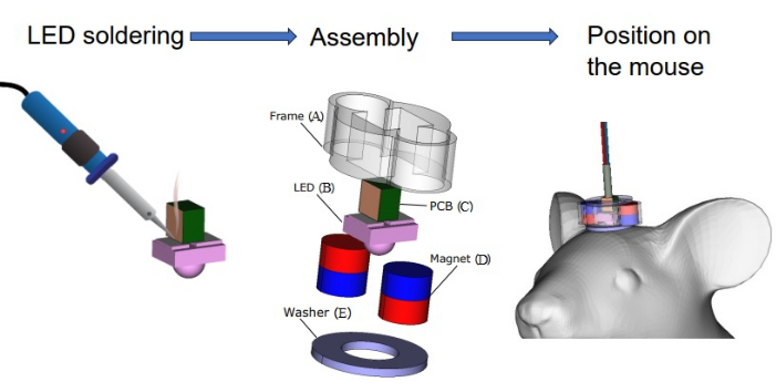
Figure 1: Hardware assembly process. (A) 3D printed frame, (B) LED, (C) LED printed circuit board, (D) magnets, (E) metallic washer. Please click here to view a larger version of this figure.
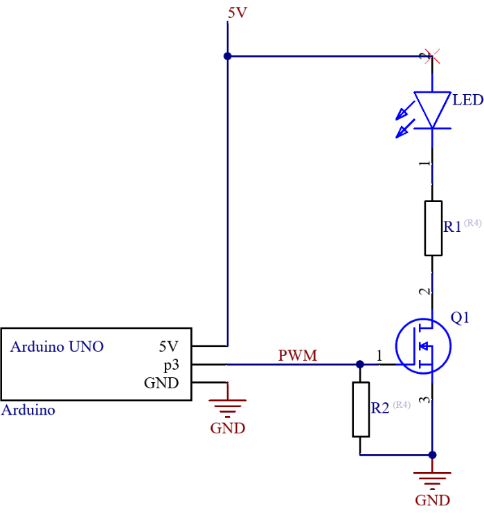
Figure 2: LED control circuit. R1 - current regulation resistor, R2 - pull-down resistor for the MOSFET gate, Q1 - n-channel MOSFET. Please click here to view a larger version of this figure.
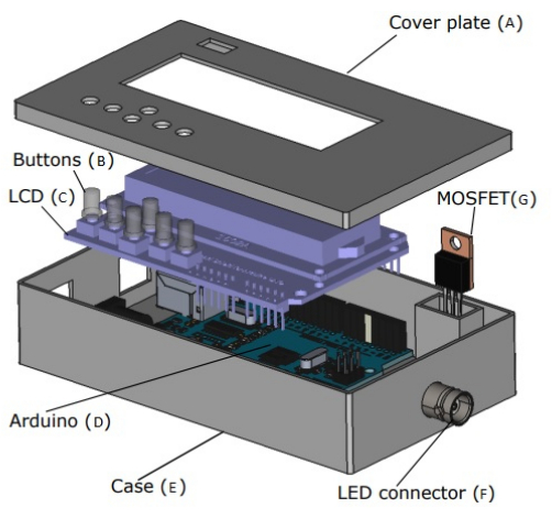
Figure 3: Case 3D view. (A) 3D printed cover plate, (B) 3D printed buttons, (C) LCD keypad shield for Arduino, (D) Arduino, (E) 3D printed case, (F) LED connector, (G) Channel MOSFET. Please click here to view a larger version of this figure.
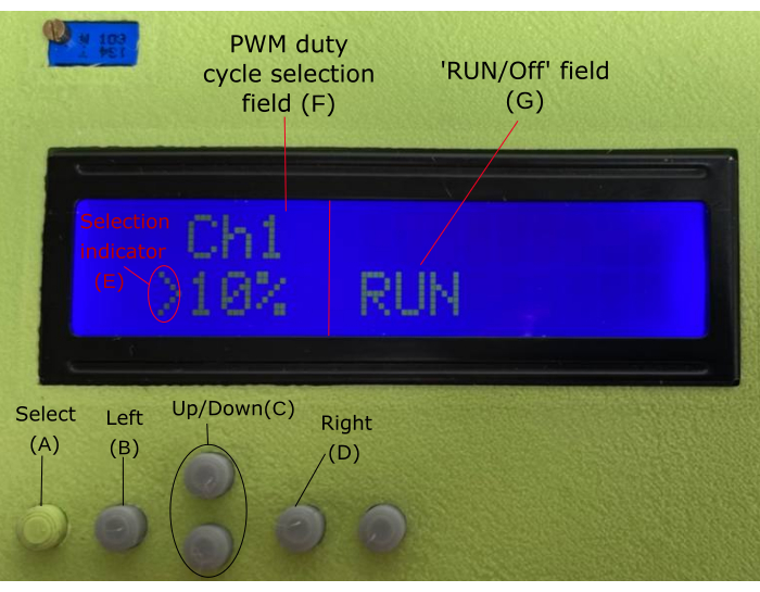
Figure 4: Software. (A) Select button, (B) Left button, (C) Up/Down button, (D) Right button, (E) Selection indicator, (F) PWM duty cycle selection field, (G) RUN/OFF field. Please click here to view a larger version of this figure.
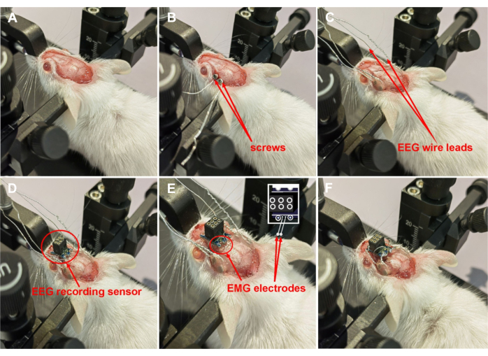
Figure 5: Implantation of an EEG recording system. (A) Skull preparation; (B) Implantation of screws; (C) Fixation of screws with the dental acrylic; (D) Fixation of the EEG recording sensor; (E) Implantation and fixation of the EMG electrodes on the back of the orbicularis oculi muscle; (F) The soldered electrodes to the silver-plated recess of the sensor. Please click here to view a larger version of this figure.
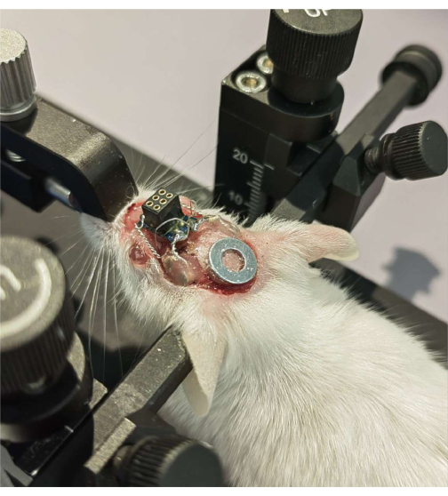
Figure 6: Fixation of a plate for PBM on the occipital bone of the skull. Please click here to view a larger version of this figure.
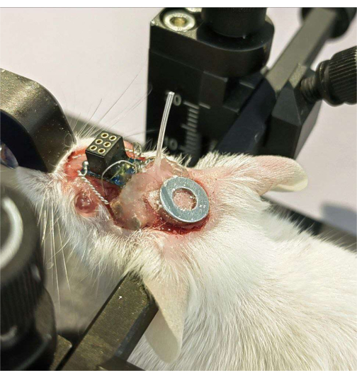
Figure 7: Implantation of a chronic catheter into the right lateral ventricle. Please click here to view a larger version of this figure.
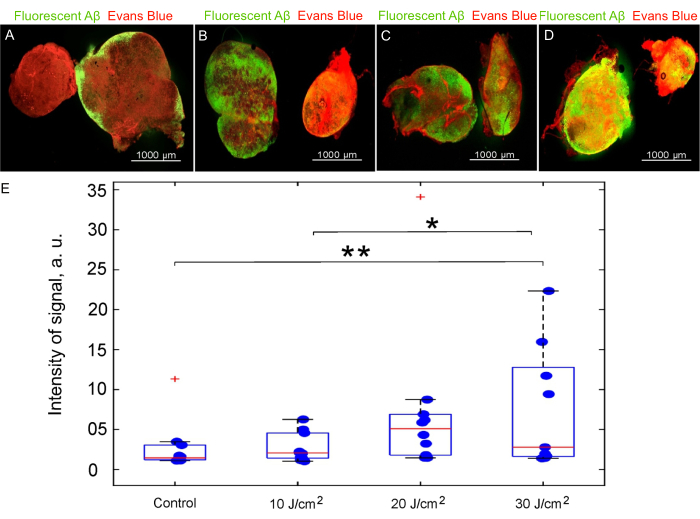
Figure 8: The effects of different LED 1050 nm doses on lymphatic removal of fluorescent Aβ from the brain to dcLNs. (A-D) Representative images of dcLNs from the (A) control group and the PBM group with (B) 10 J/cm2, (C) 20 J/cm2, and (D) 30 J/cm2; (E) Quantitative analysis of the accumulation of fluorescent Aβ in dcLNs from the tested groups (n = 8, one-way ANOVA with Duncan post hoc test, **p < 0.01, *p < 0.05). Please click here to view a larger version of this figure.
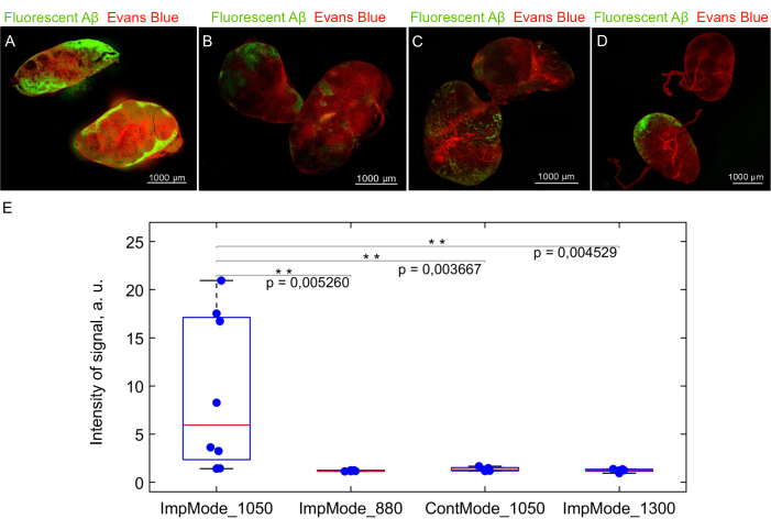
Figure 9: The effects of different LED wavelengths on lymphatic removal of fluorescent Aβ from the brain to dcLNs. (A-D) Representative images of dcLNs from the PBM group with impulse mode 1050 nm (ImpMode_1050, A), 880 nm (ImpMode_880, B), 1300 nm (ImpMode_1300, C) and continuous mode 1050 nm (ContMode_1050 nm, D); (E) Quantitative analysis of accumulation of fluorescent Aβ in dcLNs from the tested groups (n = 8, one-way ANOVA with Duncan post hoc test, **p < 0.01). Please click here to view a larger version of this figure.
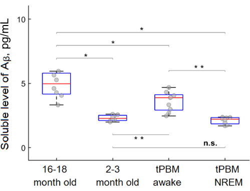
Figure 10: The PBM effects (LED 1050 nm in impulse mode, 30 J/cm2) during NREM and awake state on the level of soluble Aβ (pg/mL) in mouse brains of adult and aged mice. A one-way ANOVA with Duncan post hoc test, **p < 0.01, *p < 0.05. Please click here to view a larger version of this figure.
Supplementary Coding File 1: lcd1key.ino Please click here to download this File.
Discussion
Loading...
$$\rightleftharpoonup{xx}$$
$$\longleftharp{xx}$$,
$$\longrightharp{xx}$$,
MLVs are an important target for the development of innovative technologies for modulation of the brain's drainage and removal of cellular debris and wastes from the brain, especially in aged subjects whose MLV function declines1,22. In a homeostatic state, deep sleep is associated with the natural activation of brain tissue cleansing13,14. Therefore, it is obvious to expect that stimulation of MLVs during deep sleep will be more effective than during wakefulness15,16. The new non-invasive and portable technology of tPBM under EEG control of sleep for stimulation of lymphatic removal of soluble Aβ from the brain of aged male mice is presented here.
The small size of the photo platform (7 mm x 11 mm) and its light weight (1 g) allow it to be firmly and securely mounted on the heads of mice, maintaining their natural motor activity. This also eliminates the need to use anesthesia during tPBM. The tPBM during deep sleep can be performed using any commercial device for an EEG control of sleep stages. Thus, our technology allows performing the study in a home cage, preserving the most natural conditions for animals.
In the first step, an LED wavelength of 1050 nm was randomly selected for the study of the effective dose of photo-effects on lymphatic removal of fluorescent Aβ from the brain to dcLNs in awake adult (2-3 month old) mice. The choice of this wavelength is related to the previous data on effective stimulation of MLVs with a 1267 nm laser8,10,27,28,29,30,31,32,33,34. This wavelength promotes the direct generation of the singlet oxygen in small quantities in brain tissue and in its meninges, which is one of the mechanisms underlying the PBM of MLVs8,28,34. However, the biological effects of both a 1267 nm laser and an LED 1050 nm are related to the generation of singlet oxygen excitation35. Indeed, the bands of a 1267 nm ± 20 nm laser and a 1065 nm ± 15 nm LED are related to formation of the singlet oxygen from the ground state of an oxygen molecule. A band of a 1267 nm laser pumps an oxygen molecule directly into the first excited singlet state, while a band of 1065 nm LED corresponds to additional vibrational energy, which decays very fast, releasing heat to the environment. Moreover, water absorption in the 1065 nm wavelength is tenfold lesser than in the 1267 nm wavelength, which makes it more favorable for biological research35. Furthermore, a 1050 nm LED is commercially available in the market and much cheaper compared with the rare and expensive 1267 nm laser. Note that the emission band of a used 1050 nm LED in this study was 40 nm wide, and it only partially overlaps with a singlet oxygen band. However, we demonstrated the significant effect of tPBM on the lymphatic removal of fluorescent Aβ from the brain to dcLNs using a 1050 nm LED (30 J/cm2). The light dose 30 J/cm2 was determined to be the most effective compared to the light doses 10 J/cm2 and 20 J/cm2 based on the higher intensity of a fluorescent signal from Aβ in dcLNs. Thus, a 1050 nm LED can be very useful for the application of the tPBM technique to humans, which is consistent with the common trend to use LEDs instead of lasers for PBM36,37.
In the second step, the research aimed to answer the question of which light wavelengths are most effective for lymphatic elimination of fluorescent Aβ from the brain of adult awake mice. This step of study was performed using a portable photo platform with different light wavelengths (880 nm, 1050 nm, 1300 nm) and modes (1050 nm in continuous and pulse regimes) with the same dose (30 J/cm2). These wavelengths were chosen due to the widespread use in clinical practice for tPBM (880 nm) and new expensive LEDs (1300 nm), which could potentially be clinically significant. In addition, the pulsed and continuous modes only for the 1050 nm LED as a light resource were compared. These data clearly demonstrate that only a 1050 nm LED in pulse mode vs. other wavelengths and the 1050 nm in continuous mode causes a significant lymphatic removal of Aβ from the brain to dcLNs. These results are consistent with the findings of other researchers, who also indicate the advantage of using a pulsed PBM to effectively achieve biological effects38,39,40.
In the final step, when the effective wavelength, dose and mode were selected, the study of the effects of a 1050 nm LED in pulse mode on the level of soluble Aβ in the brain of aged mice before and after the 10 day-course of tPBM under an EEG control of NREM and wakefulness was carried out. The immunoassay analysis revealed that the soluble Aβ content in the brain was significantly higher in aged mice (16-18 months old) compared with adult mice (2-3 months old). It is interesting to note that the 10-day course of tPBM during deep sleep, but not during awake, effectively reduced the soluble Aβ level in the brains of aged mice to the level of soluble Aβ in the brains of adult mice. An increase in the Aβ content in brain tissue of healthy mice and rats with age has also been noted in other studies25,26, which may be associated with an age-related decrease in the MLV functions leading to ineffective cleansing of brain tissue from this toxic protein1,22. The more effective tPBM of removal of soluble Aβ from brain tissue in sleeping mice compared to awake ones can be explained by the natural activation of brain tissue drainage during sleep. The MLV morphology changes significantly with age, leading to reduced lymphatic removal of metabolites from the aged brain22. However, tPBM during sleep helps restore the lymphatic removal of soluble Aβ from the brain tissue of aging mice to the level of adult animals.
Considering the scattering of light energy when passing through the skull, tPBM limits its effects only on MLVs without penetrating deep into the brain tissues. However, despite the fact that the lymphatic vessels have not yet been found directly in the brain tissues of humans and animals, growing evidence is emerging indicating the presence of the cerebral lymphatic system41,42,43,44. This explains the results showing the removal of toxins (blood and Aβ) from the deep structures of the brain (the ventricular system, the hippocampus) to MLVs and further to the periphery (dcLNs)5,7. Over the course of a century, knowledge has accumulated, indicating a close connection between the brain's drainage and the peripheral lymphatic system45. Even in the absence of a generally recognized lymphatic network in the central nervous system, there are facts indicating lymphatic removal of macromolecules from the deep parts of the brain to the periphery1,2,3,4,5,6,7 This also explains why tPBM, acting only on MLVs, stimulates the clearance of blood and Aβ from the brain5,7.
In summary, this study presents a portable technology of tPBM under EEG control of sleep designed to photo-stimulate the removal of toxins using the example of soluble Aβ from the brain of aged mice with the ability to compare the therapeutic effectiveness of different optical resources. The technology can be used in the natural condition of a home cage without anesthesia, maintaining the natural motor activity of mice. These data open up new prospects for the development of non-invasive and clinically promising photo-technologies for correcting age-related changes in the MLV functions and brain's drainage processes and for the effective cleansing of brain tissues from metabolites and toxins. The proposed technology for photostimulation of the brain's drainage and lymphatic removal of toxins from the brain has limited effects on MLVs due to the scattering of light energy when passing through the skull. Therefore, the technology can be used to develop new methods for treating brain diseases associated with MLV dysfunction. The most pronounced stimulating photo-effects on MLVs are observed in deep (NREM) sleep, which requires the use of PBM under EEG control. This creates certain difficulties (technical, programming) for simultaneous control of the onset of deep sleep and the supply of light exposure.
Disclosures
Loading...
$$\rightleftharpoonup{xx}$$
$$\longleftharp{xx}$$,
$$\longrightharp{xx}$$,
The authors have nothing to disclose.
Acknowledgements
Loading...
$$\rightleftharpoonup{xx}$$
$$\longleftharp{xx}$$,
$$\longrightharp{xx}$$,
This research was supported by a grant from the Russian Science Foundation (No. 23-75-30001).
Materials
| Name | Company | Catalog Number | Comments |
|---|---|---|---|
| 0.1% Tween20 | Helicon, Russia | SB-G2009-100ML | |
| Catheter | Scientific Commodities Inc., USA | PE-10, 0.28 mm ID × 0.61 mm OD | |
| CO2 chamber | Binder, Germany | CB-S 170 | |
| Confocal microscop | Nikon, Japan | A1R MP | |
| Dental acrylic | Zermack, Poland-Russia | Villacryl S, V130V4Z05 | |
| Drill | Foredom, Russia | SR W-0016 | |
| Dumont forceps | Stoelting, USA | 52100-07 | |
| Evans Blue dye | Sigma-Aldrich, St. Louis, MO, USA | 206334 | |
| Hamilton | Hamilton Bonaduz AG, Switzerland | 29 G needle | |
| Ibuprofen | Sintez OJSC, Russia | N/A | Analgesic drug |
| Insulin needle | INSUPEN, Italy | 31 G, 0.25 mm x 6 mm | |
| Levomekol antibacterial ointment | Nizhpharm | D06C | For external use at a dose of 40 mg/g, 1 time per day |
| Micro forceps | Stoelting, USA | 52102-02P | |
| Microcentrifuge | Gyrozen, South Korea | GZ-1312 | |
| Microinjector | Stoelting, USA | 53311 | |
| Non-sharp tweezer | Stoelting, USA | 52108-83P | |
| PINNACLE system | Pinnacle Technology, USA | 8400-K3-SL | System for recording EEG (2 channels) and EMG (1 channel) of mice |
| Shaving machine | Braun | Series 3310s | |
| Single and multi-channel pipettes | Eppendorf, Austria | Epp 3120 000.020, Epp 3122 000.019 | |
| Sodium chloride | Kraspharma, Russia | N/A | |
| Soldering station | AOYUE, China | N/A | |
| Stereotaxic frame | Stoelting, USA | 51500 | |
| Straight dissecting scissors | Stoelting, USA | 52132-10P | |
| Tetracycline | JSC Tatkhimfarmpreparaty, Russia | N/A | Eye ointment |
| Tweezer | Stoelting, USA | 52100-03 | |
| Ultrasonic cell disrupter | Biobase, China | USD-500 | |
| Wound retractor | Stoelting, USA | 52125 | |
| Xylanit | Nita-Farm, Russia | N/A | Muscle relaxant |
| Zoletil 100 | Virbac Sante Animale, France | N/A | General anesthesia |
References
Loading...
$$\rightleftharpoonup{xx}$$
$$\longleftharp{xx}$$,
$$\longrightharp{xx}$$,
- Functional aspects of meningeal lymphatics in ageing and Alzheimer's disease. Nature. 560 (7717), 185-191 (2018).">Da Mesquita, S., et al. Functional aspects of meningeal lymphatics in ageing and Alzheimer's disease. Nature. 560 (7717), 185-191 (2018).
- Meningeal lymphatics clear erythrocytes that arise from subarachnoid hemorrhage. Nat Commun. 11, 3159(2020).">Chen, J., et al. Meningeal lymphatics clear erythrocytes that arise from subarachnoid hemorrhage. Nat Commun. 11, 3159(2020).
- Blocking meningeal lymphatic drainage aggravates Parkinson's disease-like pathology in mice overexpressing mutated α-synuclein. Transl Neurodegener. 8, 7(2019).">Zou, W., et al. Blocking meningeal lymphatic drainage aggravates Parkinson's disease-like pathology in mice overexpressing mutated α-synuclein. Transl Neurodegener. 8, 7(2019).
- Meningeal lymphatic vessels regulate brain tumor drainage and immunity. Cell Res. 30 (3), 229-243 (2020).">Hu, X., et al. Meningeal lymphatic vessels regulate brain tumor drainage and immunity. Cell Res. 30 (3), 229-243 (2020).
- Photostimulation of brain lymphatics in male newborn and adult rodents for therapy of intraventricular hemorrhage. Nat Comm. 14 (1), 6104(2023).">Dong-Yu, L., et al. Photostimulation of brain lymphatics in male newborn and adult rodents for therapy of intraventricular hemorrhage. Nat Comm. 14 (1), 6104(2023).
- Meningeal lymphatic dysfunction exacerbates traumatic brain injury pathogenesis. Nat Commun. 11 (1), 4524(2020).">Bolte, A., et al. Meningeal lymphatic dysfunction exacerbates traumatic brain injury pathogenesis. Nat Commun. 11 (1), 4524(2020).
- Mechanisms of phototherapy of Alzheimer's disease during sleep and wakefulness: the role of the meningeal lymphatics. Front Optoelectron. 16, 22(2023).">Semyachkina-Glushkovskaya, O., et al. Mechanisms of phototherapy of Alzheimer's disease during sleep and wakefulness: the role of the meningeal lymphatics. Front Optoelectron. 16, 22(2023).
- Photostimulation of lymphatic clearance of β- amyloid from mouse brain: new strategy for the therapy of Alzheimer's disease. Front Optoelectron. 16, 45(2023).">Dongyu, L., et al. Photostimulation of lymphatic clearance of β- amyloid from mouse brain: new strategy for the therapy of Alzheimer's disease. Front Optoelectron. 16, 45(2023).
- Mechanisms of phototherapy of Alzheimer's disease during sleep and wakefulness: the role of the meningeal lymphatics. Front Optoelectron. 16, 22(2023).">Semyachkina-Glushkovskaya, O., et al. Mechanisms of phototherapy of Alzheimer's disease during sleep and wakefulness: the role of the meningeal lymphatics. Front Optoelectron. 16, 22(2023).
- Intranasal delivery of liposomes to glioblastoma by photostimulation of the lymphatic system. Pharmaceutics. 15 (1), 36(2023).">Semyachkina-Glushkovskaya, O., et al. Intranasal delivery of liposomes to glioblastoma by photostimulation of the lymphatic system. Pharmaceutics. 15 (1), 36(2023).
- Night photostimulation of clearance of beta-amyloid from mouse brain: New strategies in preventing Alzheimer's disease. Cells. 10 (12), 3289(2021).">Semyachkina-Glushkovskaya, O., et al. Night photostimulation of clearance of beta-amyloid from mouse brain: New strategies in preventing Alzheimer's disease. Cells. 10 (12), 3289(2021).
- Technology of the photobiostimulation of the brain's drainage system during sleep for improvement of learning and memory in male mice. Biomed Opt Express. 15 (1), 44-58 (2024).">Semyachkina-Glushkovskaya, O., et al. Technology of the photobiostimulation of the brain's drainage system during sleep for improvement of learning and memory in male mice. Biomed Opt Express. 15 (1), 44-58 (2024).
- Coupled electrophysiological, hemodynamic, and cerebrospinal fluid oscillations in human sleep. Science. 366 (6465), 628-631 (2019).">Fultz, N., et al. Coupled electrophysiological, hemodynamic, and cerebrospinal fluid oscillations in human sleep. Science. 366 (6465), 628-631 (2019).
- Sleep drives metabolite clearance from the adult brain. Science. 342 (6156), 373-377 (2013).">Xie, L., et al. Sleep drives metabolite clearance from the adult brain. Science. 342 (6156), 373-377 (2013).
- Phototherapy of Alzheimer's disease: Photostimulation of brain lymphatics during sleep: A systematic review. Int J Mol Sci. 24 (13), 10946(2023).">Semyachkina-Glushkovskaya, O., et al. Phototherapy of Alzheimer's disease: Photostimulation of brain lymphatics during sleep: A systematic review. Int J Mol Sci. 24 (13), 10946(2023).
- Brain waste removal system and sleep: Photobiomodulation as an innovative strategy for night therapy of brain diseases. Int J Mol Sci. 24 (4), 3221(2023).">Semyachkina-Glushkovskaya, O., et al. Brain waste removal system and sleep: Photobiomodulation as an innovative strategy for night therapy of brain diseases. Int J Mol Sci. 24 (4), 3221(2023).
- Increased glymphatic influx is correlated with high EEG delta power and low heart rate in mice under anesthesia. Sci Adv. 5 (2), 5447(2019).">Hablitz, L. M., et al. Increased glymphatic influx is correlated with high EEG delta power and low heart rate in mice under anesthesia. Sci Adv. 5 (2), 5447(2019).
- Primary cultures of neuronal and non-neuronal rat brain cells secrete similar proportions of amyloid beta peptides ending at A beta40 and A beta42. Neuroreport. 10 (14), 2965-2969 (1999).">Fukumoto, H., et al. Primary cultures of neuronal and non-neuronal rat brain cells secrete similar proportions of amyloid beta peptides ending at A beta40 and A beta42. Neuroreport. 10 (14), 2965-2969 (1999).
- Large quantities of Abeta peptide are constitutively released during amyloid precursor protein metabolism in vivo and in vitro. J Biol Chem. 286 (16), 15989-15997 (2011).">Moghekar, A., et al. Large quantities of Abeta peptide are constitutively released during amyloid precursor protein metabolism in vivo and in vitro. J Biol Chem. 286 (16), 15989-15997 (2011).
- The role of amyloid oligomers in neurodegenerative pathologies. Int J Biol Macromol. 181, 582-604 (2021).">Wells, C., Brennan, S., Keon, M., Ooi, L. The role of amyloid oligomers in neurodegenerative pathologies. Int J Biol Macromol. 181, 582-604 (2021).
- Turnover of amyloid beta-protein in mouse brain and acute reduction of its level by phorbol ester. J Neurosci. 18 (5), 1743-1752 (1998).">Savage, M., et al. Turnover of amyloid beta-protein in mouse brain and acute reduction of its level by phorbol ester. J Neurosci. 18 (5), 1743-1752 (1998).
- Meningeal lymphatic vessels at the skull base drain cerebrospinal fluid. Nature. 572 (7767), 62-66 (2019).">Ahn, J., et al. Meningeal lymphatic vessels at the skull base drain cerebrospinal fluid. Nature. 572 (7767), 62-66 (2019).
- Regional amyloid correlates of cognitive performance in ageing and mild cognitive impairment. Brain Commun. 4 (1), 016(2022).">Stevens, D., et al. Regional amyloid correlates of cognitive performance in ageing and mild cognitive impairment. Brain Commun. 4 (1), 016(2022).
- Amyloidosis in Alzheimer's disease: Pathogeny, etiology, and related therapeutic directions. Molecules. 27 (4), 1210(2022).">Ma, C., Hong, F., Yang, S. Amyloidosis in Alzheimer's disease: Pathogeny, etiology, and related therapeutic directions. Molecules. 27 (4), 1210(2022).
- Intracellular amyloid-β in the normal rat brain and human subjects and its relevance for Alzheimer's disease. J Alzheimers Dis. 95 (2), 719-733 (2023).">Kobro-Flatmoen, A., Hormann, T., Gouras, G. Intracellular amyloid-β in the normal rat brain and human subjects and its relevance for Alzheimer's disease. J Alzheimers Dis. 95 (2), 719-733 (2023).
- Endogenous murine amyloid-β peptide assembles into aggregates in the aged C57BL/6J mouse suggesting these animals as a model to study pathogenesis of amyloid-β plaque formation. J Alzheimers Dis. 61 (4), 1425-1450 (2018).">Ahlemeyer, B., Halupczok, S., Rodenberg-Frank, E., Valerius, K., Baumgart-Vogt, E. Endogenous murine amyloid-β peptide assembles into aggregates in the aged C57BL/6J mouse suggesting these animals as a model to study pathogenesis of amyloid-β plaque formation. J Alzheimers Dis. 61 (4), 1425-1450 (2018).
- Pilot study of transcranial photobiomodulation of lymphatic clearance of beta-amyloid from the mouse brain: Breakthrough strategies for nonpharmacologic therapy of Alzheimer's disease. Biomed Opt Express. 10 (8), 4003-4017 (2019).">Zhinchenko, E., et al. Pilot study of transcranial photobiomodulation of lymphatic clearance of beta-amyloid from the mouse brain: Breakthrough strategies for nonpharmacologic therapy of Alzheimer's disease. Biomed Opt Express. 10 (8), 4003-4017 (2019).
- Transcranial photobiomodulation of clearance of beta-amyloid from the mouse brain: Effects on the meningeal lymphatic drainage and blood oxygen saturation of the brain. Adv Exp Med Biol. 1269, 57-61 (2021).">Semyachkina-Glushkovskaya, O., et al. Transcranial photobiomodulation of clearance of beta-amyloid from the mouse brain: Effects on the meningeal lymphatic drainage and blood oxygen saturation of the brain. Adv Exp Med Biol. 1269, 57-61 (2021).
- Photobiomodulation of lymphatic drainage and clearance: Perspective strategy for augmentation of meningeal lymphatic functions. Biomed Opt Express. 11 (2), 725-734 (2020).">Semyachkina-Glushkovskaya, O., et al. Photobiomodulation of lymphatic drainage and clearance: Perspective strategy for augmentation of meningeal lymphatic functions. Biomed Opt Express. 11 (2), 725-734 (2020).
- Photostimulation of extravasation of beta-amyloid through the model of blood-brain barrier. Electronics. 9 (6), 1056(2020).">Zhinchenko, E., et al. Photostimulation of extravasation of beta-amyloid through the model of blood-brain barrier. Electronics. 9 (6), 1056(2020).
- Photostimulation of cerebral and peripheral lymphatic functions. Transl Biophotonics. 2 (1-2), e201900036(2020).">Semyachkina-Glushkovskaya, O., et al. Photostimulation of cerebral and peripheral lymphatic functions. Transl Biophotonics. 2 (1-2), e201900036(2020).
- Photomodulation of lymphatic delivery of liposomes to the brain bypassing the blood-brain barrier: New perspectives for glioma therapy. Nanophotonics. 10 (12), 3215-3227 (2021).">Semyachkina-Glushkovskaya, O., et al. Photomodulation of lymphatic delivery of liposomes to the brain bypassing the blood-brain barrier: New perspectives for glioma therapy. Nanophotonics. 10 (12), 3215-3227 (2021).
- Photomodulation of lymphatic delivery of Bevacizumab to the brain: The role of singlet oxygen. Adv Exp Med Biol. 1395, 53-57 (2022).">Semyachkina-Glushkovskaya, O., et al. Photomodulation of lymphatic delivery of Bevacizumab to the brain: The role of singlet oxygen. Adv Exp Med Biol. 1395, 53-57 (2022).
- Transcranial photosensitizer-free laser treatment of glioblastoma in rat brain. Int J Mol Sci. 24 (18), 13696(2023).">Semyachkina-Glushkovskaya, O., et al. Transcranial photosensitizer-free laser treatment of glioblastoma in rat brain. Int J Mol Sci. 24 (18), 13696(2023).
- Direct 1O2 optical excitation: A tool for redox biology. Redox Biol. 13, 39-59 (2017).">Blázquez-Castro, A. Direct 1O2 optical excitation: A tool for redox biology. Redox Biol. 13, 39-59 (2017).
- Comparison of laser and diode sources for acceleration of in vitro wound healing by low-level light therapy. J Biomed Opt. 19 (3), 038001(2014).">Spitler, R., Berns, M. Comparison of laser and diode sources for acceleration of in vitro wound healing by low-level light therapy. J Biomed Opt. 19 (3), 038001(2014).
- Comparative effectiveness of light emitting diodes (LEDs) and Lasers in near infrared photoimmunotherapy. Oncotarget. 7 (12), 14324-14335 (2016).">Sato, K., Watanabe, R., Hanaoka, H., Nakajima, T., Choyke, P., Kobayashi, H. Comparative effectiveness of light emitting diodes (LEDs) and Lasers in near infrared photoimmunotherapy. Oncotarget. 7 (12), 14324-14335 (2016).
- Photobiomodulation with pulsed and continuous wave near-infrared laser (810 nm, Al-Ga-As) augments dermal wound healing in immunosuppressed rats. PLoS One. 11 (11), e0166705(2016).">Keshri, G., Gupta, A., Yadav, A., Sharma, S., Singh, S. Photobiomodulation with pulsed and continuous wave near-infrared laser (810 nm, Al-Ga-As) augments dermal wound healing in immunosuppressed rats. PLoS One. 11 (11), e0166705(2016).
- Pulse frequency dependency of photobiomodulation on the bioenergetic functions of human dental pulp stem cells. Sci Rep. 7 (1), 15927(2017).">Kim, H., et al. Pulse frequency dependency of photobiomodulation on the bioenergetic functions of human dental pulp stem cells. Sci Rep. 7 (1), 15927(2017).
- The pulse light mode enhances the effect of photobiomodulation on B16F10 melanoma cells through autophagy pathway. Lasers Med Sci. 38 (1), 71(2023).">Chen, Z., et al. The pulse light mode enhances the effect of photobiomodulation on B16F10 melanoma cells through autophagy pathway. Lasers Med Sci. 38 (1), 71(2023).
- An immunohistochemical study of lymphatic elements in the human brain. Proc Natl Acad Sci U S A. 118 (3), e2002574118(2021).">Mezey, E., et al. An immunohistochemical study of lymphatic elements in the human brain. Proc Natl Acad Sci U S A. 118 (3), e2002574118(2021).
- Characteristic features of deep brain lymphatic vessels and their regulation by chronic stress. Research. 6, 0120(2023).">Chang, J., et al. Characteristic features of deep brain lymphatic vessels and their regulation by chronic stress. Research. 6, 0120(2023).
- Multiple sclerosis: Presence of lymphatic capillaries and lymphoid tissue in the brain and spinal cord. Science. 203 (4385), 1123-1125 (1979).">Prineas, L. W. Multiple sclerosis: Presence of lymphatic capillaries and lymphoid tissue in the brain and spinal cord. Science. 203 (4385), 1123-1125 (1979).
- Pilot identification of the Live-1/Prox-1 expressing lymphatic vessels and lymphatic elements in the unaffected and affected human brain. bioRxiv. , 458990(2021).">Semyachkina-Glushkovskaya, O., et al. Pilot identification of the Live-1/Prox-1 expressing lymphatic vessels and lymphatic elements in the unaffected and affected human brain. bioRxiv. , 458990(2021).
- Blood-brainbarrier, lymphatic clearance, and recovery: Ariadne's thread in labyrinths of hypotheses. Int J Mol Sci. 19 (12), 3818(2018).">Semyachkina-Glushkovskaya, O., Postnov, D., Kurths, J. Blood-brainbarrier, lymphatic clearance, and recovery: Ariadne's thread in labyrinths of hypotheses. Int J Mol Sci. 19 (12), 3818(2018).
Reprints and Permissions
Request permission to reuse the text or figures of this JoVE article
Request Permission