This study presents a simple, user-friendly ratiometric FRET assay for the detection and quantitative assessment of DNase activity, and demonstrates its application in the analysis of a weak nuclease.
A subscription to JoVE is required to view this content. Sign in or start your free trial.
Method Article
Assessment of DNase Activity by Ratiometric Fluorescence Resonance Energy Transfer
In This Article
Summary
Abstract
The present protocol describes a simple and sensitive ratiometric Förster resonance energy transfer (FRET) assay for the detection and quantitative assessment of DNase activity. Ratiometric FRET measurements make use of the ratio of donor and acceptor emission signals. The assay detects single-stranded DNA breaks using a staple-shaped, dual-tagged 38-mer FRET oligoprobe, employed as a real-time DNA cleavage sensor. The main application of the described approach is the quantitative assessment of the effects of reaction conditions, such as pH, temperature, and buffer composition, on DNase activity. Due to its ability to detect even minor and slow DNA cleavage, the assay is particularly well suited for investigating weak nucleolytic activity requiring extended periods of observation and for studying the effects of pH on DNase activity. These specific advantages of this ratiometric FRET protocol are illustrated by its application to the detection and analysis of the DNase activity of leukocyte elastase inhibitor (LEI).
Introduction
The ratiometric fluorescence approach is an analytical method that uses ratios of fluorescence signals instead of relying solely on the intensity of a single signal1,2. The advantage of using the ratios is that they provide internal calibration and compensate for many random factors, such as variations in sample and probe concentrations, instrument parameters, etc. This enhances the accuracy and sensitivity of fluorescent probes.
In the specific context of Förster resonance energy transfer (FRET) probes, the term "ratiometric" refers to the fact that the measurement is based on the ratio of two emission signals from the pair of FRET donor and acceptor fluorophores. To quantify the activity of the target analyte, ratiometric FRET probes use the ratio of acceptor/donor emission intensities (or, in some cases, the inverted donor/acceptor ratio). The ratio is directly related to the FRET efficiency. It provides a self-normalizing mechanism that minimizes background noise and accounts for variations in illumination conditions and probe concentration3.
Recent interesting applications of the ratiometric method include its use in ratiometric electrochemical biosensors for the detection of circulating tumor DNA (ctDNA)4,5, in Brownian motion-powered bio-nanomachines for FRET detection of the phagocytic phase of apoptosis6, and for express FRET labeling and analysis of phagocytic clearance7.The ratiometric FRET approach described in this work is useful for the quantitative assessment of DNase activity. The assay measurements are based on the ratios of the FRET pair emissions. Due to its ability to detect even minor and slow DNA cleavage, the assay is particularly well suited for the investigation of weak nucleolytic activity requiring longer observation periods and for experiments at different pH levels.
Here, some specific advantages of the ratiometric FRET assay are illustrated by its application to study the DNase activity of a weak nuclease-leukocyte elastase inhibitor (LEI). The report includes a detailed protocol with step-by-step instructions and a description of the ratiometric data analysis.
Ratiometric FRET probe description
The ratiometric FRET probe is a self-complementary 38-mer DNA oligo carrying the donor-acceptor FRET pair of FAM-TAM (Fluorescein-Tetramethylrhodamine). Its sequence is 5'-AAGGGT(TAM) CCTGCTGCAGGACCCTTAACGCATTATGCGT(FAM)T-3'. The self-hybridizing 38-mer assumes a staple-shaped conformation, comprising two connected hairpins of 23 and 15 nucleotides, which position its two fluorophores 23.8 Å from each other. This is significantly closer than the Förster radius for this pair, R0 = 55 Å8. Such positioning corresponds to a very high efficiency of energy transfer from donor to acceptor7: EFRET = 0.993477. As a result, when illuminated at the FAM excitation wavelength (488 nm), the FRET probe emits fluorescence at the emission wavelength of the acceptor TAM (580 nm), whereas FAM emission (525 nm) is suppressed.
Figure 1A shows the UNAFold-predicted secondary structure of the probe, with small loops at both hairpin apexes due to steric hindrance from tight curvature. The schematic of DNA break detection by the FRET probe is presented in Figure 1B. It shows that breakage of the oligoprobe by a DNase separates its FRET pair. This separation produces drastic changes in the emission spectra of both fluorophores in the probe. Figure 1C presents the probe emission spectra before and after its cleavage and demonstrates that the split of the FRET pair after DNase II probe cleavage increases donor emission at 525 nm (ID 525 nm) and simultaneously decreases acceptor emission at 580 nm (IA 580 nm). Figure 1D shows the PAGE gel and FRET ratios corresponding to the emission spectra. DNase II probe cleavage produced two wide bands corresponding to 12-16-mer FAM (green) and 20-24-mer TAM (red) fragments, indicating random cuts of the 38-mer on either side of T23 in the vicinity of the connection area between the hairpins.
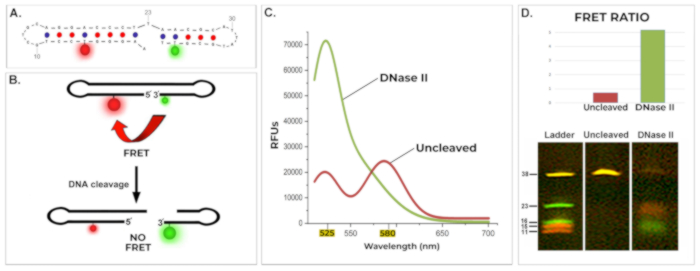
Figure 1: Structure and operation of the ratiometric FRET probe. (A) Predicted secondary structure of the FRET probe generated by UNAFold. (B) Schematic representation of the working FRET probe. The ratiometric FRET probe is a staple-shaped 38-mer oligonucleotide labeled with a FRET donor (FAM) and acceptor (TAM) at an effective FRET distance of 23.8 Å, ensuring >99% FRET efficiency. Cleavage of the oligoprobe by a DNase separates the FRET pair, abolishing FRET. (C) Emission spectra of the FRET probe before and after DNase II-mediated cleavage. Prior to cleavage, FAM emission is suppressed, and TAM emission is prominent. DNase-mediated cleavage separates FAM and TAM, restoring FAM emission (525 nm peak) and abolishing TAM emission (580 nm drop). λexcitation = 488 nm. (D) Ratiometric and electrophoretic assessment of the FRET probe before and after DNase II cleavage. Upper panel: FRET ratios (RD/A = ID 525 nm / IA 580 nm), representing a quantitative measure of the probe state. Red: uncleaved; green: DNase II-cleaved. Lower panel: Denaturing PAGE gel corresponding to fluorometric data. DNase II cleavage yields FAM-labeled fragments (12-16 nt, green) and TAM-labeled fragments (20-24 nt, red), indicating cleavage near the connection between hairpins, flanking T23. Uncleaved control shows yellow fluorescence. Ladder: FAM- and TAM-labeled oligonucleotides corresponding to the 5′ and 3′ segments of the 38-mer probe, with fluorescent labels in their original positions. Reaction conditions: 10 mM sodium acetate buffer (pH 5.2), 1 pmol/µL FRET probe, 0.0033 U/µL DNase II, 24 h at 37 °C. RD/A = ID 525 nm/IA 580 nm; λexcitation = 488 nm. UNAFold conditions: 1 µM FRET probe, 10 mM Na+, 0 mM Mg2+, 37 °C. Please click here to view a larger version of this figure.
While the changes in the individual test emission spectra are obvious, quantitative comparisons between large numbers of emission spectra require a more user-friendly and compact quantitative parameter. This role is played by the FRET ratio. The presented fluorescence method belongs to a group of ratiometric approaches that rely on the direct (two-channel) ratios of fluorescence emissions of FRET pair members for their quantitative assessments2,9.
The approach uses the ratio of donor/acceptor emissions (RD/A = ID 525 nm / IA 580 nm), which is related to the FRET efficiency and quantitatively characterizes the condition of the 38-mer (broken or intact). The ID / IA ratio provides a self-normalizing mechanism that inherently accounts for variations in factors such as probe concentration, excitation intensity, and photobleaching3. The approach is suitable only for FRET probes that have donor and acceptor linked within the same probe, where their ratio is known and constant. For this reason, the approach does not need to consider either the direct excitation of the acceptor or donor crosstalk2. The fixed donor-acceptor stoichiometry of the probe ensures that the donor/acceptor emission ratios are not affected by these variations and directly reflect the probe's condition. As a result, the ratio of donor-acceptor emissions provides a quantitative DNA cleavage parameter with built-in signal normalization2,3.
The assay employs the ID / IA ratio (rather than the opposite IA / ID ratio) because this ratio describes FRET cessation after probe cleavage and thus changes in the same direction as DNase activity-increasing with increased cleavage. This is illustrated by Figure 1D, which shows the FRET ratio of the ratiometric probe and its PAGE electrophoresis before and after its cleavage by DNase II, both corresponding to the spectra in Figure 1C. To demonstrate the ratiometric FRET assay's convenience, time ecomomy, and ease of use, it was applied to uncover and study the DNase activity of a weak nuclease-leukocyte elastase inhibitor (LEI).
Weak nuclease LEI
Protease inhibitor LEI is naturally metastable, i.e., topologically unbalanced. This feature is essential for its inhibitory mechanism. When its target protease cleaves the LEI reactive site loop (RSL), the protease inhibitor molecule undergoes a drastic conformational change, mechanically stretching the protease into an inactive form10,11. Remarkably, this also breaks and distends the LEI molecule itself and unmasks a cryptic DNase site that is covered in its native fold, thus converting LEI into an active endonuclease L-DNase II (LEI-derived DNase II)11,12.The cryptic DNase site can also be exposed by the denaturation and breakage of LEI caused by its overnight incubation at pH 2 in sulfuric acid10,13. The utilization of this FRET assay to study LEI demonstrated that harsh chemical denaturation treatments and targeted enzymatic cleavage are not strictly required to unmask LEI's latent nucleolytic activity.
The presented application and protocol of the ratiometric FRET approach describe the uncovering of weak DNase activity inherently present in normal, non-denatured LEI. The discovery of the intrinsic nucleolytic ability of uncleaved LEI and its analysis was possible due to the assay's suitability for extended periods of observation at different acidic pH levels.
Access restricted. Please log in or start a trial to view this content.
Protocol
Assessment of DNase Activity by Ratiometric Fluorescence Resonance Energy Transfer
The method is suitable for the detection and analysis of the nucleolytic properties of a soluble protein. To examine the pH dependence of the investigated DNase, the procedure simultaneously assesses DNase activity across a range of pH values. The technique can be used to study weak and slow nuclease activities. The details of the reagents and the equipment used in this study are listed in the Table of Materials.
1. Preparing incubation mix components for the fluorometric assessment system
- Prepare the buffer solutions at desired pH ranges. For pH range 6.8 to 8.0, use Tris-HCl or phosphate buffers. Use Tris-HCl buffer for physiological pH conditions (pH 7.2 to 8.0) due to its stability and minimal interference with enzyme activity.
NOTE: Phosphate buffers are appropriate for this pH range but are less suitable for higher pH values (>7.5) due to their buffering capacity limitations.
For a pH range of 4.2 to 7.2, use acetate or citrate buffers. Acetate buffer is effective for lower pH conditions, down to pH 4.2.
NOTE: Citrate buffer is usable at slightly higher pH values (pH 5.6 to 7.2) and offers good buffering capacity. These buffers are suitable for studying DNase activity under acidic to neutral conditions.The presented study of the nucleolytic activity of LEI used 10 mM sodium acetate buffers with pH ranging from 4.8 to 6.5. - Dilute the protein sample to a suitable concentration in each buffer solution. When activity in the sample is completely unknown, prepare a range of dilutions of the protein. The optimal dilution depends on the strength of DNase activity and must be determined experimentally by testing several protein concentrations.
NOTE: The presented study of the ultra-weak nucleolytic activity of LEI used it at a concentration of 1 µM.- Always check the pH of the final solution when using highly concentrated buffers, because the pH of concentrated buffers can change upon dilution.
- Prepare the ratiometric FRET probe solution in nuclease-free water. Use FRET probe concentrations close to 1 µM (1 pmol/µL). Avoid using very high probe concentrations.
Rapidly heat and cool the probe solution before application to ensure complete dissociation of undesirable base pairings.
NOTE: The presented study of the nucleolytic activity of LEI used a concentration of 1 µM (1 pmol/µL) of FRET probe in nuclease-free water.
2. Induction of the DNase reaction using the ratiometric FRET probe as the substrate
- Prepare three wells in a 96-well plate for each pH condition to be tested. Include three additional wells, each for the positive and negative controls.
- In each well for the pH series, combine the following:
70 μL of Nuclease-Free Water
10 μL of 10X Buffer (buffer of your choice)
10 μL of sample with protein
10 μL of ratiometric probe solution (10 pmol/μL stock concentration)
NOTE: The presented study of the nucleolytic activity of LEI in acidic pH used the following 10X Buffer: 100 mM Na Acetate pH 5.2 - In each positive control well, combine the following:
79 μL of Nuclease-Free Water
10 μL of 10X DNase II Buffer (100 mM Na Acetate pH 5.2)
0.33 μL of DNase II (1 U/μL stock)
10 μL of ratiometric probe solution (10 pmol/μL stock concentration)
NOTE: For studies at acidic pH, use DNase II as a positive control. When assessing DNase activity near pH 7 (specifically pH 6.5-8), switch to DNase I and its associated buffer (e.g., 10 mM Tris-HCl, 2.5 mM MgCl2, 0.5 mM CaCl2, pH 7.6) instead of DNase II and acetate buffer. - In each negative control well add nuclease-free water in place of the protein sample.
- In each well for the pH series, combine the following:
- Induce the DNase reaction by adding the probe solutions to the reaction mixtures containing DNase protein at different pH values.
NOTE: Mix all samples thoroughly while avoiding bubble formation. For active DNase samples, start all reactions as close to simultaneously as possible. For ultra-weak activity, such as that of LEI, strict timing is less critical.
NOTE: To ensure consistent incubation times, especially when dealing with active nucleases, stagger the addition of samples based on the time required for each measurement. For example, if each measurement takes 10 s, add the second sample 10 s after the first.
3. Incubation
- Incubate the studied DNase with the ratiometric FRET probe at 37°C in a water bath or temperature-regulated incubator for various time periods. Use multiple time-points with regular sampling intervals to accurately assess enzyme turnover rates and minimize artifacts. The presented study of the ultra-weak nucleolytic activity of LEI used 24 h incubations to register the slow nucleolytic reaction.
NOTE: Determine the optimal incubation time in the preliminary experiments, because it depends on the strength of the studied DNase. An active nuclease would require a very short time to complete the probe cleavage, so start with a 15-30 min incubation. If the sample is too concentrated, and the reaction is too fast, the protein concentration should be reduced. If the sample protein is too weak, the incubation time can increase as needed.
4. Signal normalization to the same pH conditions
- Stop all incubation reactions by adding 100 μL of 250 mM Tris-HCL (pH 8.0). This step also equalizes the pH across all samples.
NOTE: The pH equalization step is important when comparing reactions initiated under different pH conditions. The procedure ensures complete separation of the probe fragments maximizing its FRET-based signal. In addition, the probe’s FAM fluorophore emission is reduced in acidic pH and is optimal at pH 8.0. For samples with higher ionic strength than those used in this study, a more concentrated (>250 mM) and higher pH (>8.0) equalization solution might be required to achieve effective pH adjustment.
NOTE: Alkaline pH effectively stops activity of acid nucleases such as LEI, as demonstrated in the current study. For neutral or alkaline nucleases, appropriate specific inactivation methods should be identified and applied prior to signal assessment.
5. Fluorometric signal detection and calculation of FRET ratios
- Immediately after the pH equalization step, use a spectrofluorometer to record the emission spectra of both the donor and acceptor fluorophores. Perform measurements directly in the 96-well plate by exciting the donor fluorophore at 488 nm and simultaneously detecting emissions at 525 nm (donor) and 580 nm (FRET-derived acceptor signal).
- Use the recorded fluorescence intensities to evaluate DNase activity in each sample. Apply appropriate statistical methods to analyze the data and assess significance.
NOTE: Calculate the FRET ratio using the formula: RFRET = Donor emission at 525 nm/Acceptor emission at 580 nm. In this assay, DNA cleavage disrupts FRET by separating the fluorophores, resulting in an increased donor emission and a decreased acceptor emission. This shift raises the FRET ratio (RFRET), which serves as an indicator of DNase activity.
6. PAGE-based verification of cleavage
- Verify the cleavage of the FRET probe by performing denaturing polyacrylamide gel electrophoresis (PAGE)14. The FRET probe and its cleavage products are fluorescently labeled and visible without additional staining.
NOTE: In the representative study, LEI-mediated cleavage of the 38-mer FRET probe was confirmed by denaturing PAGE (see Figure 2). The analysis revealed that LEI produced 6-8-mer FAM-labeled fragments and 15-16-mer TAM-labeled fragments, consistent with cleavage near both hairpin apexes. This pattern differed from the fragment profile generated by DNase II.- Load 15 µL of each sample onto a 20% denaturing PAGE gel.
- Run the gel for 1 h at 100 V.
- Acquire gel images using a documentation system equipped with a high-resolution color camera.
NOTE: The images in the representative study were captured using a digital SLR camera.
Access restricted. Please log in or start a trial to view this content.
Results
This representative study highlights specific advantages of the ratiometric FRET approach for investigating slow enzymatic reactions that require extended periods of observation. In particular, the application demonstrates the detection and analysis of the inherent DNase activity of LEI using a ratiometric FRET system. The results of this study are shown in Figure 2.
Using this approach, we demonstr...
Access restricted. Please log in or start a trial to view this content.
Discussion
This study demonstrates the application of ratiometric FRET for the detection and evaluation of DNase activity using a weak nuclease - leukocyte elastase inhibitor (LEI). The approach provides a convenient and quantitative method for assessing the DNase properties of proteins and biomolecules. Its protocol is straightforward and its data analysis procedure is direct and simple. The assay uses a 38-mer FRET probe which tolerates long incubations at various acidic pH. This property, as we demonstrated, enables the assay to...
Access restricted. Please log in or start a trial to view this content.
Disclosures
The authors have nothing to disclose.
Acknowledgements
This research was supported by grant R01 GM148812 from the National Institute of General Medical Sciences, NIH; by grants R21 CA255979 from the National Cancer Institute, NIH; and grants R21 AG073887 and R21 AG071978 both from National Institute on Aging, NIH (all V.V.D.).
Access restricted. Please log in or start a trial to view this content.
Materials
| Name | Company | Catalog Number | Comments |
|---|---|---|---|
| 96 well plate | Falcon | 353219 | |
| DNA FRET Probe | IDT DNA | 5’-AAG GGT(TAM)CCT GCT GCA GGA CCC TTA ACG CAT TAT GCG T(FAM)T- 3’; FAM – Fluorescein-dT; TAM – Tetramethylrhodamine-dT. | |
| DNase II | Sigma Aldrich | D8764 | |
| EVOLT digital SLR Camera | Olympus | E500 | |
| Gel Electrophoresis Apparatus with Power supply | Bio-Rad | ||
| LEI | Novoprotein | CJ01 | |
| LiCOR Gel Imaging System | LiCOR | ||
| Nuclease-free water | Invitrogen | 10977015 | |
| Pre-Cast PAGE Gel | Invitrogen | EC68852 | |
| Sodium Acetate Buffer Solution pH 5.2 | Sigma Aldrich | S2899-100ML | |
| Spectrofluorometer | Tecan | Saffire 2 |
References
- A guide to ratiometric fluorescence. , AZoM. https://www.azom.com/article.aspx?ArticleID=16081 (2018).
- Müller, S. M., Galliardt, H., Schneider, J., Barisas, B. G., Seidel, T. Quantification of Förster resonance energy transfer by monitoring sensitized emission in living plant cells. Front Plant Sci. 4, 413(2013).
- Lakowicz, J. R. Principles of fluorescence spectroscopy. , Springer Science & Business Media. (2006).
- Chai, H., Tang, Y., Guo, Z., Miao, P. Ratiometric electrochemical switch for circulating tumor DNA through recycling activation of blocked DNAzymes. Anal Chem. 94 (6), 2779-2784 (2022).
- Liu, G., Ma, X., Tang, Y., Miao, P. Ratiometric fluorescence method for ctDNA analysis based on the construction of a DNA four-way junction. Analyst. 145 (4), 1174-1178 (2020).
- Minchew, C. L., Didenko, V. V. Nanoblinker: Brownian motion powered bio-nanomachine for FRET detection of phagocytic phase of apoptosis. PLoS One. 9 (9), e108734(2014).
- Didenko, V. V. Express FRET labeling and analysis of phagocytic clearance. Methods Mol Biol. 1644, 3-11 (2017).
- Domingo, B., Sabariegos, R., Picazo, F., Llopis, J. Imaging FRET standards by steady-state fluorescence and lifetime methods. Microsc Res Tech. 70 (12), 1010-1021 (2007).
- Lee, M. H., Kim, J. S., Sessler, J. L. Small molecule-based ratiometric fluorescence probes for cations, anions, and biomolecules. Chem Soc Rev. 44 (13), 4185-4191 (2015).
- Torriglia, A., Leprêtre, C., Padrón-Barthe, L., Chahory, S., Martin, E. Molecular mechanism of L-DNase II activation and function as a molecular switch in apoptosis. Biochem Pharmacol. 76 (11), 1490-1502 (2008).
- Padron-Barthe, L., Leprêtre, C., Martin, E., Counis, M. F., Torriglia, A. Conformational modification of serpins transforms leukocyte elastase inhibitor into an endonuclease involved in apoptosis. Mol Cell Biol. 27 (11), 4028-4036 (2007).
- Torriglia, A., Martin, E., Jaadane, I. The hidden side of SERPINB1/leukocyte elastase inhibitor. Semin Cell Dev Biol. 62, 178-186 (2017).
- Torriglia, A., et al. L-DNase II, a molecule that links proteases and endonucleases in apoptosis, derives from the ubiquitous serpin leukocyte elastase inhibitor. Mol Cell Biol. 18 (6), 3612-3619 (1998).
- Summer, H., Grämer, R., Dröge, P. Denaturing urea polyacrylamide gel electrophoresis (Urea PAGE). J Vis Exp. (32), e1485(2009).
- Lauková, L., Konečná, B., Janovičová, Ľ, Vlková, B., Celec, P. Deoxyribonucleases and their applications in biomedicine. Biomolecules. 10 (7), 1036(2020).
- Minchew, C. L., Didenko, V. V. Dual detection of nucleolytic and proteolytic markers of lysosomal cell death: DNase II-type breaks and cathepsin D. Methods Mol Biol. 1554, 229-236 (2017).
- Nucleases (DNases and RNases). , Sigma-Aldrich. https://www.sigmaaldrich.com/US/en/products/protein-biology/proteins-and enzymes/nucleases (2025).
- Altairac, S., Zeggai, S., Perani, P., Courtois, Y., Torriglia, A. Apoptosis induced by Na+/H+ antiport inhibition activates the LEI/L-DNase II pathway. Cell Death Differ. 10 (5), 548-557 (2003).
- Li, J. J., Geyer, R., Tan, W. Using molecular beacons as a sensitive fluorescence assay for enzymatic cleavage of single-stranded DNA. Nucleic Acids Res. 28 (11), E52(2000).
- Ma, C., Tang, Z., Huo, X., Yang, X., Li, W., Tan, W. Real-time monitoring of double-stranded DNA cleavage using molecular beacons. Talanta. 76 (2), 458-461 (2008).
Access restricted. Please log in or start a trial to view this content.
Reprints and Permissions
