This methodology allows applying a dental appliance on any specimen at any angle with standardized force and stability. This approach might be extensively used in health sciences to standardize the impacts of dental equipment with hand-holding elements such as micromotors, turbines, and ultrasonic scalers on varied surfaces.
Method Article
Roughness Impact of Piezoelectric Dental Scaler on Two Distinct Flowable Composite Filling Materials
In This Article
Summary
Abstract
Dental ultrasonic scalers are commonly employed in periodontal treatment; however, their ability to roughen tooth surfaces is a worry since roughness may increase plaque production, a key cause of periodontal disease. This research studied the influence of a piezoelectric ultrasonic scaler on the roughness of two distinct flowable composite filling materials. To do this, 10 disc-shaped samples were generated from each of the two flowable composite materials. After standardized polishing, samples were submerged in water for 24 h before the first surface examination using electron microscopy and profilometry. The ultrasonic scaler was applied to a specified location of each sample for 60 s under water cooling and regulated force. Post-scaler surface parameters were again examined. Following the application of the scaler, both composite materials exhibited a notable increase in surface roughness, as determined by profilometry (p < 0.01). Additionally, the observed surface roughness was also qualitatively visualized with scanning electron microscopy. While initial roughness levels were comparable across the two composites (p = 0.143) after scaler application, no substantial discrepancy in surface texture was noticed between them (p = 0.684). The use of a high-power piezoelectric ultrasonic scaler on routinely used flowable composite restorations might generate considerable surface roughness, possibly leading to increased plaque accumulation. Nevertheless, it might be postulated that nanohybrid flowable composite materials having conventional monomer ingredients may demonstrate comparable surface alterations within the limitations of this experiment.
Introduction
Maintaining oral health is a cornerstone of comprehensive dental care, and the role of hygiene in the prevention and treatment of periodontal diseases is well-established. One tool employed during the hygiene phase is the dental ultrasonic scaler, which is used to remove dental calculus and plaque1. However, while the efficacy of the scaler in cleaning tooth surfaces is critical, its impact on restorative materials is a subject of ongoing research and interest within dental materials science. Surface roughness in particular has been shown to be a contributor to the accumulation and retention of plaque2, highlighting the need for an understanding of how commonly used dental instruments affect restorative materials.
Recent studies have conducted comparative analyses on the roughness effects of piezoelectric dental scalers on teeth or composite filling materials3,4,5. Mittal et al.5 found that root surfaces scaled with a piezoelectric scaler were less rough than those scaled with a magnetostrictive scaler, although the former lost more material and had more noticeable scratches. Arabacı et al.3 examined the influence of tip wear on root surface roughness using piezoelectric ultrasonic scalers and found differences in erosion ratio based on tip wear. Goldstein et al.4 reported that a magnetostrictive ultrasonic scaler had more adverse effects on the surface roughness of resin-based restorative materials compared to a sonic scaler. Recent research has shown that the use of ultrasonic scaling and air polishing can significantly increase the surface roughness of composite filling materials6,7. These findings are important as increased surface roughness can lead to bacterial adhesion and compromise the longevity of dental restorations. Therefore, it is crucial for dental professionals to consider the potential impact of these procedures on the surface roughness of composite filling materials.
This study seeks to expand the body of knowledge by investigating the roughness effect induced by piezoelectric ultrasonic dental scalers on restorative materials, specifically two different flowable composite filling materials. Given the prevalence of composite materials in restorative dentistry and their differentiation in terms of monomer content and technology, such as conventional composites versus giomer-based composites, it is imperative to assess whether the use of ultrasonic scalers differently affects these materials6,8,9,10. Flowable composites are defined by a reduced filler content, which ultimately results in diminished mechanical properties. Consequently, these materials are unsuitable for use in high-stress-bearing locations such as cervical tooth regions11. In recent decades, manufacturers have launched a new generation of flowable materials with increased mechanical and physical qualities. These materials are stated to be appropriate for use in a wide variety of direct anterior and posterior restorations, including those exposed to extreme stress. Consequently, it is of clinical value to examine the mechanical and physical qualities of several commercially available high-strength flowable dental composites12. By meticulously comparing the roughness effect of scalers on two distinct flowable composite filling materials, the study aims to inform clinical practice, ensuring that procedures optimize both oral health outcomes and the longevity and aesthetics of these recent restorative materials. In assessing the impact of dental instruments on various surfaces, standardization of application across all groups is crucial for ensuring the accuracy of the data obtained. Standardizing characteristics such as tip type, angulation, wear, applied force, movement in dental scaler applications, and similar initial surface characteristics would enhance the quality of these investigations3,13,14,15,16. The configurations established for similar investigations mostly include elements that feature a scale to quantify the applied force, an item to provide the requisite weight for the handpiece, and a limb or individual to carry and apply the hygiene equipment. Standardizing the setup for ultrasonic dental scalers enhances consistency, minimizes variability due to different individual parameters, and improves diagnostic accuracy for assessing surface alterations. The setup configuration revealed similar initial surface properties established in this study to reduce discrepancies in individual-specific applications and provide better outcomes. Additionally, it is distinctive regarding the various items utilized. Furthermore, the method is straightforward and can be readily adopted by a wide range of medical practitioners.
This investigation, through a standardized and controlled in vitro approach, strives to delineate the effects of ultrasonic dental scaler application that result in significant roughness, which is crucial for refining dental hygiene protocols and enhancing the sustainable health of restored teeth.
Protocol
NOTE: This research employed two distinct kinds of flowable dental composite materials: nanohybrid Group P, and nanohybrid Group B manufactured using unique giomer technology. Casarin et al.'s study17 parameters (mean defect depth difference (Ra; µm): 15, standard deviation (µm): 10, alpha error: 0.05, beta error: 0.90) were utilized in a power analysis to estimate sample size.
1. Creation of composite specimens with similar initial surface roughness
- Obtain a piece of transparent glass, a rubber gasket, and a piece of transparent tape to make a composite sample with a thickness of 2 mm and a diameter of 7 mm in accordance with ISO specifications18 (Figure 1A).
- After placing the gasket on the transparent tape, apply the composite sample to the gasket, condense it, and close the transparent glass over the gasket and the composite sample (Figure 1B, C).
- Polymerize the composite material using a light curing system for 20 s from the top and 20 s from the bottom (Figure 1D, E). Use the same system for the other sample as well.
- Use a polishing system for the same length of time and, in the same way, to achieve similar levels of roughness on the surfaces of the composite samples to be analyzed (Figure 2A, B). Immerse in distilled water for 24 h after polishing.
- Prepare an additional composite sample from both composite groups and record the initial electron microscope images at different magnifications (1000x, 2000x, 5000x magnification (Figure 3A,B). Coat the samples with gold for 90 s at 18 mA using a sputter coater and examine the samples using scanning electron microscopy (SEM) at an accelerating voltage of 10 kV.
2. Stabilization of the samples into acrylic blocks
- Find a plastic l-connection hanger element that is used to fix the kitchen terraces to the wall (Figure 4A). The product consists of two parts. The outer part is made of plastic, and the inner part consists of a metal cover. Just use the plastic part.
- Fill the bottom and back of the plastic part with cold-curing pink acrylic and allow the acrylic to polymerize and harden on a flat surface. Subsequently, excise a section of the holder using a diamond cutting disk, and use a monster lab bur to create a groove that will accommodate the fixation of any specimen (Figure 4B).
- Use the silicone impression material to make a copy of the plastic and acrylic mixture holder prototype. Then, make a negative version of the prototype. This will allow to make enough holders to hold all the specimens separately (Figure 4C).
- Use cold-curing acrylic to fill in the negative and then create sufficient holders. Next, use cold-curing acrylic to stabilize the composite specimens and mark the region where the instrument will be applied (Figure 4D).
- Collect the profilometric measurements. Go to the measure condition tab in the menu of the profilometer. Click on the Settings tab and make the relevant numerical settings as follows: λc = 0.8, λs = 2.5, and Opt length = 2. These settings perform the read-out of the surface roughness at over 2 mm with a cut-off value of 0.8 mm at a speed of 0.25 mm/s (Figure 5A).
- Mark 2 mm below the top center of the composite specimen with the help of a caliper (Figure 5B). Adjust the sensitive tip of the profilometer to that point. Then, start the profilometric measurement. Measure each sample's average roughness (Ra) using a contact profilometer.
- Verify the readings using the designated locations of each specimen (middle top). For each reading, move the device's needle 2 mm inside the indicated region (Figure 5C, D). Measure the surface properties of each specimen 3x and compute the average before and immediately after the instrumentation operations.
3. Creating the setup for the scaler application
- Obtain a parallelometer (Figure 6A). Fix the acrylic block on the table of the parallelometer (Figure 6B, C).
- Obtain a rubber pipe clamp with a triphone, which is used for fixing pipes by mounting them to a wall, ceiling, or floor. Use this to attach the handpiece of the device to the holder arm of the parallelometer (Figure 7A). Increase the number of clamp parts and the amount of rubber in accordance with the thickness of the handpiece. Thicken the screw part of the clamp with cold-curing acrylic (Figure 7B) so that it can be inserted into the holding arm of the parallelometer (Figure 7C).
- Apply the ultrasonic scaler, which is designed for supragingival deposit removal, to each composite material for 60 s using this unique setup at an angle of 0°, at maximum power, under water cooling, and with equal force on a specified designated region (Figure 8A, B, C, D).
- Following the scaler application, repeat the profilometric measurements and the scanning electron microscope imaging for each sample (Figure 9A,B).
Results
The statistical analyses were done using statistical analysis software. The Wilcoxon Signed Rank Test was performed to assess changes within the group. The Mann Whitney-U Test was employed to undertake intergroup comparisons. The significance level was determined at p < 0.05.
In the intragroup profilometric comparison of both groups, it was noted that the scaler application resulted in a considerable roughness, which can be qualitatively visualized by electron microscope images (Group P, p = 0.005; Group B, p = 0.007; Table 1). In the intergroup profilometric comparison, analysis of the roughness changes after scaler application to both composite types whose beginning roughness levels were determined to be comparable (p = 0.143) showed no significant difference (p = 0.684; Table 2).
The efficacy of the innovative technique in standardizing the use of dental appliances is well shown by the typical outcomes. The notable rise in surface roughness seen in both flowable composite materials after ultrasonic scaler application underscores the potential effects of such devices on dental restorations. This finding highlights the need for meticulous evaluation of treatment parameters and material choice to reduce iatrogenic harm. The methodology developed in this study has the potential to be applied for further investigation of the effects of a variety of dental instruments on a variety of materials and under a variety of clinical conditions, thereby offering valuable insights to both dental practitioners and researchers.
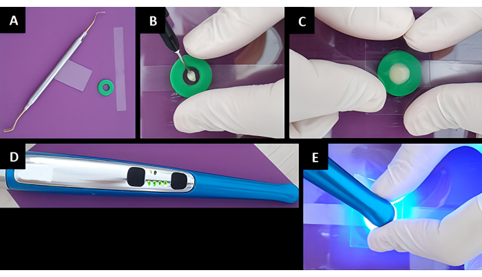
Figure 1: Preparation and polymerization of composite disks. (A) The equipment required for composite disk preparation. (B) The application of the composite sample into the gasket. (C) The condensation of the composite sample between the transparent tape and the transparent glass. (D) The light curing device. (E) The polymerization of the composite sample. Please click here to view a larger version of this figure.
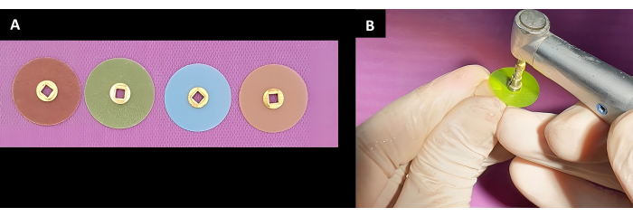
Figure 2: Polishing application. (A) The disks used for polishing. (B) The polishing disk application on the composite sample. Please click here to view a larger version of this figure.
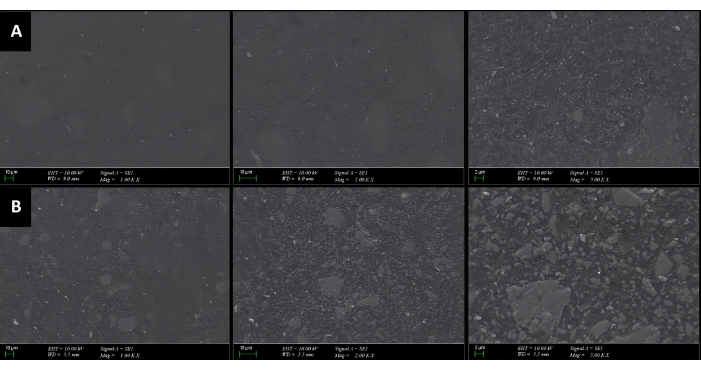
Figure 3: SEM images before scaling application. The images are from (A) Group P, (B) Group B. From left to right, 1000x, 2000x, and 5000x magnification are shown. Please click here to view a larger version of this figure.
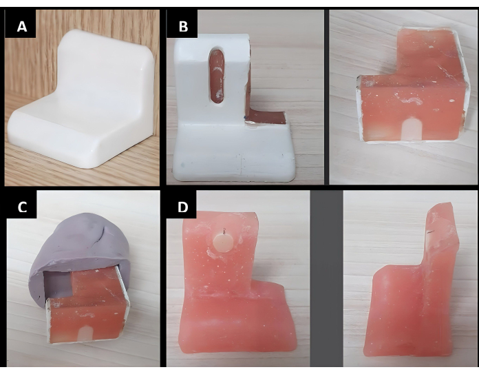
Figure 4: Stabilization of the samples into acrylic blocks. (A) An example illustrating theplastic l-connection hanger element. (B) Cold-curing acrylic application into the plastic l-connection hanger element and preparation of the prototype of the specimen holder. (C) Using silicon-based impression material in order to create the negative of the holder prototype. (D) Insertion of the composite specimens on the acrylic holder. Please click here to view a larger version of this figure.
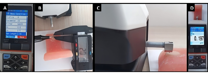
Figure 5: Steps for profilometric measurement. (A) Profilometer settings, (B) profilometer tip adjustment, (C) profilometer tip contact with the specimen, (D) reading screen of the profilometer. Please click here to view a larger version of this figure.
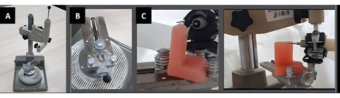
Figure 6: Fixation of the acrylic block with the composite specimen. (A) An illustration of the parallelometer used for the special setup. (B) The table of the parallelometer. (C) Exact positioning of the acrylic block. Please click here to view a larger version of this figure.
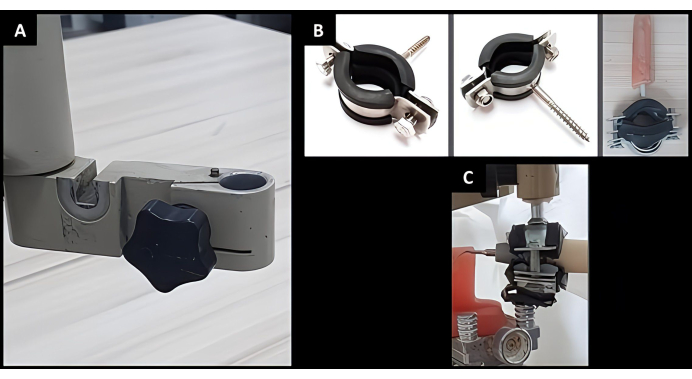
Figure 7: Preparation for the stabilization of the handpiece of the scaler. (A) Holder arm of the parallelometer. (B) Modification of a rubber pipe clamp with triphone. (C) Fixation of the scaler handpiece to the holder arm. Please click here to view a larger version of this figure.
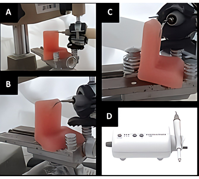
Figure 8: Ultrasonic scaler application with equal force to a marked area with the help of a specially prepared apparatus. (A) Frontal view, (B) diagonal view, (C) lateral view of the application, (D) ultrasonic dental scaler. Please click here to view a larger version of this figure.
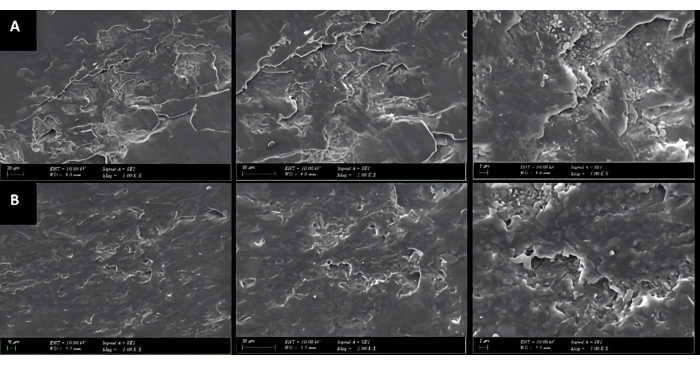
Figure 9: SEM images after scaling application. The images are from (A) Group P and (B) Group B. From left to right, 1000x, 2000x, and 5000x magnification are shown. Please click here to view a larger version of this figure.
| GROUPS | Before µm | After µm | P value* |
| (Mean± SD) | (Mean ± SD) | ||
| Group P (n =10) | 0.16 ± 0.17 | 0.44 ± 0.28 | 0.005 |
| Group B (n =10) | 0.25 ± 0.26 | 0.49 ± 0.26 | 0.007 |
| P value# | 0.143 |
Table 1: Intragroup roughness change. SD: Standard Deviation; * Wilcoxon Signed Rank Test, p ˂ 0.01, # Mann Whitney-U Test, p ˂ 0.05.
| Groups | Roughness change, µm |
| (Mean ± SD) | |
| Group P (n = 10) | 0.27 ± 0.29 |
| Group B (n = 10) | 0.23 ± 0.18 |
| P value* | 0.684 |
Table 2: Evaluation of the roughness change between the groups. SD: Standard Deviation; * Mann Whitney-U Test, p ˂ 0.05.
Discussion
Research consistently shows that both sonic and ultrasonic scaling can increase the surface roughness of tooth-colored restorative materials, with ultrasonic scaling having a more detrimental effect8,9. Ultrasonic scaling and air-powder polishing can further increase the roughness of composite resin and restoration margins, and the extent of damage is material-dependent6. The type of restorative material used can also impact the extent of surface damage, with flowable and silorane-based composites showing smoother surfaces and lower bacterial count1. The current study's findings contribute to the existing body of literature on the effects of ultrasonic scaling on the surface roughness of composite fillings.
A comparative analysis of the two groups in this study, nanohybrid Group P (monomer content: Bis-EMA, TEGDMA; filler content: pre-polymerized filler, barium glass, silica filler) and patented giomer nanohybrid Group B (monomer content: Bis-GMA, TEGDMA; filler content: glass filler, fluoroboroaluminosilicate glass), revealed no significant difference in roughness alteration following scaler application. This finding is noteworthy as it indicates that advancements in composite technology, specifically giomer-based formulations, do not inherently convey resistance to scaler-induced roughness alterations. The lack of statistically significant difference in the degree of roughness added to both composite types might be discussed considering the material's filler content and particle size. While the technology and monomer content vary, the mechanical impact of scaling could be independent of these factors, affecting only the most superficial layer where similarities between the composites may be more pronounced. The results obtained here suggest that while technologically advanced composite materials may offer several clinical benefits, their resistance to mechanical alterations induced by scaling must be critically evaluated. Dental practitioners should consider the implications of these findings when performing scaling on patients with composite restorations and possibly favor the use of less abrasive instruments or techniques to maintain surface integrity.
In order to conduct a comparison by examining the surface change of any material, it is necessary that the initial surface characteristics of the materials being compared are comparable. The minimum amount of energy required for the polymerization of the composite materials used in the present study was reported as 10 J/cm2 by the manufacturers19. According to the manufacturer's instructions, the light curing system used in the present investigation delivers between 1 J and 3 J of energy per cm2 per second. Checci et al.20 proved that extended curing duration enhances the mechanical characteristics of the composites. In accordance with the aforementioned facts, the light system was applied for the polymerization of the samples for a period of 20 s from the top and 20 s from the bottom. Flowable composites are distinguished by a reduced amount of filler material and a higher concentration of diluent monomers in the composition. Traditionally, these composites were made by maintaining the tiny particle size of conventional hybrid composites but lowering the amount of filler. This allowed for an increased amount of resin, which in turn decreased the viscosity of the mixture1,21. Nevertheless, the mechanical qualities of these composites, such as flexural strength and wear resistance, have been consistently shown to be worse when compared to traditional composites. Flowable composites have been proposed as suitable filler materials for low-stress applications and scenarios that involve challenging access or need effective penetration. In order to acquire similar initial surface roughness values in the present investigation, the tested flowable composite materials were polymerized and polished in the same manner, and similar surface roughness values were validated by analyzing the surface characteristics obtained by a profilometer. No manufacturer recommendation was available regarding the rpm value and polishing time for the polishing system employed in the present study22. The samples were polished to similar initial roughness values by the same individual using a 4-step polishing system, as illustrated in Figure 2.
The evidence regarding the effects of various ultrasonic devices commonly employed by experts is inconclusive23,24,25,26. According to Yousefimanesh et al.26, when the same forces were applied, piezoelectric scalers produced smoother root surfaces than magnetostrictive scalers. Flemmig et al.24 also reported that a magnetostrictive device was more aggressive on root surfaces than a piezoelectric device. Nevertheless, Busslinger et al.23 discovered that a piezoelectric device produced a coarser root surface than a magnetostrictive device following instrumentation. Singh et al.25 found that magnetostrictive and piezoelectric ultrasonic instruments created equivalent surface roughness levels and demonstrated comparable capabilities for creating a physiologically harmonious surface, which differed from the previous investigations. The inconsistent findings emphasize the need for greater study in this area. A recent study examined the effects of several ultrasonic scalers and polishing devices on the wear and roughness of root surfaces10. As a result, it is demonstrated that the application settings might have a significant impact on the roughness of the root surface. Furthermore, using the piezoelectric ultrasonic scaler at maximum intensity at a 0° angle resulted in the biggest wear and roughness levels on the root surfaces. As a result, comparable application conditions were applied to the samples in the current investigation, where the most significant impact was found.
In surface analysis research, it is of great importance to be able to perform reproducible applications and establish standard exposures for all samples in order to avoid bias. Moreover, the capacity to precisely disclose the examined surface after the application is also extremely beneficial to the findings. Therefore, when conducting such studies, it is necessary to establish a setup to standardize individualized application and evaluation parameters. In terms of ultrasonic scaler application, the methodological factors that might affect the findings can be mentioned as follows: adjusted lateral force, tip angulation, power settings, and consistency of the application site to be examined10,24,26,27,28. The current study demonstrates that several critical methodological parameters can be acquired using a parallelometer, an acrylic block for securing the sample, and a custom holder that allows the handpiece to be applied to the sample at the desired angle with minimal contact force. In this methodological approach, the inability to measure and alter the force applied to the specimen represents a potential limitation of the constructed setup. On the other hand, it should be remembered that ultrasonic instruments should be applied to dental and restorative surfaces only in contact with the surface without any force in order to minimize any surface damage. Also, in the current setup, it is possible to clearly define the application zone, which is crucial for the exact analysis of the results revealed after the application.
The surface texture of dental composites is critical for their longevity and performance, as rougher surfaces may harbor more plaque and bacteria, leading to an increased risk for caries and periodontal disease. It has been shown that dental plaque accumulation is influenced by surface roughness, with a threshold of 0.2 µm below which bacterial adherence is limited2. This is supported by in vivo studies that have found a correlation between surface roughness and plaque accumulation29,30. The present study demonstrates that the application of a piezoelectric ultrasonic scaler can indeed increase the roughness of composite material surfaces to a level that leads to plaque deposition. This effect was consistent across both tested material types.
It is also important to consider the limitations of this study. The in vitro nature of the experiment may not precisely replicate clinical conditions, particularly concerning the scaler's angle and the presence of saliva, which could modulate the effects seen. Long-term studies in a clinical setting would confirm these findings and provide a clearer understanding of the scaler's impact over time.
In conclusion, the present study underscores the importance of considering the physical effects of hygiene procedures on dental composites. Although the clinical significance of the observed roughness increase requires further investigation, the potential for increased plaque retention should inform clinical practices, emphasizing the need for careful scaling techniques around composite restorations.
Disclosures
The author declares no conflict of interest.
Acknowledgements
I express my gratitude to Prof. Dr. Oğuzhan Gündüz from Marmara University Nanotechnology and Biomaterials Application and Research Center/Marmara University Faculty of Technology Department of Metallurgy and Material Engineering; Prof. Dr. Pınar Yılmaz Atalı from Marmara University Faculty of Dentistry, Department of Restorative Dentistry; and Dr. Semra Ünal Yildirim from Marmara University Genetic and Metabolic Diseases Research and Investigation Center who provided valuable insights and expertise that seriously supported the investigation.
Materials
| Name | Company | Catalog Number | Comments |
|---|---|---|---|
| Beautifil Flow Plus | Shofu | United States | |
| Evo MA10 Scanning Electron Microscope | Zeiss | Germany | |
| EWO Typ 990 Paralellometer | Kavo | Germany | |
| Finishing Discs | Bisco | United States | |
| G4 Scaler Tip | Woodpecker | China | |
| Premise Flowable | Kerr | United States | |
| SC 7620 model sputter coater | Quorum Technologies | UK | |
| Surftest SJ-210 | Mitutoyo | Japan | |
| UDS-A-LED Dental Scaler | Woodpecker | China | |
| Valo LED Cordless Curing Light | Ultradent | United States | |
| Zetaplus Silicon Impression Material | Zhermack | Italy |
References
- Hossam, A. E., Rafi, A. T., Ahmed, A. S., Sumanth, P. C. Surface topography of composite restorative materials following ultrasonic scaling and its impact on bacterial plaque accumulation. An in vitro sem study. J Int Oral Health. 5 (3), 13-19 (2013).
- Zissis, A. J., Polyzois, G. L., Yannikakis, S. A., Harrison, A. Roughness of denture materials: A comparative study. Int J Prosthodont. 13 (2), 136-140 (2000).
- Arabaci, T., et al. Influence of tip wear of piezoelectric ultrasonic scalers on root surface roughness at different working parameters. A profilometric and atomic force microscopy study. Int J Dent Hyg. 11 (1), 69-74 (2013).
- Goldstein, R. E., et al. Microleakage around class v composite restorations after ultrasonic scaling and sonic toothbrushing around their margin. J Esthet Restor Dent. 29 (1), 41-48 (2017).
- Mittal, A., Nichani, A. S., Venugopal, R., Rajani, V. The effect of various ultrasonic and hand instruments on the root surfaces of human single rooted teeth: A planimetric and profilometric study. J Indian Soc Periodontol. 18 (6), 710-717 (2014).
- Babina, K., et al. The effect of ultrasonic scaling and air-powder polishing on the roughness of the enamel, three different nanocomposites, and composite/enamel and composite/cementum interfaces. Nanomaterials (Basel). 11 (11), (2021).
- Demirci, F., Birgealp Erdem, M., Tekin, M., Caliskan, C. Effect of ultrasonic scaling and air polishing on the surface roughness of polyetheretherketone (peek) materials. Am J Dent. 35 (4), 200-204 (2022).
- Erdilek, D., Sismanoglu, S., Gumustas, B., Efes, B. G. Effects of ultrasonic and sonic scaling on surfaces of tooth-colored restorative materials: An in vitro study. Niger J Clin Pract. 18 (4), 467-471 (2015).
- Lai, Y. L., Lin, Y. C., Chang, C. S., Lee, S. Y. Effects of sonic and ultrasonic scaling on the surface roughness of tooth-colored restorative materials for cervical lesions. Oper Dent. 32 (3), 273-278 (2007).
- Mahiroglu, M. B., Kahramanoglu, E., Ay, M., Kuru, L., Agrali, O. B. Comparison of root surface wear and roughness resulted from different ultrasonic scalers and polishing devices applied on human teeth: An in vitro study. Healthcare (Basel). 8 (1), (2020).
- Cadenaro, M., et al. Flowability of composites is no guarantee for contraction stress reduction. Dent Mater. 25 (5), 649-654 (2009).
- Basheer, R. R., Hasanain, F. A., Abuelenain, D. A. Evaluating flexure properties, hardness, roughness and microleakage of high-strength injectable dental composite: An in vitro study. BMC Oral Health. 24 (1), 546(2024).
- Brine, E. J., Marretta, S. M., Pijanowski, G. J., Siegel, A. M. Comparison of the effects of four different power scalers on enamel tooth surface in the dog. J Vet Dent. 17 (1), 17-21 (2000).
- Kuka, G. I., Kuru, B., Gursoy, H. In vitro evaluation of the different supragingival prophylaxis tips on enamel surfaces. Photobiomodul Photomed Laser Surg. 41 (5), 212-217 (2023).
- Parashar, A., Bhavsar, N. Assessing the effect of piezoelectric ultrasonic scaler tip wear on root surface roughness under influence of various working parameters: A profilometric and atomic force microscopic study. J Indian Soc Periodontol. 27 (6), 583-589 (2023).
- Vengatachalapathi, H., Naik, R., Rao, R., Venugopal, R., Nichani, A. S. The effect of piezoelectric ultrasonic scaler tip wear on root surface roughness at different working parameters: An atomic force microscopic and profilometric study. J Int Acad Periodontol. 19 (1), 15-21 (2017).
- Casarin, R. C., et al. Root surface defect produced by hand instruments and ultrasonic scaler with different power settings: An in vitro study. Braz Dent J. 20 (1), 58-63 (2009).
- International Organization for Standardization. Part 12: Sample preparation and reference materials in Biological evaluation of medical devices. ISO 10993-12:2021. , (2021).
- Sabatini, C. Color stability behavior of methacrylate-based resin composites polymerized with light-emitting diodes and quartz-tungsten-halogen. Oper Dent. 40 (3), 271-281 (2015).
- Checchi, V., et al. Wear and roughness analysis of two highly filled flowable composites. Odontology. , (2024).
- Poggio, C., Dagna, A., Chiesa, M., Colombo, M., Scribante, A. Surface roughness of flowable resin composites eroded by acidic and alcoholic drinks. J Conserv Dent. 15 (2), 137-140 (2012).
- Tepe, H., Erdilek, A. D., Sahin, M., Efes, B. G., Yaman, B. C. Effect of different polishing systems and speeds on the surface roughness of resin composites. J Conserv Dent. 26 (1), 36-41 (2023).
- Busslinger, A., Lampe, K., Beuchat, M., Lehmann, B. A comparative in vitro study of a magnetostrictive and a piezoelectric ultrasonic scaling instrument. J Clin Periodontol. 28 (7), 642-649 (2001).
- Flemmig, T. F., Petersilka, G. J., Mehl, A., Hickel, R., Klaiber, B. Working parameters of a magnetostrictive ultrasonic scaler influencing root substance removal in vitro. J Periodontol. 69 (5), 547-553 (1998).
- Singh, S., Uppoor, A., Nayak, D. A comparative evaluation of the efficacy of manual, magnetostrictive and piezoelectric ultrasonic instruments-an in vitro profilometric and sem study. J Appl Oral Sci. 20 (1), 21-26 (2012).
- Yousefimanesh, H., Robati, M., Kadkhodazadeh, M., Molla, R. A comparison of magnetostrictive and piezoelectric ultrasonic scaling devices: An in vitro study. J Periodontal Implant Sci. 42 (6), 243-247 (2012).
- Flemmig, T. F., Petersilka, G. J., Mehl, A., Hickel, R., Klaiber, B. The effect of working parameters on root substance removal using a piezoelectric ultrasonic scaler in vitro. J Clin Periodontol. 25 (2), 158-163 (1998).
- Oliveira, G., Macedo, P. D., Tsurumaki, J. N., Sampaio, J. E., Marcantonio, R. The effect of the angle of instrumentation of the piezoelectric ultrasonic scaler on root surfaces. Int J Dent Hyg. 14 (3), 184-190 (2016).
- Bergstrom, J. Photogrammetric registration of dental plaque accumulation in vivo. Acta Odontol Scand. 39 (5), 275-284 (1981).
- Quirynen, M., et al. The influence of surface free energy and surface roughness on early plaque formation. An in vivo study in man. J Clin Periodontol. 17 (3), 138-144 (1990).
Reprints and Permissions
