We present a method for a fungal pathogenesis model that preserves the natural positioning of fungal spores in the lung airways for analysis via fluorescent microscopy.
A subscription to JoVE is required to view this content. Sign in or start your free trial.
Method Article
Efficient Method for Imaging Murine Lungs that Preserves Spatial Dynamics of Fungal Spores in the Airways
In This Article
Summary
Abstract
Fungi infect humans when environmental spores are inhaled into the lungs. The lung is a heterogeneous organ. Conducting airways, including bronchi and bronchioles, branch until terminating in the alveolar airspace where gas exchange occurs. Infections originating in the bronchioles or alveoli elicit distinct host responses and disease manifestations. Therefore, understanding precisely where spores naturally localize in the lungs, particularly soon after infection, expands opportunities for investigation of host-pathogen interactions. Herein, we detail an in-situ analysis of lungs from mice infected with Coccidioides posadasii cts2/ard1/cts3Δ arthroconidia. Conventional methods for histological preservation involve liquid inflation of the airways with a fixative solution, which displaces the natural location of aspirated fungal particles, pushing spores from proximal bronchioles to terminal airspaces.
Conversely, this method of air-inflation with blood vasculature perfusion-fixation preserves the physiologic position of fungal spores within the bronchioles. Moreover, we describe a simple approach to cryopreserving, embedding, and imaging lung specimens. We also share high-throughput computational techniques via the open-source QuPath program to analyze the spatial distribution of fungal spores within the lung. The method presented here is simple and quick, requires minimal equipment to perform, and can be easily adapted for use with many respiratory fungal infection models.
Introduction
Humans can inhale up to billions of spores per day from a variety of environmental fungi1. To understand our barrier defenses against these inhaled spores, we must appreciate the precise microanatomical environments where these spores land within the airways and lung parenchyma. The cellular composition of the airways (i.e., epithelial cells) significantly transforms along the trachea, bronchi, bronchioles, and alveoli. Each of these distinct regions is composed of different cell types with discrete functions which avails an arsenal of defenses to prevent pathologic infection.
The precise location of pulmonary fungal spore deposition can vary between the airway lumens of columnar epithelial-lined bronchioles, alveolar ducts, or alveoli2. Most clinically relevant fungal species produce spores between 1 µm and 10 µm in diameter3. The deposition of these spore particles in the lung depends on several factors, such as aerodynamics, density, electrical charge, and phoretic forces, which can influence the mechanism of sedimentation after inhalation4. Generally, large particles (> 6 µm) deposit in the upper airway, medium-sized particles (2-6 µm) can deposit in smaller airways, and small particles (<2 µm) reach the alveolar region5. Aspergillus spores (2-3 µm) have been reported to reach alveolar spaces, but clinical pathology reports also indicate a significant burden of bronchial and bronchiolar disease6. There is also increasing recognition of endobronchial fungal infections of Aspergillus fumigatus, Coccidioides immitis, Candida species, Cryptococcus neoformans, Histoplasma capsulatum, and Zygomycetes due to the increasing popularity of flexible bronchoscopy7. Recent advances in microscopic imaging of Aspergillus infections in mice have also revealed that more proximal airspaces such as bronchi and bronchioles may bear the highest burden for pathologic fungal proliferation8. Research on the host response to pulmonary fungal infections has shown that both bronchiolar epithelial cells and alveolar epithelial cells play important roles as immune sentinels, so elucidating the exact sites of spore deposition and epithelial interaction will be vital for future work2,9,10.
Studying these proximal airway pathogenesis dynamics is difficult because standard lung fixation and sectioning preparation techniques can displace spores from these proximal positions in epithelial-lined airways and push them toward distal terminal alveolar regions. Commonly, 10% formalin or 4% paraformaldehyde is used to inflate the lungs and rapidly expose the entire lung to fixative. When preparing lungs for cryosectioning, some groups administer optimal cutting temperature (OCT) compound into the lungs to improve cryosectioning performance11. These practices are useful in the right context but have been found by our lab and other groups to displace spores and particles from proximal locations, thereby interfering with interpretations about the spore-exposed cell types and the subsequent host response12.
To accurately establish the microanatomical localization of inhaled fungal spores, we have developed a quick, low-resource method for the preservation of the location of fungal spores in the airways of mice. We adapted a murine air-inflation vascular perfusion-fixation method from Thomas et al. (2021), where we reduced the amount of equipment, time, and technical skill required to achieve a satisfactory result13. From the method described here, we have observed that Coccidioides posadasii cts2/ard1/cts3Δ arthroconidia (3-5 µm in size) accumulate more proximally than previously shown, namely in distal bronchioles and broncho-alveolar junctions rather than terminal alveoli. This information can focus biological questions concerning the critical cell types associated with these regions of the lung and their influence on the early responses to inhaled fungal spores.
Access restricted. Please log in or start a trial to view this content.
Protocol
All methods described in this protocol have been approved by the Institutional Animal Care and Use Committee (IACUC) of Rutgers Biomedical Health Sciences.
1. Label spores with intracellular fluorescence
- Stain 1 x 10⁶ spores in carboxyfluorescein succinimidyl ester (CFSE), Cell Tracker Orange, or Cell Tracker Red (or intracellular dye of choice) at 5 µM for 30 min at 30 °C, then wash with PBS, and spin the spores at 12,000 x g. Repeat this wash once.
- Prepare the spore inoculum at the appropriate concentration so that 25 µL contains the appropriate spore dose for one mouse.
2. Inoculating mouse with spores through aspiration inhalation using isoflurane anesthesia
CAUTION: Isoflurane is a volatile anesthetic agent and must be used within a ducted biosafety cabinet or fume hood.
- To prepare the isoflurane exposure jar, place a folded napkin into a 1 L screw-top Nalgene jar and place a plastic mesh circle over the napkin as a platform for the mice. Add 2 mL of isoflurane to the napkin, close the jar, and wait 1-2 min for the gas to equilibrate in the container.
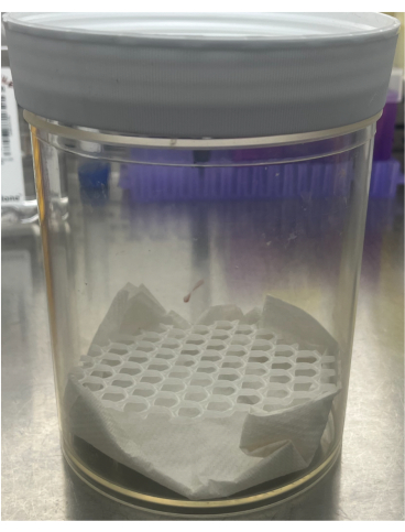
Figure 1: Apparatus for isoflurane open drop method of spore inoculation. A folded napkin is placed in the bottom of a 1 L screw top jar, and then a circular plastic mesh is inserted to act as a platform for the mice. Please click here to view a larger version of this figure.
- Prior to exposing the mouse to anesthesia, prepare a pipet with 25 µL of the inoculum. Place the mouse in the isoflurane container and tilt the container to monitor for loss of the righting reflex, where the mouse has lost consciousness and is upside down. This usually takes about 10 s.
- Monitor the respiration rate (RR) until it slows to a consistent 50-80 breaths per minute (bpm), about 50% of resting RR. This usually takes another 10 s. At this time, remove the mouse from the isoflurane jar.
NOTE: This is a high concentration of isoflurane, which will produce rapid induction of anesthesia, and the mouse must be very closely monitored. Lethal isoflurane overdose is a risk, as the concentration of isoflurane can be variable, and the dose-response curve to isoflurane is very steep. If the breathing rate dips below 50 bpm and/or is irregular, remove the mouse from isoflurane, allow it to recover for 3-5 min, and try again (a maximum of two repeated attempts). - Confirm the anesthetic depth by lack of response to toe pinch. Then, position the mouse in a supine position in one hand, and allow the mouse to take 3-5 regular gasping breaths with increasing rate (beginning at 50-60 bpm) and vigor. As this begins, place the pipet tip at the posterior of the mouse oropharynx and release the inoculum during an inhalation.
NOTE: These gasps through the mouth jolt the head inferiorly due to the recruitment of accessory muscles for breathing. When the pipet is inserted during these gasps, the mouth should open with each gasp. If the mouth does not open, the mouse is insufficiently anesthetized and will likely not inhale contents from the oropharynx. The pipet should depress the tongue, pushing it towards the bottom and front of the mouth to prevent swallowing of the inoculum. When the pipet is in the mouth and the mouse is adequately anesthetized, the mouse will open its mouth with each gasp. - With the hand and thumb holding the mouse, feel for crackles on the posterior and anterior aspects of the mouse thorax to confirm inhalation of the inoculum.
3. Euthanasia of mice
- Inject the mouse intraperitoneally with a cocktail of 200 mg/kg ketamine and 24 mg/kg xylazine 10 min prior to the sacrifice time point.
- Wait 10 min post-injection until the mouse enters a surgical plane of anesthesia, confirmed by lack of response to toe pinch.
4. Perfusion of lungs with PBS and formalin
- Spray the anesthetized mouse with 70% ethanol. Use scissors to snip back the skin of the mouse and pull apart the skin to expose the peritoneum on all sides. Pull the skin from the mouse's superior half over its head, pulling out the arms from the skin.
- Cut the peritoneal membrane at the sternum and along the inferior aspect of the ribcage, exposing the peritoneum. Displace the liver inferiorly, revealing the diaphragm. Use scissors to carefully puncture the diaphragm away from the lung parenchyma to avoid puncturing a hole in the lungs themselves.
NOTE: Inadvertently puncturing the lungs will make subsequent air inflation impossible. - After the diaphragm is punctured, air enters the thorax, collapsing the lungs. Cut the ribcage on the left side to reveal the heart. Inject 5-10 mL of PBS into the right ventricle with a 30 G needle at a rate of 1 mL per 5 s (to avoid damaging vessels), rapidly whitening the lungs.
CAUTION: Formalin is a hazardous volatile substance that should be handled within a ducted biosafety cabinet or fume hood. - When finished, remove the needle and use a new needle to inject 5 mL of 10% neutral-buffered formalin (NBF) into the right ventricle at the same rate.
5. Air-inflating the perfused lung
- Remove the anterior half of the ribcage. To expose the trachea, cut the superior ribs and collarbones to the right and left of the neck. Take care to avoid cutting near the midline where the trachea will lie. With forceps, clasp the remaining top ribs and collarbones on the neck midline and pull superiorly until the trachea is exposed.
- Prepare a suture thread of 10 cm in length, pull it under the trachea, and pre-tie a loose knot at the inferior end of the exposed trachea. Prepare a 1 mL syringe of air with an 18 G catheter. Use an 18 G needle to make a hole in the trachea at the superior end of the exposed region.
- Place the catheter into that hole, ensuring a relatively tight fit to prevent air from escaping. Slowly inject the 1 mL of air into the lungs over the course of 10 s, watching for lung inflation of all lobes. Full inflation results in the lungs wrapping slightly around the heart and filling the volume they occupy in the unpunctured diaphragm.
- Pull the knot tight around the trachea, and remove the catheter. Hold the trachea and use blunt scissors to remove the lungs from the mouse, being careful not to puncture the lung.
6. Immersion fixation and dehydration
- Place the lungs on the top edge of a 50 mL conical tube filled with 20 mL of 10% NBF. Keep the suture threads outside of the conical, screw down the cap, and invert the conical so that the lungs are suspended upside down in 20 mL of NBF. Place it at 4 °C for 24-48 h.
- Rinse the lungs in PBS, then place in a 30% sucrose-PBS (w/v) solution for 72-96 h at 4 °C to dehydrate the lungs in preparation for cryopreservation. Remove the lungs from sucrose, place the lungs in a cryomold with optimal cutting temperature (OCT) medium, and freeze the specimen at -80 °C.
7. Cryosectioning
- Equilibrate the specimens in OCT cryoblocks to the -20 °C temperature of the cryostat for 1 h prior to sectioning. Section the blocks at thicknesses of 20-100 µm.
NOTE: Air inflation can lead to increased fragility of the specimen, and cutting at increased thickness can prevent tissue breakage during sectioning. - Collect sections onto glass slides and let dry for 30 min to 1 h to ensure tissue adherence to the slide.
NOTE: Slides may be held at -80 °C at this stage.
8. Slide preparation and blocking
- Place slides in PBS bath (in a dish or Coplin jar) for 30 min at room temperature (RT) to remove OCT from the slides.
NOTE: Thicker sections (40-100 µm) may not adhere to glass slides as well as thinner sections, so it is best to keep them flat and upright in a dish rather than placed on their side in a Coplin jar. - Dry the slides after OCT has dissolved away from the tissue. Use a wipe to dry excess droplets from the slide's perimeter around the tissue specimen.
- Use a hydrophobic Pap pen to draw a perimeter around the specimen, and let it dry for 5 min.
- Prepare a blocking buffer of animal-free blocking solution with 0.3% Tween-20 and 1:100 Fc Block in 300 mL per slide.
NOTE: For intracellular targets, 1% Tween-20 may be used. A volume of 300 µL is usually sufficient, but the necessary volume to cover the tissue fully depends on the perimeter size of the hydrophobic marker. The tissue should not dry from this point onwards. - Leave the blocking solution on the slides for 1 h at RT.
9. Immunostaining
- Wash the slide by pipetting off the fluid and adding 300-500 µL of PBS. Repeat the wash once.
- Prepare primary AlexaFluor-647 conjugated rat anti-mouse EpCAM (clone G8.8) antibody (airway epithelial marker) in animal-free blocker solution with 0.33% Tween-20 (or 1% for intracellular targets). Add 300 µL per slide of this solution and stain overnight at 4 °C.
NOTE: Antibodies should be tested empirically for staining concentration, time, and temperature. Thin sections (8-20 µm) are stained for 30 min at 37 °C and thicker sections (20-100 µm) at 4 °C overnight using 1:100 dilutions for most antibodies. If using unconjugated antibodies, wash 2x (as above) and apply a secondary fluorescent antibody for an empirically determined time and temperature. Generally, secondary antibody staining is done at RT for 1 h at 1:1000 dilution. - Remove the antibody solution from the specimen, and wash the slide with 300 mL of PBS for 5 min. Repeat this wash once. Dry the slides, removing all excess fluid from the specimen and surrounding glass.
- Add one drop of RT soft set mounting media (e.g., SlowFade Glass) to each piece of tissue. Take care to avoid bubbles by letting the bottle settle while upside down and slowly dropping each drop onto the tissues. Place an appropriately sized cover slip to extend beyond the sample and hydrophobic marker perimeter.
10. Imaging via fluorescent microscopy
- Use a multichannel fluorescent microscope to scan whole lung sections in an unbiased manner using an objective with sufficient resolution to resolve individual spores and host cells (e.g., Zeiss Axioscan 7 using 20x oil objective with NA = 0.8 or similar).
NOTE: Ensure that laser power and PMT voltage gain are set to maximize the signal-to-noise ratios of the target over the background determined with a fluorescence minus one (FMO) control. - Collect sufficient image tiles to capture the entire lung lobe (e.g., ~200 tiles of size 400 μm x 400 μm). Use the microscope software program (e.g., Zeiss ZEN 3.7 or similar) to export the merged tile image for downstream processing.
11. Spatial analysis via QuPath
- In QuPath, a free open-source software14, create a project file and add the fluorescent images to the file.
- Classify the EpCAM+ pixels as epithelium by clicking the top menu option Classify, then Pixel classification > Create Thresholder. Select the resolution, channel, smoothing sigma, and threshold value that best identifies the target region. Save the classifier under a unique name, and select Create Objects.
- In the new Create objects window, select New object type as Annotation. For lung epithelium, set the minimum object size and minimum hole size to 100 mm2. There is no need to select Split Objects here. After selecting OK, the annotations will be created, and they can be modified by eye using the Brush tool in the upper left-hand area.
- To classify spores, repeat the steps from 11.3-11.4 using the channel in which the spores were captured and make the new object type Detection rather than Annotation. Set minimum object size and minimum hole size to 0 mm2, and select Split Objects.
- To conduct spatial analysis of the spores, select the top menu option Analyze > Spatial Analysis > Calculate signed distance to annotations 2D. The distances to EpCAM+ epithelium will be calculated for each detected spore. Export these via the top menu option Measure > Export Measurements.
Access restricted. Please log in or start a trial to view this content.
Results
This method ultimately produces immunofluorescent images of mouse lungs using physiologic air inflation to leave the airways undisturbed. Importantly, multiple checkpoints along the way will confirm that components of the protocol have been performed successfully. During the inoculation, it is important to confirm that the inoculum was aspirated by feeling for "crackles" on the posterior chest wall of the mouse that indicate the liquid has entered the airways. If there is no sensation of crackles, it is possible that the...
Access restricted. Please log in or start a trial to view this content.
Discussion
We have established a pipeline for spore inhalation and analysis of the spatial deposition of the inhaled spores. This pipeline provides valuable information to determine the relevant stromal regions of the lung affected by inhalation of Coccidioides posadasii cts2/ard1/cts3Δ arthroconidia. We have observed that Coccidioides spores and similarly sized inert particles (data not shown) accumulate in distal bronchiolar regions and broncho-alveolar junctions rather than fully dispersed throughout alve...
Access restricted. Please log in or start a trial to view this content.
Disclosures
The authors have nothing to disclose.
Acknowledgements
Funding and support were acquired through NIH grant K22 AI153678-01 and Rutgers School of Graduate Studies. We thank Fawad Yousufzai and Luke Fritzky from the Rutgers Biomedical Health Sciences Cellular Imaging and Histology Core for their work and expertise in obtaining immunofluorescent images.
Access restricted. Please log in or start a trial to view this content.
Materials
| Name | Company | Catalog Number | Comments |
|---|---|---|---|
| 18 G, 1 1/2 needle (305185) | Fisher | 305185 | |
| 1 L Screwtop Jar (Nalgene) | Fisher Scientific | 11-823-33 | |
| Air-Tite Bulk Unsterile Syringes 10 mL Luer Lock | Fisher | 14-817-175 | |
| AnaSed Injection (xylazine sterile solution) | Akorn | 59399-110-20 | |
| Animal-Free Blocker and Diluent, R.T.U. | Vector | SP-5035-100 | |
| BD Insyte Autoguard Winged Shielded IV Catheter with BD Vialon Catheter Material 18 G x 1.88 in | BD | 381547 | |
| BD Pharmingen Purified Rat Anti-Mouse CD16/CD32 (Mouse BD Fc Block™) | BD | 553142 | |
| CellTracker Orange CMRA Dye | Fisher Scientific | NC0873640 | |
| CFSE | Labviva | 75003 | |
| Coccidioides posadasii cts2/ard1/cts3Δ | BEI Resources | NR-166 | |
| Corning 70 micron strainers | VWR | 10054-456 | |
| EpCAM AlexaFluor647 monoclonal antibody | Biolegend | 118211 | |
| Exel International HYpodermic Needles 30 G x 1/2" | Labviva | EN3012 | |
| Fisherbrand Sterile Syringes for Single Use (1mL, Leur Slip) | Fisher | 14-955-462 | |
| Glucose Monohydrate | Azer Scientific | ES17530-500G | |
| High Vacuum Grease | VWR | 59344-055 | |
| Hoechst 33342 Solution 20 mM (5 mL) | ThermoFischer | 62249 | |
| Isoflurane USP | Covetrus | 29405 | |
| Ketamine Hydrochloride | Dechra | 1000001250 | |
| KIMWIPES Delicate Task Wipers (4.4'' x 8.4") | VWR | 21905-026 | |
| Lexer Baby Scissors | FST | 14078-10 | |
| Micro-Adson Forceps | FST | 11018-12 | |
| Neutral Buffered Formalin (10%) (Azer Scientific) | Fisher | 22-026-350 | |
| Nunc EasYFlask tissue culture flasks, T75, filter caps | VWR | 15708-134 | |
| PBS | VWR | 45000-446 | |
| Peel-A-Way embedding molds | Sigma | E6032-1CS | |
| QuPath 0.5.1 Software | Open-source | https://qupath.github.io/ | |
| Silk Suture thread size 3/0 | FST | 18020-30 | |
| SlidesMicro Slides Superfrost Plus | VWR | 48311-703 | |
| SlowFade Glass Soft-set Antifade Mountant (2 mL) | ThermoFischer | S36917 | |
| Sucrose | Sigma | S0389-500G | |
| Tissue-Tek O.C.T. Compound, Sakura Finetek | VWR | 25608-930 | |
| Tween 20 | ThermoFischer | J20605.AP | |
| Vector Laboratories ImmEDGE Hydrophobic Barrier Pen Set Of 2 | Fisher Scientific | NC9545623 | |
| VWR Micro Cover Glasses, Rectangular (24 mm x 40 mm #1.5) | VWR | 48393-230 | |
| White Plastic Wire Mesh | MAPORCH | 789862904922 | |
| Yeast Extract | Fisher | BP9727-500 | |
| Zeiss AxioScan 7 | Carl Zeiss Microscopy GmbH | https://www.zeiss.com/microscopy/us/products/imaging-systems/axioscan-for-biology.html | multichannel fluorescent microscope |
| ZEN 3.7 Software | Carl Zeiss Microscopy GmbH | https://www.zeiss.com/microscopy/us/products/software/zeiss-zen.html | microscopy software |
References
- American Society for Microbiology. One Health: Fungal Pathogens of Humans, Animals, and Plants. Report on an American Academy of Microbiology Colloquium. , Washington, DC. (2019).
- Crossen, A. J., et al. Human airway epithelium responses to invasive fungal infections: A critical partner in innate immunity. J Fungi (Basel). 9 (1), 40(2022).
- Yamamoto, N., et al. Particle-size distributions and seasonal diversity of allergenic and pathogenic fungi in outdoor air. ISME J. 6 (10), 1801-1811 (2012).
- Thakur, A. K., Kaundle, B., Singh, I. Mucoadhesive Drug Delivery Systems in Respiratory Diseases.Targeting Chronic Inflammatory Lung Diseases Using Advanced Drug Delivery Systems. , Elsevier, Academic Press. (2020).
- Darquenne, C. Aerosol deposition in health and disease. J Aerosol Med Pulm Drug Deliv. 25 (3), 140-147 (2012).
- Kradin, R. L., Mark, E. J. The pathology of pulmonary disorders due to Aspergillus spp. Arch Pathol Lab Med. 132 (4), 606-614 (2008).
- Karnak, D., Avery, R. K., Gildea, T. R., Sahoo, D., Mehta, A. C. Endobronchial fungal disease: An under-recognized entity. Respiration. 74 (1), 88-104 (2006).
- Amich, J., et al. Three-dimensional light sheet fluorescence microscopy of lungs to dissect local host immune-Aspergillus fumigatus interactions. mBio. 11 (1), e02752-e02819 (2020).
- Wiesner, D. L., et al. Club cell TRPV4 serves as a damage sensor driving lung allergic inflammation. Cell Host Microbe. 27 (4), 614-628.e6 (2020).
- Evans, S. E., Hahn, P. Y., McCann, F., Kottom, T. J., Pavlovic', Z. V., Limper, A. H. Pneumocystis cell wall β-glucans stimulate alveolar epithelial cell chemokine generation through nuclear factor-κB-dependent mechanisms. Am J Respir Cell Mol Biol. 32 (6), 490-497 (2005).
- Bauer, C., Krueger, M., Lamm, W. J. E., Glenny, R. W., Beichel, R. R. lapdMouse: associating lung anatomy with local particle deposition in mice. J Appl Physiol. 128 (2), 309-323 (2020).
- Srirama, P. K., Wallis, C. D., Lee, D., Wexler, A. S. Imaging extra-thoracic airways and deposited particles in laboratory animals. J Aerosol Sci. 45, 40-49 (2012).
- Thomas, S. M., Bednarek, J., Janssen, W. J., Hume, P. S. Air-inflation of murine lungs with vascular perfusion-fixation. J Vis Exp. 168, e62215(2021).
- Bankhead, P., et al. QuPath: Open source software for digital pathology image analysis. Sci Rep. 7 (1), 16878(2017).
- Davenport, M. L., Sherrill, T. P., Blackwell, T. S., Edmonds, M. D. Perfusion and inflation of the mouse lung for tumor histology. J Vis Exp. 162, e60605(2020).
- Hsia, C. C. W., Hyde, D. M., Ochs, M., Weibel, E. R. An official research policy statement of the American Thoracic Society/European Respiratory Society: Standards for quantitative assessment of lung structure. Am J Respir Crit Care Med. 181 (4), 394-418 (2010).
- Gil, J., Weibel, E. R. Extracellular lining of bronchioles after perfusion-fixation of rat lungs for electron microscopy. Anat Rec. 169 (2), 185-199 (1971).
- Bachofen, H., Ammann, A., Wangensteen, D., Weibel, E. R. Perfusion fixation of lungs for structure-function analysis: credits and limitations. J Appl Physiol Respir Environ Exerc Physiol. 53 (2), 528-533 (1982).
- Evans, C. M., et al. The polymeric mucin Muc5ac is required for allergic airway hyperreactivity. Nat Commun. 6, 6281(2015).
- Galati, D. F., Asai, D. J. Immunofluorescence microscopy. Curr Protoc. 3 (8), e842(2023).
- Hickey, S. M., et al. Fluorescence microscopy-An outline of hardware, biological handling, and fluorophore considerations. Cells. 11 (1), 35(2021).
- Risling, T. E., Caulkett, N. A., Florence, D. Open-drop anesthesia for small laboratory animals. Can Vet J. 53 (3), 299-302 (2012).
- Bodnar, M. J., Ratuski, A. S., Weary, D. M. Mouse isoflurane anesthesia using the drop method. Lab Anim. 57 (6), 623-630 (2023).
Access restricted. Please log in or start a trial to view this content.
Reprints and Permissions
