Here we present a protocol for performing a low-dose intradermal inoculation of Mycobacterium avium in a mouse ear to reflect exposure-like conditions.
Method Article
Intradermal Inoculation of Mycobacterium avium in the Mouse Ear
In This Article
Summary
Abstract
In contrast to natural nontuberculous mycobacterial (NTM) exposures or infections whose pathology is founded on a slow-replicating organism, the aerosol infection in a mouse model can cause an elevated proliferation of bacterial growth in the lungs. Chronic aerosol and systemic infections can be treated with anti-mycobacterial drugs, which could also reflect a prior exposure, but these procedures are lengthy and less representative of natural exposures. The goal of this protocol is to develop a less invasive mouse model of NTM exposure-like conditions that are more translational. Here, we describe a method to intradermally challenge preclinical mice through ear tissue using Mycobacterium avium (M. avium) to mimic an exposure and enable immune responses to the bacteria without obvious disease. In this article, we describe the intradermal ear injection method and confirm the presence of sustained and contained mycobacterial infection, which provides further opportunities to interrogate the immune responses in this in vivo model.
Introduction
Nontuberculous mycobacteria (NTM) are an expanding list of species that include a subset of pathogenic bacteria that are often difficult to treat1,2. NTM can be found in environmental sources such as soil, water, and water aerosols3,4,5. Despite reported trends suggesting rising NTM infections, as a group, they are routinely reported in less than 20 states across the U.S.6,7. Individuals with specific comorbidities, including persons with cystic fibrosis or individuals on immunosuppressive drugs, are at increased risk of productive infection with pathogenic NTM species2. Healthy individuals, however, are presumed to have repeated and continual exposures to NTM through environmental sources without leading to robust or chronic infections. There is some evidence that NTM exposure may 'precondition' the overall immune response to mycobacterial antigens in vaccinations8, outcomes after exposure to other mycobacteria, and diagnostic testing9, but to date, preclinical modeling of this kind of exposure has been difficult.
Chronic pulmonary infections10 and treatments that are leveraged for pathogen-specific vaccine efficacy studies do not represent relatively innocuous environmental exposures, and the presumably different resulting immune responses. Chronic infections, for example, can induce severely damaged tissue and disseminated disease10. There has been recent momentum to address this research gap, including a mouse model providing NTM exposure via drinking water, from the Henao-Tamayo lab11, where the immune response to this exposure was further interrogated at length. Latent tuberculosis (TB) is another disease state of mycobacterial infection that has been difficult to model in preclinical animals. The Kaufmann lab pioneered intradermal ear infections with M. tuberculosis in C57BL/612 and NOS2-/-13 mice to mimic latent TB infection, reactivation, and to characterize the mechanisms of hypoxic granulomas. Nemeth et al. leveraged the contained intradermal ear model to mimic prior exposure to M. tuberculosis, characterizing distinct immune responses, in order to determine how this affected subsequent challenge outcomes14. The intradermal ear model has been successfully reported using 104 colony-forming units (CFU) of M. tuberculosis strains H37Rv and Erdman, but to date, it has not been tested using NTM isolates.
The overall goal of this method is to establish a reproducible NTM ear challenge model that limits the disease caused by other challenge routes, which can be further studied with regard to the immune response to exposure. Exposing susceptible C57BL/6 mice to high doses of M. avium in drinking water results in more infection-like conditions than exposures11. Therefore, the rationale for developing this technique includes prior success using M. tuberculosis as a challenge organism to the ear dermis with a relatively higher challenge dose12,13,14. Here, we aimed to evaluate whether the model can be successfully adapted for an M. avium isolate at half the bacterial concentration previously reported12,13,14 (102 CFU). We expect that advancing new models of NTM exposure, and not infection, will help address outstanding biological questions about this influence on health and future infection/vaccination dynamics.
Protocol
Female C57BL/6 were purchased from a research model supply laboratory at 6-7 weeks of age. Mice were housed at the Seattle Children's Research Institute (SCRI) biosafety level 2 (BSL-2) animal facility under pathogen-free conditions and were handled in accordance with experimental protocols that were approved by the SCRI Institutional Animal Care and Use Committee (IACUC). All methods were carried out in accordance with relevant guidelines and regulations with respect to animal welfare. The institute operates under USDA Certificate #91-R-0061 and PHS Assurance #A4337-01. The details of the reagents and equipment used in the study are listed in the Table of Materials.
1. Preparation of bacterial stocks for inoculation
- In a biosafety cabinet conducive for bacterial work, remove a stock vial of Mycobacterium avium (ATCC 25291) from -80 °C storage and thaw at room temperature.
NOTE: Stock concentrations of M. avium are determined in advance, as best practice with all stock materials, by plating serial dilutions on 7H10 agar plates and counting colony-forming units (CFU). - Using a 26 G size, blunt-tipped needle, resuspend the stock sample by passing the liquid up and down the 1.0 mL syringe 10x times or more to create a uniform suspension.
- Check the concentration of M. avium by plating 100 µL of serial 1:10 dilutions of the suspension on 7H10 agar plates. Incubate the plates for 3 weeks at 37 °C and count colonies to ensure inoculum concentrations are accurate.
- Adjust the concentration of M. avium to the preferred dose by diluting the sample with sterile PBS. Keep at room temperature until ready to use.
NOTE: In this experiment, 102 CFU per 10 µL dose was used.
2. Preparation and anesthetization of animals
- Before administering anesthesia, prepare a warm surface, such as a heating pad, on which the cages of inoculated mice can be kept in an effort to regulate their body temperature during recovery.
- Inject Ketamine (100 mg/mL) and Xylazine (20 mg/mL) cocktail into mice via the intraperitoneal route.
- Once anesthetized, perform a toe pinch to confirm mice are sufficiently anesthetized for procedure manipulations.
- Apply eye lubricant bilaterally.
3. Inoculation platform and materials preparation
- Use a foam board to create a ledge upon which the mouse can become stabilized while being manipulated.
NOTE: Double-sided tape will help hold the ear down onto the platform in a flat orientation. The adhesive must not be too strong to preserve ear tissue health. - Arrange a lighted magnifying glass to view the platform.
- Gather a sterile Hamilton syringe and a working M. avium preparation nearby.
4. Ear dermis inoculation
- Place the mouse in a supine position atop the ledge in such a way that the ear can be positioned on double-sided tape.
- Use gentle pressure to flatten the ear onto the tape and ensure the ear will not move.
NOTE: Alcohol wipes can be used on the ears prior to the injection to help limit cross-contamination and infection with skin commensals post-infection. However, such infections have not been observed in pilot studies without using alcohol wipes. - Draw up 10 µL of M. avium preparation containing 100 CFU into the sterile Hamilton syringe.
NOTE: Swirl M. avium suspension before drawing up 10 µL volume to ensure it is homogenous. - Hold the platform in one hand and the Hamilton syringe in the other, ensuring the bevel side of the needle is pointed up (Figure 1).
- Position the Hamilton syringe under the ear dermis using very light pressure. Ensure the needle doesn't go completely through the thin ear tissue.
- Once the needle is in place, depress the syringe plunger at a gradual and slow pace.
- Observe the color of the ear during administration.
NOTE: Successful administration will appear as an opaque elevation at the site of injection, like a bubble. - Once the dosage is fully administered, check the mouse ear and tape for any signs of leakage.
NOTE: If leakage does occur, an estimated lost dose can be administered to the opposite ear. At the minimum, the animal should be marked and recorded as possibly having been given a below target dose. - When the dosage is successful, ensure that the mouse is placed back in their home cage to be exposed to the warm surface while being monitored until it has successfully woken up from anesthesia.
5. Bacterial burden evaluation
- Euthanize animals according to Institutional Animal Care and Use Committee-approved protocols.
- Here, euthanasia is completed with a minimum of two methods per sacrifice. For this procedure, the two methods chosen are the use of CO2 asphyxiation and cervical dislocation.
- First, extend a tube to dispense CO2 gas into the mice's home cage and administer the gas until there are no more signs of movement from the mice. Be careful to use a cage that will not allow the gas to leak out.
- Next, dislocate the cervical vertebrae through manual manipulation.
- Dissect the ears from M. avium challenged animals four weeks post-exposure (28 days, matching prior studies12). Dissect the ears of 2 mice using sterile tools. Insert the ears into conical tubes containing CFU buffer (PBS + 0.05% Tween80) and use a tissue homogenizer to homogenize until a uniform suspension is formed. Unchallenged ears are also processed and served as controls.
NOTE: Any post-exposure timepoints and organs of interest can be used. For this experiment, the lung, spleen, draining lymph node, and brain organs were also collected. - Add 100 µL of homogenate to 700 µL of CFU buffer in a deep well block as a 1/8-fold dilution. Repeat across the block so that a minimum of 5 serial dilutions are generated. Add 50-100 µL of neat homogenate or serial dilutions (dilutions 0 through 5 used here) to 7H10 agar tri-plates, spread evenly using T-spreaders, and incubate at 37 °C with 5% CO2 for 2-3 weeks.
- Count the colony-forming units (CFU) and transform them into log10 for analysis purposes in spreadsheet software.
Results
We used the protocol detailed above to determine whether a reproducible NTM challenge model using the ear dermis as a route of infection or exposure could be established. Ears were collected from two animals at 28 Day post-challenge timepoint, homogenized, and plated as described on the mycobacteria-selective 7H10 media. We observed that M. avium persisted to 28 days in ears that were challenged intradermally and an absence of M. avium in the unchallenged ear (Figure 2).
While we did not observe dissemination to the unchallenged ear by 28-days post infection, others using this ear dermis model for latent TB modeling have observed dissemination to other distal organ sites14. We hypothesized that a lower dose of challenge and using M. avium isolate may reduce dissemination out to later timepoints that may better reflect exposure versus chronic infection. Therefore, euthanasia was performed on cohorts of n=5 M. avium challenged mice at 28-, 56-, 90- and 132-days post-infection. The following organs were homogenized with the same protocol as described for the ear tissues: lung, spleen, draining lymph node (DLN), and brain. This route of challenge resulted in a sporadic dissemination over time, where fewer than half of the animals demonstrated countable CFU in organs tested by 56- or 90-day post-infection (Table 1). By 132 days post-challenge, all mice had detectable M. avium in their spleens (n = 5/5 animals evaluated) but seemed to harbor minimal (n = 1/5 in DLN) or no (n = 0/5 in lungs or brain) bacteria in other organs by this timepoint (Table 1).
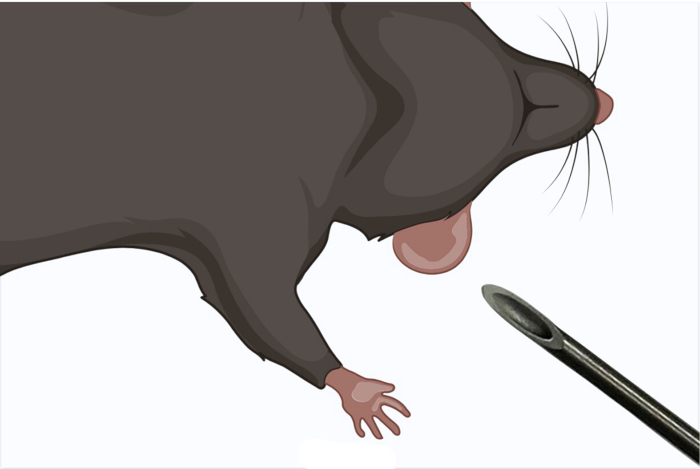
Figure 1: Orientation of a needle with the bevel side up. Prior to insertion of the needle, the bevel side must be facing upwards to properly administer the inoculum. Please click here to view a larger version of this figure.
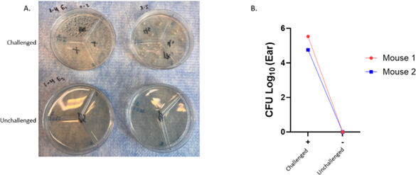
Figure 2: Ear tissue colonization by M. avium 28 days post challenge. Female C57BL/6 mice were challenged in the ear with 102 CFU of Mycobacterium avium. The challenged ear (+) was compared to the unchallenged ear (-) at 28 days, n=2 individual animals from a single experiment shown, with each animal in a different color and symbol. Please click here to view a larger version of this figure.
| Time (day) | Organ Log10 CFU | |||
| Lung | Spleen | DLN | Brain | |
| 28 | 3.08, ND, ND, ND, ND | 2.81, ND, ND, ND, ND | ND, ND, ND, ND, ND | 0.78, 0.90, ND, ND, ND |
| 56 | 2.30, ND, ND, ND, ND | 1.53, 3.43, ND, ND, ND | 2.78, ND, ND, ND, ND | 1.20, ND, ND, ND, ND |
| 90 | ND, ND, ND, ND, ND | 0.60, 3.35, 3.26, ND, ND | ND, ND, ND, ND, ND | 0.60, ND, ND, ND, ND |
| 132 | ND, ND, ND, ND, ND | 4.03, 3.58, 3.58, 4.03, 3.69 | 2.41, ND, ND, ND, ND | ND, ND, ND, ND, ND |
| DLN = draining lymph node, ND = not detected | ||||
Table 1: Dissemination of M. avium from the ear over time post challenge. Lungs, spleen, draining lymph node (DLN), and brain were harvested from n=5 mice per timepoint post challenge. Homogenized tissues were plated and evaluated for bacterial growth. Organ CFU presented as log10 transformed values where bacteria were counted and not detected (ND) where no CFU were observed at any concentration plated. Time course data representative of one experiment.
Discussion
Here we have demonstrated how to reproducibly challenge mice with mycobacteria in a contained intradermal ear model. It is vital, when performing this procedure, to follow sterility precautions and to avoid needle-stick injuries. With this inoculation method, it is critical to have a proper setup station and good lighting to ensure that the dermis space is targeted and not punctured through the ear entirely. Should that happen, depending on the study, a second attempt can be made in the other ear. Untreated ears within the same mouse can also be used as per-mouse control. This method should be accessible to researchers with minimal needs for equipment or time. Dedicating mice and time for training helps with the protocol's success rate of inoculation. The most common errors in this technique are not entering the intradermal space with the syringe (and puncturing through the ear) or depressing the syringe too rapidly, causing an inconsistent delivery or slippage of the needle from the intradermal space. Good lighting and magnification can help with the proper positioning of the needle for inoculation.
We are leveraging the methodology for 'exposure-like' conditions of NTM. The vast majority of M. avium is contained within the ear or the DLN. However, a limitation of this methodology is that dissemination to systemic organs was observed, namely the spleen, in all animals evaluated by 132 days post-challenge. There may be a threshold of bacterial inoculation that could reduce the amount or frequency of dissemination, which could be accomplished with a titration study. This procedure is amenable to modifying both the organism, for example, other NTM species, and the dose delivered, depending on the research question.
Human behavior (lack of appropriate potable water treatment, pollution, smoking), increased numbers of people with deteriorating health due to age15 and other comorbidities, lack of appropriate diagnostic tests, and climate change16 may play a role in the rising rates of NTM infections, and this trend is likely to increase in the near future. We propose that models like mucosal delivery via drinking water11 or intradermal contained ear infections with NTM in mice provide an opportunity to determine what influence this rising exposure will have on subsequent vaccination and vaccine efficacy. Although we have adapted this methodology of a contained ear model for M. avium, we propose that it may be used successfully for other NTM species as an exposure technique.
Disclosures
The authors declare they have no conflicts of interest.
Acknowledgements
The authors express their gratitude to the committed vivarium personnel for their exceptional care of the animals throughout this study. The authors also wish to thank the Henao-Tamayo Lab at Colorado State University for sharing the M. avium strain used in this study. The authors would like to thank the members of the Aderem and Urdahl labs at Seattle Children's Research Institute for training in the original methodology. This project has been funded in whole or in part with federal funds from the National Institute of Allergy and Infectious Diseases, National Institutes of Health, Department of Health and Human Services, under Contract No. 75N93021C00029 (to RNC), and Grants R01AI125160 and R01AI183642 (to RNC and SLB).
Materials
| Name | Company | Catalog Number | Comments |
|---|---|---|---|
| 0.05% Tween80 | Sigma | P6224 | |
| 1mL syringe | Fisher | 15-823-434 | |
| 7H10 agar triplates | Moltox | 21-40S234 | |
| 26G blunt needle | Fisher | NC0981254 | |
| Biosafety cabinet | Nuaire | NU-543-400 | |
| CO2 incubator | Fisher Scientific | 15-103-0513 | |
| Double-sided tape | Scotch | 3136 | |
| Hamilton 701N microliter syringe | Fisher | 14-824-16 | |
| Hard tissue omni tips | Fisher | 15-340-102 | |
| Heating pad | Kent Scientific | HTP-1500 | |
| Insulin needle | Fisher | NC1551333 | |
| Ketamine | Patterson Veterinary | 07-890-8598 | |
| Lighted magnifying glass | Brightech | B07MMZ27QK | |
| PBS | ThermoFisher | 10010072 | |
| Tissue homogenizer | Omni | TH115 | |
| Tissue homogenizer stand | Omni | S1000 | |
| Xylazine | Patterson Veterinary | 07-808-1947 |
References
- Abdelaal, H. F. M., et al. Mycobacterium abscessus: It's Complex. Microorganisms. 10 (7), (2022).
- Baldwin, S. L., et al. The complexities and challenges of preventing and treating nontuberculous mycobacterial diseases. PLoS Negl Trop Dis. 13 (2), p. e0007083(2019).
- Falkinham, J. O. 3rd Environmental sources of nontuberculous mycobacteria. Clin Chest Med. 36 (1), 35-41 (2015).
- Honda, J. R. Environmental sources and transmission of nontuberculous mycobacteria. Clin Chest Med. 44 (4), 661-674 (2023).
- Pfaller, S., et al. Occurrence revisited: Mycobacterium avium and Mycobacterium intracellulare in potable water in the USA. Appl Microbiol Biotechnol. 106 (7), 2715-2727 (2022).
- Mercaldo, R. A., et al. Environmental risk of nontuberculous mycobacterial infection: Strategies for advancing methodology. Tuberculosis (Edinb). 139, 102305(2023).
- Marshall, J. E., et al. Nontuberculous mycobacteria testing and culture positivity in the United States. BMC Infect Dis. 24 (1), 288(2024).
- Ghasemi, F., et al. Effects of non-tuberculous mycobacteria on BCG vaccine efficacy: A narrative review. J Clin Tuberc Other Mycobact Dis. 36, 00451(2024).
- Njagi, L. N., et al. Attenuated tuberculin skin test responses associated with Mycobacterium intracellulare sputum colonization in an adolescent TB prevalence survey in Western Kenya. Tuberculosis (Edinb). 147, 102514(2024).
- Larsen, S. E., et al. Subunit vaccine protects against a clinical isolate of Mycobacterium avium in wild type and immunocompromised mouse models. Scientific Reports. 11 (1), 9040-9040 (2021).
- Dutt, T. S., et al. Mucosal exposure to non-tuberculous mycobacteria elicits B cell-mediated immunity against pulmonary tuberculosis. Cell Rep. 41 (11), 111783(2022).
- Kupz, A., et al. A Mouse Model of Latent Tuberculosis Infection to Study Intervention Strategies to Prevent Reactivation. PLoS One. 11 (7), e0158849(2016).
- Reece, S. T., et al. Serine protease activity contributes to control of Mycobacterium tuberculosis in hypoxic lung granulomas in mice. J Clin Invest. 120 (9), 3365-3376 (2010).
- Nemeth, J., et al. Contained Mycobacterium tuberculosis infection induces concomitant and heterologous protection. PLoS Pathogens. 16 (7), e1008655(2020).
- Jarchow-MacDonald, A., et al. Changing Incidence and Characteristics of Nontuberculous Mycobacterial Infections in Scotland and Comparison With Mycobacterium tuberculosis Complex Incidence (2011 to 2019). Open Forum Infect Dis. 10 (1), ofac665(2023).
- Thomson, R. M., et al. Influence of climate variables on the rising incidence of nontuberculous mycobacterial (NTM) infections in Queensland, Australia 2001-2016. Sci Total Environ. 740, 139796(2020).
Reprints and Permissions
