This protocol presents how optimally positioning a mouse and inserting a camera-based otoscope transorally enables visualization of the glottis, rapid intubation with minimal tissue trauma, and consistent liquid delivery to the lungs.
Method Article
Improved Methodology for Liquid Delivery to the Mouse Lung: Intubation using a Consumer Otoscope
In This Article
Summary
Abstract
Consistent delivery of liquids, including potential therapies, to the lungs is critical for modeling pulmonary disease and pre-clinical therapeutic development. However, current methods used in mice -such as oropharyngeal aspiration by the tongue-pull method- can result in variable delivery. Here, we describe the use of an affordable, mobile phone-linked, lighted otoscope to visualize the glottis and permit direct inoculation into the airway. We detail an optimized staging technique with adequate lighting and positioning aids. How intubation can be reliably conducted using a 20 G flexible catheter with or without a fiberoptic light is also described. This technique greatly reduces variance in intrapulmonary delivery and increases the volume of delivered liquid that reaches the lungs. Due to reduced variance, smaller animal cohort sizes can be used when employing this method instead of oropharyngeal instillation by the tongue-pull method. What follows is a method for effective, rapid, and consistent intubation of mice and delivery to the lungs, including robust confirmation of endotracheal tube placement. With this improved methodology, consistent, safe intubation and replicable, efficient delivery to the lungs are within reach.
Introduction
Globally, respiratory diseases are the number one killer of children under 5 years of age and are a major cause of morbidity and mortality in adults1. In 2021, SARS-CoV2 infection was the third leading cause of death in the US2. Murine models are often used to study lung diseases, including asthma, pneumonia, chronic obtrusive pulmonary disease, and pulmonary fibrosis. Nearly all these models require the delivery of liquid agents to the lungs. This can be done by direct canulation of the trachea (typically under direct visualization using a surgical cut-down technique), intranasal delivery, or oropharyngeal aspiration by the well-established tongue-pull method3,4,5. However, these techniques all have drawbacks. Scar tissue formation after the surgical cut-down technique renders it inappropriate for studies that require repeated administration of agents to the lung, and the latter two techniques can lead to variable lung delivery. Moreover, when using the tongue-pull method, tissue trauma to the tongue can be pronounced, and the delivery efficiency is dependent on the researcher performing the procedure, see Figure 1.

Figure 1: Guided intubation and IT instillation results in improved delivery to the lung and less variability when compared to tongue pull. (A) Three different investigators delivered 50 µL of dilute India ink by tongue pull, the established method for oropharyngeal (OP) administration. Two researchers also delivered ink to the lung using intubation as described. (B) The lungs were removed 1-4 h after delivery and photographed under 2x magnification. Photographs were assessed for area stained, using ImageJ. (n=6 (intubation) or 9 (op administration), results from 2 or 3 independent replicates by 2 -3 different researchers, p<0.0001 Welch's T Test) (C) The variability in lung administration was also calculated as a percent deviation from the mean. OP administration was far more variable. (p = 0.0317, Welsh's T-Test, GraphPad Prism 10.2.3 all statistics and graphing) (C57BL/6J Male and Female mice were used, they were ~10 weeks of age.) Please click here to view a larger version of this figure.
Murine anatomy makes intubation difficult and time-intensive. The mouth is small, and the buccal tissue may obstruct a researcher's view of the rima glottidis6. Further, the vocal folds are very small, and it can be challenging to view them as they lie close to the base of the tongue. Finally, confirmation of proper placement of the endotracheal tube is difficult as the small tidal volume often fails to fog a mirror7. These difficulties have encouraged a range of innovative techniques to intubate mice. These improved methods include the use of a neonatal laryngoscope and wire to advance a catheter into the trachea8,9, the use of a fiber optic light source to guide and stiffen the catheter10,11, and the use of a live video feed6. More recently, the use of a clinical otoscope has aided in orotracheal intubation12. Others have employed transillumination to visualize the glottis and intubate mice7,13,14. However, there are many downsides to these methods. The use of depilatory agents on the mouse's neck and animal proximity to a hot light source make transillumination a less-than-ideal approach. The use of the needle within the catheter or insertion of sharp instruments into the mouth means some methods to stiffen the catheter place animals at undue risk. Finally, making an incision over the trachea to confirm placement presents significant downsides, including the need for sterility and the buildup of scar tissue. To this point, intratracheal instillation by intubation has proven too difficult for widespread adoption15 and the advances in methodologic development have remained siloed and unused by many laboratories15. Further studies have focused on anesthetic choice during delivery, finding that ketamine may have advantages over isoflurane5. However, isoflurane, not being a controlled substance and rapidly wearing off upon procedure completion, presented attractive benefits, so we have used this form of anesthesia while optimizing other aspects of this methodology16,17.
The method we describe overcomes the visualization challenges others have attempted to address, mitigates risks to experimental animals, decreases the time needed to instill small volumes intratracheally via an endotracheal tube, increases the volume of liquid reaching the lung, and offers an improved method for endotracheal tube placement confirmation. Using a suspension stand, magnetic helping hands (alligator clips on flexible arms secured to a base), and brighter lighting allows an individual researcher to perform efficient intratracheal delivery. Visualization is made easier using an 'at home' otoscope with WIFI connectivity. This device is more affordable than commercial intubation instruments. Moreover, the WIFI connectivity allows for a simultaneous video feed on a portable device (such as a mobile phone), making it suitable for training. Intubation can utilize either a fiberoptic light source threaded through a 20 g x 1" flexible (PTFE) catheter (needle removed) or the catheter without the light. Depth is controlled by two methods - the addition of an intubation safety wedge (a conical guard on the catheter) and depth observation in the video feed. Endotracheal tube placement is confirmed using the "bubble method" to visualize tidal volume more clearly than the traditional mirror-fog method18. This is less stressful to the animal than relying on tracheal obstruction to modify respiratory urgency10 to confirm placement. The combined use of extension tubing coupled to the catheter to confirm successful intubation (hereafter referred to as the bubble-method), simplified from Watanabe et. al7, and a consumer video otoscope in a single procedure makes intubation a practical method for therapeutic delivery to the lung and a clear improvement to the tongue-pull method (see Figure 1). If seeking a reliable, consistent, and affordable method for nonsurgical liquid delivery into mouse lungs, especially without the use of controlled substances (like ketamine) or expensive equipment, the following method may be ideal.
Protocol
It is imperative that these procedures are reviewed and approved by the institutional animal care and use committee (IACUC), listed on the protocol, and veterinary staff are consulted before proceeding. The IACUC at our institution approved the protocol prior to starting the experiment and reviewed the video content prior to submission. Support and training were received through the Tulane Department of Comparative Medicine to ensure the highest standard of care for all our laboratory animals.
1. Set up
NOTE: See Figure 2 for the setup.

Figure 2: The station used for guided intubation is prepared as shown. From Left: Pipettes for delivery, Bubble tube for confirmation of appropriate endotracheal tube placement, forceps, spatula, pediatric nasal speculum (discussed but not needed for this procedure), helping hands, magnifying light, mouse stand with nose cone, fiberoptic probe, anesthesia induction chamber. Please click here to view a larger version of this figure.
- Clean the safety wedge provided with the intubation kit with an isopropanol prep pad. Slide the clean safety wedge (green in the video) onto the 20 G flexible canula and discard the provided needle safely.
NOTE: The rodent intubation kit (Table of Materials) used includes intubation safety wedges. These are small, conical sleeves that fit over a 20 G x 1-inch flexible catheter. They prevent the catheter from damaging the carina. - Gather the other tools needed, including a mobile phone, commercial otoscope, mouse stand with a nose cone, finely serrated bent forceps, the prepared endotracheal tube (from 1.1), and the fiber optic light if using it. Clean all of these with isopropanol prep pads before use.
- Take approximately 20 cm of flexible tubing. Bend it in a U shape, add a Luer lock connection on one end, and secure it with a twist tie. Add 200-300 µL of sterile filtered water or India ink to the tube. Hereafter, this assembly is referred to as a "bubble tube."
- Make a suture loop by tying ~13 cm of size 0 braided silk suture in a circle. Place this loop at the top of the stand and ensure both sides of the suture are secured, and the stand is clean. Place the mouse stand on top of the metal magnifying light base. Position the helping hands around where the mouse's mouth will be.
NOTE: Many vendors make interchangeable stands (see Table of Materials), and one can be made within the lab cheaply4,7,14. Size 0 braided silk suture was used to suspend the animal. However, the use of other materials for the same purpose has been described4,13. - Place a mouse in the anesthesia induction chamber. Use constant flow rate O2 at 0.8 L/min and 2% isoflurane. Anesthetize the animal sufficiently (see note) so that it does not awaken during the placement of the nose cone. Do not significantly suppress the mouse's respiratory rate. Regular breaths aid in the appreciation of the vocal folds.
NOTE: The mouse's respiratory rate should be normal. This criterion is used to establish the appropriate depth of anesthesia. This may vary between animals and may exceed 200 breaths/min. As such, it is not practical or necessary to count respirations or use a toe pinch.
CAUTION: This procedure can be aborted when necessary. However, animals should not remain in the induction chamber any longer than is strictly necessary. - Connect the otoscope to a mobile phone and ensure the video feed is stable.
CAUTION: To minimize the researchers' isoflurane exposure during this procedure, use a chemical respirator if performing multiple intubations or training in this method. Alternatively, perform the procedure in a fume hood.
2. Animal staging
- As soon as the first animal is asleep, remove it from the induction chamber, scruff it, and place its top teeth on the length of the suture. Apply light tension to the animal to maintain the suture's positioning while laying the mouse back onto the stand.
NOTE: On average, animals are anesthetized for a total of 2 min including induction. This greatly decreases the risk to the animal, removes the need for ophthalmic lubrication, and prevents saliva buildup. Work to keep animals sedated for as little time as possible. - Adjust the neck angle slightly back (placing the neck in minor extension, 5°-15°). Gently push the nose cone over the nares. Do not push the cone down firmly, as it can interfere with the suture, suspending the mouse.
- Firmly grasp the tongue as close to the bottom lip as possible, using large, finely serrated bent forceps. Pull it down and away from the top teeth to one side of the bottom teeth.
CAUTION: Prolonged traction, excessive tension, or crushing the tongue can compromise animal health. Please monitor the tongue for signs of trauma (bruising, bleeding, swelling, or desiccation), and if observed, abort the procedure and alert veterinary staff. - If using helping hands, support the forceps in this position. Depending on the researcher's experience and visual acuity, a lighted magnifying glass may help with positioning the animal appropriately.
- Feed the fiberoptic light (if using) through the flexible catheter with the intubation safety wedge (green in the video) in place.
NOTE: Do not secure the fiber optic cable to the light source too tightly. Pushing too hard makes it difficult to remove the light while leaving the endotracheal tube in place.
3. Visual appreciation of the vocal cords and intubation
- With the otoscope in the non-dominant hand, lightly press it on the tongue. Then, angle the camera and its light source to visualize the vocal cords near the base of the tongue.
NOTE: Slight variations in anatomy and tissue color can make orienting oneself difficult. Use the movement associated with breathing to confirm structural identification. - With the fiberoptic light on (if using), feed the cannula alongside the top of the otoscope, making light contact with the base of the tongue or the lip portion of the otoscope for reference. Advance it into the mouth of the mouse between the soft pallet and the lip of the otoscope.
NOTE: If the fiberoptic light is not being used, the "bubble tube" described in step 4 can be attached to the catheter at this stage. Bright, focused lighting in the mouse's mouth is necessary for this modification to the procedure. - Adjust the angle of approach so that the catheter is very shallow. Immediately after inserting the catheter (less than 1 mm beyond the vocal folds), raise the tip of the catheter, so it is nearly parallel to the benchtop. This prevents the epiglottis from forcing the catheter dorsally into the esophagus. Especially during training, when having difficulty, or early in one's experience, the use of the video feed is invaluable in placing the endotracheal tube between the vocal folds.
NOTE: Saliva production does not obstruct viewing of the vocal folds while completing this procedure. However, the use of a spear-tip cotton applicator to remove obstructing saliva is described, in previous publications, when intubating rats19. This method may prove useful if saliva becomes a problem.- If resistance is encountered, immediately remove the catheter and allow respirations to return to normal. If bleeding is observed, consult veterinary staff.
NOTE: There should be almost no resistance when advancing the catheter.
- If resistance is encountered, immediately remove the catheter and allow respirations to return to normal. If bleeding is observed, consult veterinary staff.
4. Confirmation of endotracheal tube placement
- Visually confirm that the endotracheal tube is in the correct location using the otoscope. Further, ensure the correct placement of the tube by considering that the trachea provides less resistance during placement than the esophagus. Observe that in the appropriate position, the endotracheal tube is ~5 mm beyond the vocal folds. Look for light from the fiberoptic source shining through the neck skin to again validate that placement is correct.
NOTE: The intubation safety wedge should prevent inserting the endotracheal tube too far. The safety wedge prevents trauma to the carina; however, it need not be preventing advancement. It should be used as a safety feature and optimal depth is often shallower than allowed by the wedge. - Then, while holding the endotracheal tube in place, remove the fiberoptic light. Ensure appropriate placement of the catheter using the "bubble method" (detailed below) rather than mirror fogging.
- Retrieve the bubble tube prepared in Step 1.3. Prepare it in advance of the procedure and reuse it for all animals.
CAUTION: The 200-300 µL of water or ink in the bubble tube should never be aspirated. To avoid this, make sure the liquid is far from the Luer lock. - With the endotracheal tube in place, secure the bubble tube in place using the Luer lock. The tidal volume should be immediately apparent. If not, the endotracheal tube is misplaced. Remove it quickly and allow the mouse's normal respiratory rate to return.
CAUTION: Misplacing the catheter in the esophagus will compress the trachea, preventing sufficient respiration. Misplacement will often be accompanied by labored breathing (including appreciable contribution from the intercostal muscles) and/or a dramatically decreased respiratory rate. Furthermore, canulating the esophagus may lead to a muscle spasm making subsequent intubation more difficult; we advise the use of multiple animals while training for this reason.
5. Intratracheal delivery
CAUTION: Much like the tongue-pull method for oropharyngeal delivery, delivery via intubation risks drowning the animal. In adolescent and adult mice, 50 µL is a generally safe volume of fluid to deliver and is sufficient to spread throughout the lungs.
- If using a syringe for administration: Detach the bubble tubing and Luer lock from the catheter, attach a loaded Luer slip syringe, and depress the plunger to deliver the desired volume (50 µL maximum).
- If using a pipette for administration: Pipette the desired volume (50 µL maximum) directly into the catheter and allow the mouse to inhale the solution.
NOTE: If training, consider using an indicator such as dilute, filtered India ink, or methylene blue. - To ensure diffuse delivery to the lungs, flush the delivered liquid into the lungs with up to 500 µL of air20. This can be done either using a preloaded 0.5mL Luer lock syringe, or a P1000 pipettor fitted with a filter tip, both of which have been proven reliable. If desired, multiple depressions of the pipettor can be conducted without risking negative aspiration of the liquid delivered.
CAUTION: If inoculating the animal using this method, consider decreasing the inoculum concentration due to increased delivery efficiency relative to oropharyngeal aspiration or intranasal delivery.
NOTE: Use of more than the tidal volume (~200 µL) may be necessary when flushing the inoculum with air. However, the use of 500 µL does not risk barotrauma as it is only about half of the vital capacity21. Adult mice have an inspiratory reserve capacity of about ~800 µL, so even if the mouse has inspired immediately prior to flushing, barotrauma does not occur21. Consider if lower air-flushing volumes might suit your needs for the distribution of the inoculum (100-200 µL has been suggested).
6. Assessment of delivery
NOTE: This is done only if one is training or practicing the procedure.
- Euthanize the animal humanely according to the approved IACUC protocol. CO2 asphyxiation as a primary method of euthanasia and cervical dislocation as a confirmatory method of euthanasia were employed here.
- Open the chest wall and remove the lungs. Do so by opening the peritoneum, accessing the diaphragm from the caudal side and puncturing it with a small pair of scissors where the heart can be seen. This will cause the lungs to retract and give more space to open the chest cavity by cutting the ribs.
NOTE: If perfusion to reduce blood in the lungs is required, perform the procedure through the right ventricle of the heart at this stage. - Rinse the lungs briefly with 1 mL of sterile PBS after removing. Lay them flat on a white surface (like a surgical sponge) to expose the maximum area while maintaining relative lobe positioning.
- Photograph the lungs ex vivo (we used a mobile phone), open the images in ImageJ, and quantify the stained and unstained areas by tracing the outline of the dye front. Quantify the area stained using an independent blinded researcher to avoid bias.
NOTE: If using luminescence and the IVIS, as shown in Figure 3, in vivo live imaging should be conducted in lieu of Step 6.
Results
As seen in Figure 1 and Figure 3, intrapulmonary inoculation using the traditional tongue pull method (oropharyngeal delivery) resulted in patchy inconsistent ink distribution and a greater variability between researchers. In contrast, IT instillation following intubation led to more consistent delivery to both right and left lungs and was less variable (Figures 1C and Figure 3D). In the lungs of animals given ink by oropharyngeal aspiration using tongue pull, the ink is subtle and only present near the large airways of animals (Figure 1A). In contrast, when guided intubation was used to deliver ink, it spread much further into the tissue marking a far greater area of the lungs. When the lung surface area stained by ink was compared between methods, the intubation method proved vastly superior to tongue-pull (Figure 1B). The variability in the amount of ink delivery was also drastically lower in the intubation condition (Figure 1C). The increased lung area stained and lower variability in the amount of liquid reaching the lungs strongly suggested that IT delivery is superior to tongue pull.
As seen in Video 1, traditional endoscopes (as previously published) have too long of a focal length, to appreciate the vocal folds and guide endotracheal tube placement6,19. In contrast, video images taken using a consumer otoscope (Figure S1 and Timestamps: 6:34 and 7:05 in the video) had much better resolution leading to improved visualization of the glottis.
This intubation method includes multiple verifications of tube placement in the trachea. Mouse tidal volume was readily apparent when using the bubble method (Figure S2 and Timestamp: 8:25) and acts as a final assurance that any fluid delivered will enter the lungs.
To further assess the variance of the two delivery methods, we assessed transgene expression in lung tissue after mice were inoculated with 1 x 10¹¹ vg of AAV6.2-Luciferase using the two different methods (oropharyngeal instillation by the tongue pull method or intratracheal delivery following guided intubation). Seven days after vector administration, luminescence measurements were taken using an in vivo imaging system (IVIS) (Figure 3A). Then, we calculated the mean radiance across both lungs. 10 min prior to imaging (30 s exposure), they were injected (subcutaneously) with 200 µL of luciferin (15 mg/mL). As shown in Figure 3B, animals given the vector by oropharyngeal instillation had luminescent signal that was widely distributed in both the chest and nasal region. In contrast, animals given the vector by intratracheal delivery using guided intubation showed concentrated luminescence in the lungs. The average radiance in intubated animals was substantially greater compared to oropharyngeal delivery, indicating improved efficiency of vector delivery to the lungs and decreased variability in delivery, calculated using difference from the mean (Figure 3C and Figure 3D).
The positive outcomes of the intubation method were replicable instillation of up to 50µL to the mouse lungs (conducted in Figure 1 and Figure 3), with diffuse distribution throughout the lung (as evidenced in Figure 1) and not in other areas (like the upper airway/nose as seen in Figure 3).
Any harm done to the animal, undue stress, reduced liquid volume reaching the lungs, poor distribution of the inoculum in the lungs, or prolonged training needs would have constituted a negative outcome, but did not occur.
Video 1: A traditional endoscopic view of the mouse oropharynx and vocal folds is out of focus and hard to use effectively. The 3 cm focal length of this endoscope means the vocal folds and orientation are difficult to appreciate. Tongue at the bottom left. Please click here to download this Video.
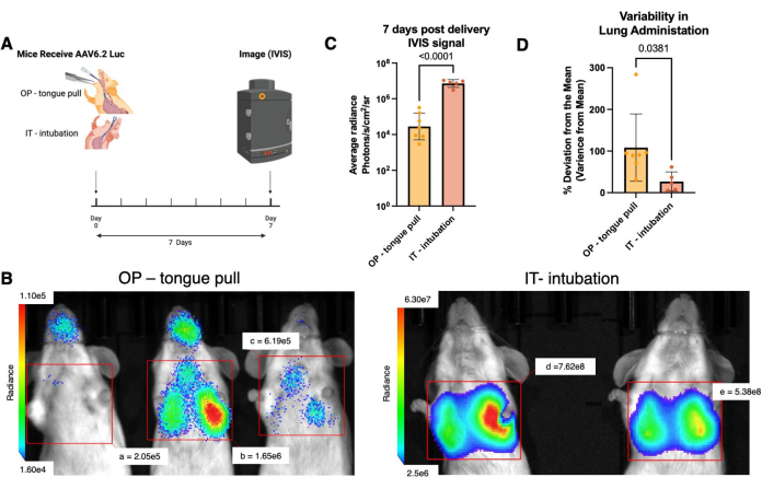
Figure 3: Intubation yields greater delivery to the lung with less variability relative to O.P. aspiration. (A) 6-10 wk old female BALB/c mice were dosed with 1x10¹¹ vg of AAV6.2-Luciferase, (Penn Vector Core), then assessed with in vivo bioluminescent imaging (IVIS) 7 days later. 10 min prior to imaging (30 s exposure), they were injected (subcutaneously) with 200 µL of luciferin (GoldBio, 15mg/mL). (Created with biorender.com) (B) Representative IVIS images of mice receiving AAV6.2-Luciferase by OP aspiration (tongue pull, left) or IT delivery (intubation, right). Regions of interest are the same size, and the average radiance within the region is shown in the image. (C) Quantification of luciferase signal in OP aspiration (n=7) versus intubation (n=5) mice. p<0.0001 by unpaired Welsh's t-test with after log-transformation of raw luciferase data. (D) Deviation from the mean as a percent of the mean average radiance. (p=0.0381, Welsh's T-test) Please click here to view a larger version of this figure.
Figure S1: Example images from the consumer otoscope used in the described protocol for guided intubation (left) and an endoscope of similar cost and availability (right) are shown. Note the clear differences in focus between the images. Please click here to download this File.
Figure S2: An example image showing the bubble tube in use. The tidal volume quickly moves the droplet in the bubble tube several centimeters. The droplet moves very little if the tube is in the esophagus. Respiratory rate, and tidal volume could be estimated by this method. Please click here to download this File.
Figure S3: A basic lighting assembly that removes the need for purchasing a costly fiberoptic lighting kit. A 20 G needle was pushed through a 50 mL conical, 0.75 mm fiber optic cable was purchased cheaply (and in bulk) and cut to length (~1m) with a razor blade and taped in place. Securing the catheter in place was unnecessary due to the snug fit on the fiber optic cable. Securing the conical lid over a mobile phone flashlight results in sufficient light transduction to perform intubation in a darkened room. This assembly further decreases the costs assumed by a laboratory incorporating this technique. Please click here to download this File.
Discussion
Intubation and intratracheal instillation are the best methods for liquid delivery to the lung, but the abundant challenges presented by murine anatomy have prevented widespread adoption in pulmonary research labs15. Though it ensures efficient delivery of an inoculum to the lung a potential downside of IT delivery by this and other intubation techniques is bypassing of the upper airway. Intranasal delivery is a proven method that does not bypass the upper airway. Though intranasal delivery is often used in pulmonary virology labs and may be more effective in some cases22, it does not reflect the natural exposure route23 for some aspirated agents, results in more variable amounts of inoculum in the lung24, and is less effective when using isoflurane as the anesthetic5. As such, extensive work has been done on aerosolizing apparatuses and comparing them with other pulmonary delivery techniques25, but these methods present aerosol-associated hazards and tend to be expensive. All of these difficulties have led to oropharyngeal instillation by tongue pull26, being the preferred method for pulmonary liquid delivery in many labs. As we have shown, this method is far more variable than intratracheal instillation following intubation. Furthermore, delivery by the tongue pull method cannot be assessed during administration, and undue trauma may be caused to the mouse tongue due to the tension needed to prevent swallowing. The tongue is also held in the guided intubation and IT instillation technique. However, because the video feed shows glottis, a researcher using our method does not need to force laryngeal positioning by placing significant tension on the tongue. This decreased traction causes less tissue trauma. However, despite the considerable advantages presented by intubation and IT instillation, challenges have stood in the way of its use in some labs. For example: compression of the trachea due to erroneous canulation of the esophagus can obstruct breathing and increase mortality; insertion of the endotracheal tube too deeply may lead to damage to the carina or mainstem bronchi; and finally, murine intubation by traditional methods takes a long time making it impractical for most researchers seeking reliable intrapulmonary delivery. Our method addresses the difficulties associated with mouse intubation, even overcoming the anatomic barriers to intubating mice.
Several methods have been proposed to overcome the anatomic challenges of murine intubation by implementing visual aids. For example, the use of a traditional endoscope has been proposed6. However the long focal depth of the instrument is poorly suited to being used for murine intubation (Figure S1). This is also readily apparent in video 1, where the vocal folds can be seen but are blurry. Clinical otoscopes have also been used for direct laryngoscopy12. However, the cost of clinical otoscopes and the lack of a video feed make them inaccessible for most labs and ill-suited to training. The use of a widely available consumer otoscope has greatly improved the practicality of guided intubation. The shorter focal length, smaller body, and extended depressor lip allow for a clearer video feed, better maneuverability, and positioning, and the otoscope lip can be used to apply pressure to the base of the tongue, which improves access to the rima glottidis. Beyond these visual aids, transillumination has also been used to successfully visualize the glottis and intubate mice7,13,14. However, transillumination-based approaches may require depilatory agents on the area overlaying the mouse neck, close proximity to a hot light source, use of the needle within the catheter, insertion of sharp instruments into the mouse mouth, and/or a small incision to be made over the trachea to confirm placement. The method described here avoids these risks and builds on the best aspects of these published methods. In addition to overcoming anatomical barriers to intubation, cost considerations have been taken into account in our method.
Together the use of helping hands, a mouse stand, the otoscope, and a fiberoptic light make intubation easier to learn and much faster to perform, at a reduced expense to labs. The cost of the commercially available intubation stand can be avoided by using a handmade one27. The cost of the fiberoptic light assembly can be defrayed by purchasing the fiberoptic cable separately (at a much lower cost, ~$9 vs $218-kent scientific- at time of writing). To complete the fiberoptic light source, a 50 mL conical lid with a pinhole in it and tape can be used to collect light from a mobile phone flashlight (see Figure S3, and Table of Materials). Das et. al describe a similar device11. For some labs, the reduced cost of this solution may make our method a much more accessible procedure. Beyond costs, difficulty confirming endotracheal tube placement has made the intubation of mice daunting.
In the past, to confirm the endotracheal tube is appropriately positioned researchers have relied on mirror fogging by holding a dental mirror above the endotracheal tube18. However, the small tidal volume and variations in room temperature often prevent mirror fogging and lead to errors in judgment. In the case of murine intubation, this poses a serious risk of injury. Others have resorted to inducing respiratory distress by obstructing the endotracheal tube10. This causes extensive stress to the animal and provides less information than the "bubble method." Further, the introduction of lung therapeutics and air into the stomach due to inadequate confirmation of endotracheal tube placement may cause distress in addition to hampering the effectiveness of the agent given. The use of the "bubble method" provides a faster, more reliable visual confirmation of endotracheal tube placement and is a slight improvement on a similar method described previously28. The "bubble method" also enables a researcher to roughly estimate the respiratory rate and tidal volume of the animal.
Animal positioning and visualization of the vocal folds are key to this technique. We found that pediatric nasal specula were too large and cumbersome to hold the mouth open and cheeks out of the way during intubation, but they were very helpful during training as they allowed a researcher to demonstrate otoscope placement. Guiding a trainee to recognize the relevant anatomy is difficult, and improved visibility in the mouse mouth was key, which was significantly aided by the use of the commercial otoscope. Staging using a lighted magnifying glass and helping hands are integral to assisting inexperienced operators in intubation, though they may not be as critical for experienced operators.
While training in this method, troubleshooting may be necessary. If the endotracheal tube is entering the esophagus repeatedly, the glottis may have spasmed, the soft palate might be inflamed, and proper placement may be more difficult. In this circumstance, allow the animal to recover for several minutes before attempting intubation again. The number of re-attempts should be specified in the IACUC protocol and always adhered to. If you are having trouble seeing the vocal folds, the respiratory rate may be suppressed due to too much anesthesia, or the mouse positioning may be inappropriate (most commonly head tilted back too far). In this case, reposition the animal and allow them to recover from anesthesia before trying again. The bubble tube should clearly demonstrate the animal's tidal volume. If the bubble is moving only slightly, gasps due to pressure placed on the trachea by a misplaced endotracheal tube may be causing this movement. In this instance, refer to Timestamp: 8:25 and Figure S3 to see how readily apparent the tidal volume is when using the "bubble-tube" on a correctly-placed endotracheal tube. If your endotracheal tube is misplaced, remove it immediately and allow the animal to recover before attempting the procedure again.
Though intubation and intratracheal instillation are far superior to other methods they have been too technically difficult to be widely adopted. The methods described here make intratracheal instillation and murine intubation accessible and practical. Though future applications of this technique are highly dependent on what is being delivered to the lungs and in what context they are given an easy and reliable method paves the way for many advances in lung biology.
Disclosures
We have no disclosures.
Acknowledgements
Abagail Boyd (University of South Alabama) provided key guidance on adapting our intubation protocol based on her experience. Larkin Kolls exposed the writers to at-home otoscope technology. Penn Vector-Core made the viral vector used in the experiment. Candice Fisher is our lab manager; she is integral to the work we do. Madison Robin maintained the working order, sterility, and organization of our vivarium throughout these experiments.
Materials
| Name | Company | Catalog Number | Comments |
|---|---|---|---|
| Fiber Optic LightiFiber Optic Lighting Kit, Mouse | Kent Scientific | ETI-12ETI-MSE-02 | A component of the fiberoptic assembly, compatible with other lights and the replacement fiberoptic cable below |
| Alternative Fiberoptic Assembly | Braintree Scientifuc | RW-A37 47 | an alternative, not discussed (but we see no reason it would not work) |
| Alternative Fiberoptic Assembly | Harvard apparatus | 72-9075 | an alternative, not discussed (but we see no reason it would not work) |
| Alternative Fiberoptic Assembly | Handmade by lab | N/A | Instructions found here using a rubber stopper: DOI 10.3791/50318 |
| Alternative Fiberoptic Assembly | Handmade by lab | N/A (materials listed below) | Made from a 50mL conical lid, tape and 0.75mm fiberoptic fiber |
| Intubation Stand, Mouse | Kent Scientific | ETI-MSE-01 | Adjustable stand for intubation, many self-made versions available, tip racks and wooden platforms used, some alternates listed many suitable alternatives available. |
| Alternative Intubation Stands | Braintree Scientifuc | RIS 100 | an alternative, not discussed (but we see no reason it would not work) |
| Alternative Intubation Stands | Natsume Seisakusho | KN-1014 | an alternative, not discussed (but we see no reason it would not work) |
| Alternative Intubation Stands | Vetland | 000A3467 | an alternative, not discussed (but we see no reason it would not work) |
| Alternative Intubation Stands | Handmade by lab | N/A | Made from a 3 ring binder here: DOI 10.3791/60844 (several alternate methods published) |
| Nonabsorbable Braided Silk Suture, Size 0 | Fine Science Tools | 18020-00 | any longer than 1 inch will damage the carina |
| Safelet IV Catheter 20G x 1" | Nipro | NPRCI+2025 | Or similar |
| RS-5358 Micro Dissecting Forceps | Fisher | RS-5358 | Or similar, includes helping hands |
| Magnifying Glass with Light and Stand, EOOKU Flexible Magnetic Helping Hand, 5X&10X 12V 108pcs LED Beads Magnifying Lamp, 3 Modes Adjustable Swivel Gooseneck Arms & Heavy Duty Base Plate | Amazon | https://www.amazon.com/Magnifying- EOOKU-Flexible-Adjustable-Gooseneck /dp/B0C5MC6QNW/ref=sr_1_5?crid=F4 PZY6UOCEE9&dib=eyJ2IjoiMSJ9. gj5cKLw4v3CDSkrQKcs73kXqVtnJl JpBKEp_FcTCIEQSO3EirxEYoH ySohcPg0ZDhmnFwGeLit22nM SJxpz1MPP0 q6-PwbPzzMT0RWB_pXkmSBiAo5xB J4QSf8_BoLZ4u8S8lfIo5WI9-VuXj75X eo0LQfygaSMgW58XO2bbasOfaw cwxZXs5cTqCUmKh5bRn7A19_ cY2a0-x_ly-27RXXPinVbBNW00Ww8jY55x fnWFCaK5lnPb4VxSV6iTCOhMnkm TopFiA3rCLNXcBDGCwfLdw22w 17T0c_rRhh-Mrew .TtsVr89Bo5YLvaXvarcbKxJ07GK zMMVSg5op4U5eyvA&dib_tag= se&keywords=magnifying+glass+with +light+EOOKU+3+in+1&qid=1709661 709&sprefix=magnifying+glass+with+ light+eooku+3+in+1%2Caps%2C98&sr=8-5 | Or similar, any bent finely serrated forceps will work |
| Ear Wax Removal - Earwax Remover Tool with 8 Pcs Ear Set - Ear Cleaner with Camera - Earwax Removal Kit with Light - Ear Camera with 6 Ear Spoon - Ear Cleaner for iOS & Android (Black) | Amazon | https://www.amazon.com/Ear-Wax- Removal-Remover-Android%EF% BC%88Black%EF%BC%89/dp/B0 9KZ8TS7L/ref=pd_lpo_sccl_1/147 -1203879-4147304?pd_rd_w=Lt8 Ta&content-id=amzn1.sym.1ad20 66f-97d2-4731-9356-36b3edf1ae 04&pf_rd_p=1ad2066f-97d2-4731 -9356-36b3edf1ae04&pf_rd_r= MDQ3AHH4AC8T2KFX1RBE& pd_rd_wg=ja4H2&pd_rd_r=52d9 372c-b34b-4f6a-93f6-974754d96 3d9&pd_rd_i=B09KZ8TS7L&psc=1 | or similar, several brands available |
| AZIMOM PMMA Plastic End Glow Fiber Optic Cable 0.75mm(0.02in) 100m(328ft)/Roll for Star Sky Ceiling All Kind Led Light Engine Driver Source | Amazon | https://www.amazon.com/AZIMOM -Plastic-Cuttable-Ceiling-1pcs0-00 98in328ft/dp/B07WC9JV5N/ref=sr _1_9?dib=eyJ2IjoiMSJ9.K7NmvB hJOux4zdNBKSPo-ac4HYPDPUS F85Qlra7o7G2FQl9MQLj1D7QtPJ cxnAeDiYm_yNWPhwj44nhSfwqM vs1AtulruHGEsfjLEAjRGSFpNLgN H-Ewc190MluERArk91lR9Od6ceB Hkz2AJad11hVUkWEOb0-emUWW b2U4fNN0upQKgyD1vE eU0DtClQ73tDe22z4CXrSLP7vC D9jF2APfz1liHou_zdiAyCSFb1Df bC8Ymce6zXV0qOD3gQFkm6j NV5bN1DG8SH_BaV4vofzj98T JVRnNElEzUzjx0rY.AMHl3h-jIz GTeGjvej0Nq8FTPLEPmfiQ UDPz_U6ctI0&dib_tag=se& keywords=fiber%2Boptic%2Blight %2Bstrands&qid=1722619884&sr =8-9&th=1 | Or similar, fiber is used to build device in line 7 |
| 50mL conical tube | Cell Treat | 229421 | any opaque plastic 50mL screw cap conical lid should function. Does not come in contact or close proximety with animal so no need for sterility |
| Isoflurane | VetOne | Consult Veternary Staff | |
| Innoculum | N/A | Specific to your Experiment | |
| Pipette Tips (filter) | N/A | Sterile Filter p100 and p1000 tips used | Brand/Supplier not relevant |
| Induction Chamber | N/A | As available at yourt institution | Brand/Supplier not relevant |
| Isopropanol Prep Pads | N/A | For instrument cleaning | Brand/Supplier not relevant |
| Tubing | N/A | small diameter flexible tubing will work | For construction of "bubble tube" (ours is ~3mm in diameter and was taken from an IV line) |
| Luer lock | N/A | to fit your tubing above | For construction of "bubble tube" |
| 3M half-facemask respirator | 3M | 17-986-9B | to limit gas exposure |
| Twist Tie | N/A | N/A | to maintain the U shape of the bubble tube |
References
- The Pneumonia Etiology Research for Child Health (PERCH) Study Group. Causes of severe pneumonia requiring hospital admission in children without HIV infection from Africa and Asia: the PERCH multi-country case-control study. Lancet. 394 (10200), 757-779 (2019).
- Shiels, M. S., Haque, A. T., Berrington de González, A., Freedman, N. D. Leading causes of death in the US During the COVID-19 pandemic, March 2020 to October 2021. JAMA Intern Med. 182 (8), 883-886 (2022).
- Egger, C., et al. Administration of bleomycin via the oropharyngeal aspiration route leads to sustained lung fibrosis in mice and rats as quantified by UTE-MRI and histology. PLOS ONE. 8 (5), e63432(2013).
- Nielsen, T. B., Yan, J., Luna, B., Spellberg, B. Murine oropharyngeal aspiration model of ventilator-associated and hospital-acquired bacterial pneumonia. J Vis Exp. (136), e57672(2018).
- Seo, Y., et al. Optimizing anesthesia and delivery approaches for dosing into lungs of mice. Am J Physiol Lung Cell Mol Physiol. 325 (2), L262-L269 (2023).
- Vergari, A., Polito, A., Musumeci, M., Palazzesi, S., Marano, G. Video-assisted orotracheal intubation in mice. Lab Anim. 37 (3), 204-206 (2003).
- Watanabe, A., Hashimoto, Y., Ochiai, E., Sato, A., Kamei, K. A simple method for confirming correct endotracheal intubation in mice. Lab Anim. 43 (4), 399-401 (2009).
- Samsamshariat, S. A., Movahed, M. R. Using a 0.035-in. straight-tip wire and a small infant laryngoscope for safe and easy endotracheal intubations in rats for cardiovascular research. Cardiovasc Revasc Med. 6 (4), 160-162 (2005).
- Hamacher, J., et al. Microscopic wire guide-based orotracheal mouse intubation: description, evaluation and comparison with transillumination. Lab Anim. 42 (2), 222-230 (2008).
- Rivera, B., Miller, S., Brown, E., Price, R. A novel method for endotracheal intubation of mice and rats used in imaging studies. Contemp Top Lab Anim Sci. 44 (2), 52-55 (2005).
- Das, S., MacDonald, K., Chang, H. Y., Mitzner, W. A simple method of mouse lung intubation. J Vis Exp. (73), e50318(2013).
- Thomas, J. L., et al. Endotracheal intubation in mice via direct laryngoscopy using an otoscope. J Vis Exp. (86), e50269(2014).
- Brown, R. H., Walters, D. M., Greenberg, R. S., Mitzner, W. A method of endotracheal intubation and pulmonary functional assessment for repeated studies in mice. J Appl Physiol. 87 (6), 2362-2365 (1999).
- Nelson, A. M., Nolan, K. E., Davis, I. C., Nelson, A. M. Repeated Orotracheal Intubation in Mice. J Vis Exp. (157), e60844(2020).
- MacDonald, K. D., Chang, H. Y., Mitzner, W. An improved simple method of mouse lung intubation. J Appl Physiol (1985). 106 (3), 984-987 (2009).
- Controlled Substance Schedules. , US Department of Justice Diversion Control Division. https://www.deadiversion.usdoj.gov/schedules/schedules.html (2025).
- Cesarovic, N., et al. Isoflurane and sevoflurane provide equally effective anaesthesia in laboratory mice. Lab Anim. 44 (4), 329-336 (2010).
- Balloy, V., Huerre, M., Latgé, J. -P. Differences in patterns of infection and inflammation for corticosteroid treatment and chemotherapy in experimental invasive pulmonary aspergillosis. Infect Immun. 73 (1), 494-503 (2005).
- Clary, E. M., O'Halloran, E. K., de la Fuente, S. G., Eubanks, S. Videoendoscopic endotracheal intubation of the rat. Lab Anim. 38 (2), 158-161 (2004).
- Silva, P. L., Scharffenberg, M., Rocco, P. R. M. Understanding the mechanisms of ventilator-induced lung injury using animal models. Intensive Care Med Exp. 11 (1), 82(2023).
- Schulz, H., et al. Respiratory mechanics in mice: strain and sex specific differences. Acta Physiol Scand. 174 (4), 367-375 (2002).
- Americo, J. L., Cotter, C. A., Earl, P. L., Liu, R., Moss, B. Intranasal inoculation of an MVA-based vaccine induces IgA and protects the respiratory tract of hACE2 mice from SARS-CoV-2 infection. PNAS. 119 (24), e2202069119(2022).
- Belser, J. A., Gustin, K. M., Katz, J. M., Maines, T. R., Tumpey, T. M. Comparison of traditional intranasal and aerosol inhalation inoculation of mice with influenza A viruses. Virology. 481, 107-112 (2015).
- Pelgrim, C. E., et al. Intratracheal administration of solutions in mice; development and validation of an optimized method with improved efficacy, reproducibility and accuracy. J Pharmacol Toxicol Methods. 114, 107156(2022).
- Kunda, N. K., Price, D. N., Muttil, P. Respiratory tract deposition and distribution pattern of microparticles in mice using different pulmonary delivery techniques. Vaccines. 6 (3), 41(2018).
- Driscoll, K. E., et al. Intratracheal instillation as an exposure technique for the evaluation of respiratory tract toxicity: uses and limitations. Toxicol Sci. 55 (1), 24-35 (2000).
- Rayamajhi, M., et al. Non-surgical intratracheal instillation of mice with analysis of lungs and lung draining lymph nodes by flow cytometry. J Vis Exp. (51), e2702(2011).
- Hattori, T., et al. Gene expression profiling of IL-17A-treated synovial fibroblasts from the human temporomandibular joint. Mediators Inflamm. 2015, 436067(2015).
Reprints and Permissions
