This protocol details an in vitro model of the catheterized urinary tract, which can be used to study both planktonic and biofilm-associated bacterial cell populations in simulated catheter-associated urinary tract infections. This model can be further utilized to study the efficacy of antimicrobial products aimed at controlling urinary tract infections.
Method Article
An In Vitro Bladder Model of Catheter-Associated Urinary Tract Infection
In This Article
Summary
Abstract
Catheter-associated urinary tract infections (CAUTIs) are among the most common healthcare-associated infections. Biofilm formation on urinary catheters is a key aspect in the pathogenesis of these infections, and frequently leads to blockage of catheters and serious complications such as pyelonephritis and septicaemia. Approaches to model and study biofilm formation in this setting are essential for both a fundamental understanding of the mechanisms underpinning the pathogenesis of CAUTI, as well as the development and robust pre-clinical evaluation of effective therapeutic strategies to control CAUTI and catheter blockage. This protocol describes an in vitro model of the catheterized urinary tract, which replicates the catheter closed drainage system as used in clinical practice, facilitating the study of CAUTI and catheter biofilm formation under representative conditions. Furthermore, this protocol details the application of the in vitro bladder model to evaluate the efficacy of antimicrobial catheter maintenance products, including products currently available on the NHS supply chain.
Introduction
Urinary catheters are amongst the most commonly deployed medical devices, with an estimated prevalence of 19% in acute care facilities1,2,3. Catheters are primarily deployed to aid in urine output management in cases of bladder dysfunction, and are inserted directly into the bladder, allowing urine to void from the bladder via a central lumen2,3. Urinary catheters can either be used intermittently, where they are inserted into the bladder periodically to drain urine, or indwelling catheters can be utilized, where catheters are left in situ in the bladder to continuously drain urine3,4. Despite their widespread use, urinary catheters are the biggest risk factor for the development of urinary tract infections (UTIs), which account for an estimated fifth of healthcare-associated infections5,6. Indeed, the catheter itself often acts as the initial site of microbial colonization, providing a surface for uropathogens to adhere to and further colonize the urinary tract1,6,7,8. Virtually all patients undergoing long-term continuous catheterization (where the catheter is left in place for ≥28 days) will experience bacteriuria, with the risk increasing by ~5% for every day the catheter is in situ2,9,10,11. Commensurately, around 80% of UTIs are associated with the use of a urinary catheter6.
Catheter-associated urinary tract infections (CAUTIs) represent a significant burden to health services, and are often associated with more serious upper UTI symptoms (such as ureteritis and pyelonephritis) than uncomplicated UTIs, as well as an increased risk of secondary bloodstream infections1,7,12,13,14. Standard treatments for CAUTIs include antibiotic therapy, however, these infections are often recalcitrant to antimicrobial treatment as a result of the development of complex biofilm communities on the catheter surface15,16,17,18,19,20. Indeed, it is estimated that ~80% of CAUTIs result from biofilm-forming urinary tract pathogens19. Biofilm formation is characterized by the attachment of bacterial cells to catheter surfaces and the development of communities embedded in an extensive matrix of extracellular polymeric substances (EPS)21,22,23,24. Production of the EPS is a defining feature of biofilm communities and is responsible for many of the emergent properties displayed by biofilm-associated cells, such as elevated resistance to antimicrobial agents and immune clearance21,22,24. The EPS can play a direct role in these phenotypes by physically shielding cells and protecting them from antimicrobials or immune effectors, as well as indirectly by facilitating the development of wider aspects of biofilm communities such as the physicochemical microenvironment of the biofilm and the formation of persister cells23,24,25,26. Furthermore, some uropathogens, including Proteus spp., which account for ~11% of CAUTIs, possess potent urease enzymes, enabling them to hydrolyze the urea present in urine and resulting in an overall increase in urinary pH2,27,28,29,30. This pH increase is accompanied by the precipitation of calcium and magnesium phosphate crystals, which can be incorporated into the growing biofilm, resulting in biomineralization and the formation of dense crystalline biofilm structures that can block catheters. These crystalline biofilms not only make treatment problematic, but can also initiate serious complications such as pyelonephritis and septicemia2,28,31,32,33,34.
The complex environmental niche in the catheterized urinary tract, and the important role of biofilm formation in these infections, pose particular challenges in the study of CAUTI pathogenesis. Simple and widely used laboratory models to study and quantify biofilm formation, such as 96-well plate-based assays, are inadequate for the study of biofilm formation in CATUI, particularly crystalline biofilms. For example, such models do not account for key aspects of the CAUTI niche, such as the complex nutrient environment provided by urine, how the catheter surface affects adhesion and subsequent biofilm production, and the continual flow of urine media35,36,37,38,39,40. Additionally, standard assays used to determine antimicrobial susceptibility of uropathogens often don't reflect real-world clinical susceptibility. Minimum inhibitory concentration assays and microbial suspension tests are frequently used to infer the antimicrobial susceptibility of CAUTI pathogens, but typically do not simulate important variables of the CAUTI environment, such as the effect the presence of a urinary catheter and any subsequent biofilm formation may have on antimicrobial susceptibility41,42,43.
This study details the setup and running of an in vitro bladder model that simulates the environment of the catheterized urinary tract, and replicates a standard catheter closed drainage system as used in clinical practice, first described by Stickler et al.44 in 1999 (Figure 1). This model utilizes standard-sized Foley catheters (which are currently available on the NHS supply chain), and artificial urine media (AUM) as previously described by Nzakizwanayo et al.45to simulate CAUTI and biofilm formation. The in vitro bladder model consists of a double-walled glass chamber that acts as the "bladder" and has an outlet through which a Foley catheter can be inserted into the central glass chamber. At the top of the central glass chamber, there is an inlet through which AUM can be supplied to the "bladder" at a biologically relevant flow rate using a peristaltic pump, which will flow through the catheter via the drainage eyeholes into a waste bag. Additionally, the outer chamber is connected to a water bath circuit, which allows the models to be kept at a constant temperature. The in vitro bladder model can be inoculated via the central chamber, allowing for the study of CAUTI pathogens in a biologically representative niche. Furthermore, as models are catheterized and constantly supplied with fresh AUM, the growth and population dynamics of both planktonic and biofilm-associated cells can be studied.
Past studies have utilized in vitro bladder models for several applications, including the fundamental study of biofilm formation in prominent CAUTI pathogens, including Proteus mirabilis and Escherichia coli 43,45,46,47,48,49. Additionally, the in vitro model has been utilized to evaluate the efficacy of products intended to control CAUTI or prevent blockage50,51,52. More recently, models have been used to determine whether adaptations and mutations linked to AMR impact biofilm formation and CAUTI pathogenesis in P. mirabilis43,46. The in vitro bladder model has also been used to study novel antimicrobial treatments aimed at treating CAUTI, including newly formulated theranostic catheter coatings and phage therapy treatments47,48. This protocol encompasses the setup and running of in vitro bladder models, the enumeration of viable planktonic and biofilm-associated cells from the model and urinary catheter surface respectively, and the measurement of urinary pH to monitor urease production. Protocols are also detailed for the assessment of antimicrobial products aimed at treating or preventing CAUTI, including bladder irrigation solutions and antimicrobial lubricant gels, which are currently available on the NHS supply chain.
Protocol
A schematic representation of the in vitro bladder model setup is shown in Figure 1. In vitro bladder models are designed to be compatible with standard Foley catheters (dimensions shown in Figure 1 are compatible with standard size 14 French catheters). Sterilize all in vitro bladder model equipment, and clamp any silicone tubing used into the media circuit prior to use. Strict adherence to the aseptic technique is required throughout the setup of in vitro models, and it is recommended that the model is assembled under sterile conditions (i.e., in a category II microbial safety cabinet or laminar flow hood). A minimum of n = 5 replicates is recommended to determine statistical significance for in vitro bladder model experiments. The details on the reagents and the equipment used are listed in the Table of Materials.
1. Preparation of artificial urine media
NOTE: Artificial urine media (AUM) is recommended for use in in vitro bladder models due to the high volumes of media required for experimentation, and variability associated with human urine samples. This protocol details the preparation of AUM as described previously and optimized for use in the in vitro bladder model by Nzakizwanayo et al. (5 L final volume)43,45,46,48,53,54.
- Prepare AUM solution 1
- Weigh out the following compounds and add to a 1 L borosilicate bottle filled with 850 mL of deionized water: 3.25 g of magnesium chloride hexahydrate (MgCl2.6H2O), 23 g of sodium chloride (NaCl), 11.5 g of anhydrous sodium sulfate (Na2SO4), 3.25 g of trisodium citrate (Na3C6H5O7), 0.1 g of sodium oxalate (Na2C2O4), 14 g of monopotassium phosphate (KH2PO4), 8 g of potassium chloride (KCl), 5 g of ammonium chloride (NH4Cl), 25 g of gelatin, and 5 g of tryptone soya broth.
- Whilst stirring the AUM solution, measure the pH using a pH meter and adjust to 5.75 (± 0.02) by adding 3 M NaOH or HCl as appropriate.
NOTE: NaOH/HCl for pH adjusting should be added to AUM dropwise to prevent salt precipitation from the solution. - Once AUM has been pH adjusted, top up the solution to a final volume of 1 L with deionized water and autoclave sterilize.
NOTE: Once sterile, the solution can be stored for up to three months at room temperature (AUM solution 1 should be discarded if precipitation or cloudiness are observed). - Separately, prepare AUM solution 2, by adding 125 g of urea (CH4N2O) and 2.45 g of calcium chloride (CaCl2) to 400 mL of sterile, deionized water in a sterile 500 mL beaker.
NOTE: Preparing the calcium urea solution 2 results in an endothermic reaction, therefore, it is recommended that AUM solution 2 is stirred and heated to ~ 50 °C on a hot plate/magnetic stirrer whilst the compounds dissolve to prevent freezing. - Once the urea and calcium chloride have dissolved, filter sterilize solution 2 using a vacuum pump and 0.45 µM nitrocellulose filter units, into a sterile, 500 mL glass bottle. Once sterilized, solution 2 is stable for three months at 4 °C or one month at room temperature
NOTE: AUM solution 2 should be discarded if precipitation or cloudiness is observed. - In a laminar flow hood or Category II microbiological safety cabinet, aseptically combine AUM solutions 1 and 2 with 3.6 L of sterile deionized water in a sterile, 5 L glass aspirator bottle. AUM should be pale yellow in color and transparent.
- Using a serological pipette, remove 50 mL from the total AUM volume and aliquot into a 50 mL centrifuge tube, and reserve for later use.
- Seal the aspirator bottle using a rubber bung or aluminum foil and leave it at room temperature until in vitro models are assembled.
- Aseptically remove 10 mL of AUM from the 50 mL aliquot and reserve the rest of the solution for inoculum preparation. Using a pH meter, measure the pH of the 10 mL AUM aliquot, which should be ~pH 6.1 (±0.2). Prepared AUM is stable at room temperature for around one month, and should be discarded if precipitation or cloudiness occurs.
2. In vitro bladder model setup
- Insert a Foley catheter into the central glass chamber of the in vitro bladder model (schematic detailed in Figure 1). Secure the catheter in place by inflating the balloon with the syringe of sterile water provided, and attach the waste bag to the catheter port.
- Connect the in vitro bladder model device45 to the AUM reservoir via connecting tubing. Connect media circuit tubing to the peristaltic pump as per manufacturer's instructions, ensuring the tubing has adequate tension without being stretched.
- Attach in vitro bladder models to clamp stands, and secure waste bags in stands below the models to ensure flow through the models via gravity.
- Connect the outer chamber of the bladder model to a circulating water bath via silicone tubing (Figure 1B). Set the water bath to heat to 37 °C and turn on the tank circulators.
- Run water baths for 30 min prior to inoculating in vitro bladder models to ensure the temperature is stable at 37 °C, and ensure the water bath remains topped up to the appropriate water level for the duration of experimentation.
- Remove any clamps from media tubing and set peristaltic pumps to run at approximately 0.75 mL/min to replicate human urine production (~1080 mL/day). Actual flow rates will be calculated during experimentation.
- Run AUM through the in vitro bladder models via the peristaltic pump for 30 min prior to inoculation to confirm that media flow is at a consistent rate and that the sterile drainage system is intact.
- Drain any accumulated media from the waste bag prior to experimentation. The volume of media can be measured to calculate exact flow rates (detailed in step 4).
3. Inoculation of in vitro bladder models
- Prepare overnight cultures for in vitro bladder models by suspending a loopful of bacterial colonies grown on solid agar into 10 mL of LB broth. Incubate overnight at 37 °C with agitation.
NOTE: The in vitro bladder model can be used to study a variety of CAUTI pathogens, including urease-positive strains such as Proteus mirabilis, and the growth media used can be modified accordingly. - Centrifuge the entire volume of overnight culture at 4,500 x g for 10 min at room temperature, and discard the supernatant. Resuspend the pellet in 10 mL of AUM and measure the absorbance at OD595 nm using a spectrometer.
- Normalize the resuspended cultures to an OD595 nm of 1.0 and a final volume of 10 mL in AUM using the following equation:
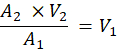
where, A1 is the absorbance at OD600 nm of the overnight culture, A2 is the desired absorbance at OD600 nm of the inoculum (1.0), V2 is the desired volume of the inoculum (10 mL), and V1 is the required volume of overnight culture for normalization.
NOTE: 10 mL of an OD595 nm 1.0 suspension will ensure models are inoculated with approximately 1010 CFU of most bacterial strains (validated for Proteus mirabilis, Escherichia coli, Enterococcus faecalis, Pseudomonas aeruginosa and Staphylococcus aureus), replicating an established infection. Starting inoculum can be modified depending on the species used or the stage of infection being modeled (i.e., a lower inoculum density can be used to simulate early-stage infection)47. - Sterilize in vitro bladder models using 70% ethanol and remove the stopper (media inlet) so the inner chamber is accessible. Remove 10 mL of AUM from the volume inside the bladder model using a serological pipette.
NOTE: When inoculating and sampling in vitro models from the central chamber, ensure work is quick and aseptic, always wear appropriate PPE, and regularly decontaminate gloves and surfaces with 70% ethanol. - Inoculate the in vitro bladder models via the central chamber using the entire normalized 10 mL suspension and mix thoroughly with a serological pipette. Remove 10 mL from the central chamber of the in vitro bladder models using a serological pipette and aliquot into a 15 mL universal for later CFU/mL calculations and pH measurement.
- Thoroughly decontaminate the external surfaces of the bladder model and bung with 70% ethanol, and replace the stopper. Resume media flow briefly to ensure that the AUM level in the central chamber reaches the eyehole, before stopping flow.
- Allow cultures to establish in the bladder model for 1 h before turning on media flow via the peristaltic pump at ~0.75 mL/min.
- Whilst the inoculum establishes, prepare dilutions from the sample taken from the central chamber.
- Serially dilute 100 µL of sample in 900 µL of sterile PBS until the sample is diluted to 10-7. Plate out 10 µL spots of the dilutions in triplicate on appropriate solid media, and incubate at 37 °C overnight.
- Following incubation, count the colonies on each dilution plate and calculate the exact CFU/mL of the initial inoculum.
NOTE: If using species which swarm, i.e., Proteus mirabilis, use MacConkey agar without salt (peptone 20.0 g/L, lactose 10.0 g/L, bile salts 5.0 g/L, neutral red 0.075 g/L and agar 12.0 g/L) to ensure single colonies are still visible for ease of counting.
- After 1 h incubation, switch on media flow via the peristaltic pump to begin running in vitro bladder models, noting the time at which the models are switched on.
4. Calculating in vitro bladder model flow rate
- Once in vitro models have been inoculated and media flow initiated, allow models to run for 4 h. Briefly pause media flow and collect the entire volume of residual AUM from the waste bag in a volumetric flask via the drainage port. Note the volume collected.
NOTE: The flow rate can be calculated from the volume of media collected in step 2.7., however more accurate flow rates can be obtained by running models for longer (i.e., recommended 4 h) as this reduces the likelihood of residual air in the media circuit tubing affecting the calculated flow rate. - Resume media flow via the peristaltic pump, ensuring that the waste bag drainage port is closed, to maintain a sterile drainage system.
- To calculate the actual flow rate of in vitro models, use the following equation:

NOTE: Flow rates in the range of 0.55-1.0 mL/min are recommended to replicate human urine production. - If the media flow rate is calculated outside of the acceptable range, discard the replicate and adjust the rpm on the peristaltic pump to fit within the acceptable range.
5. Sampling planktonic cells from the in vitro bladder model
NOTE: In vitro bladder models should be sampled routinely throughout experimentation to monitor population dynamics. All models should be sampled immediately after inoculation, at the point of AUM flow activation, 4 h post-inoculation (when the flow rate is calculated), and at the point, models are terminated (either blockage or a predetermined endpoint). If running models for >24 h, it is recommended that samples are taken every 24 h to monitor CFU/ mL, and additional samples can be taken as required by users.
- Decontaminate the bladder model stopper with 70% ethanol and remove it to access the central chamber.
- Avoiding the catheter tip and balloon, gently mix the contents of the central chamber using a serological pipette.
- Using a serological pipette, remove a 10 mL sample from the chamber, and transfer it to a sterile tube. Immediately decontaminate the stopper with 70% ethanol and replace, completing the sterile, closed, drainage circuit.
- Remove 1 mL from the sample using a pipette, and aliquot into a 1.5 mL microcentrifuge tube for serial dilutions.
- Measure the pH of the remaining sample using a pH meter, ensuring the sample is thoroughly mixed, and the pH meter is calibrated prior to use.
- Serially dilute the reserved 1 mL aliquot from the sample, and plate onto appropriate solid media as described previously in step 3.8., plating out all dilutions.
NOTE: Planktonic samples taken from the bladder model are appropriate for other analyses, including genomic or molecular biology applications (optional).
6. Sampling biofilm-associated cells from the in vitro bladder model
- Catheter removal and preparation
NOTE: Biofilm sampling of in vitro bladder models is an endpoint analysis, requiring the deactivation of models and the removal of catheters for processing.- Disconnect the waste bag from the cathete, and drain any residual urine from the model by deflating the balloon. Remove the catheter from the bottom of the model using sterile forceps.
- Place the removed catheter onto a sterile board and, using a sterile scalpel, cut the catheter at the required points for sectioning (outlined in Figure 2).
- Ensure catheter sections are cut lengthways to expose the lumen; then, using sterile forceps, dip the catheter section 3 times into sterile PBS to remove non-adherent cells.
NOTE: If imaging catheter sections downstream, i.e., using SEM, leave 1 cm sections intact (do not cut lengthways) and instead remove excess moisture from the lumen with a sterile paper towel before fixation.
- Disruption of biofilm for CFU/mL enumeration
NOTE: Protocols detailing the disruption of biofilm-associated cells may require further optimization if biofilms are extensively crystalline or mucoid.- Incubate the prepared catheter section in 0.25% trypsin at 37 °C for 30 min. The trypsin volume used should be the minimum required to cover the catheter; e.g., for a 1 cm catheter section cut perpendicularly, 1 mL of 0.25% trypsin is sufficient.
- After incubation, sonicate the solution containing the catheter for 10 min. The catheter section can then be removed and discarded. To deactivate the trypsin, the cell solution should be immediately diluted in sterile PBS and plated out for CFU counts on appropriate solid media, as described previously in step 3.8.
- Catheter biofilm quantification via crystal violet staining
- Add the prepared catheter sections to wells in an appropriately sized flat-bottomed well plate. Use an appropriate volume of 0.1% crystal violet solution to completely submerge the catheter section, and incubate statically at room temperature for 15 min. Ensure that a sterile catheter section is also included as a background control.
- Remove the catheter sections from the plate, and transfer to a new plate. Wash each catheter section twice in distilled water.
- After rinsing, transfer catheter sections to a new plate, and cover with 33% acetic acid. Incubate for 15 min at room temperature.
- Remove the catheter section from the plate, and then, using a plate reader, measure OD595 nm for each replicate, using the wells that contained the sterile catheter sections as background controls.
7. Antimicrobial susceptibility testing in the in vitro bladder model
NOTE: Most therapeutics aimed at controlling catheter-associated urinary tract infection are compatible with standard Foley catheters, check compatibility prior to beginning in vitro bladder model experiments.
- Antimicrobial lubricant gels
NOTE: To simulate the use of lubricating agents at catheter insertion, this step should be performed during step 2.1, immediately prior to insertion of Foley catheter. After inserting the catheter, the model should immediately be connected, and step 2.6. should be omitted so that the gel is not diluted and drained prior to inoculation. Inoculation should take place on the same day as catheter insertion. Lubricating agents designed for use with catheters are packaged as individual, sterile, single-use syringes of either 6 mL or 11 mL volume. Approximately 5 mL of lubricant is adequate to fill both the neck of the bladder model and coat the base of the model.- Using the aseptic technique, insert the end of the syringe containing the lubricating agent into the neck of the bladder model, and insert 5 mL of gel to fill the neck and enter the base of the chamber.
- Rotate the model so that the base is evenly coated with gel, then insert a Foley catheter into the model by pushing the catheter tip through the gel and secure by filling the balloon as normal.
- Connect the model to the rest of the setup, then fill the model lumen with urine until it starts to drain. As soon as urine begins to drain through the catheter eyehole, stop the pump to prevent loss of gel before inoculation.
- Incubate models for 30 min to allow urine to reach 37 °C, then inoculate models as described previously to ensure uniformity of diffusion time for gel components into the urine volume.
- Inoculate the model as described in step 3.5 and leave it to incubate for 1 h prior to switching on peristaltic pumps. At the point of turning on the pump, take a 1 mL planktonic sample and plate out dilutions for CFU counts as described in step 3.8.
- Irrigation solutions
NOTE: Irrigation solutions are often available in bulk or as individual, sterile, single-use "pouches". If using individual solutions, ensure they are compatible with the drainage ports of the catheters to be used prior to experimentation. If using bulk irrigation solutions, ensure triple-lumen catheters are used, and manufacturer's instructions are followed to determine volume/timeframe of product administration.- Prior to treatment, run in vitro models for at least 4 h to ensure biofilm-associated communities have had adequate time to establish. Once models have run for 4 h, pause media flow and take a pre-treatment sample from the inner chamber of the in vitro models, as detailed in step 5.
NOTE: If using bulk irrigation solutions, proceed to step 7.2.5. - If using an individual irrigation "pouch", disconnect the waste bag from the catheter and use the cap provided with the pouch to seal the waste bag. Immediately attach the irrigation solution pouch to the catheter, and using light pressure, squeeze the product into the central lumen. Apply as directed by manufacturer's instructions (i.e., 15 min application).
- Once the recommended treatment is completed, allow the residual solution to drain from the inner chamber via the catheter eye hole into the irrigation pouch, then disconnect the pouch. The remaining contents of the pouch can be discarded or used for further downstream applications.
- Sterilize the catheter drainage port and the waste bag connection port with 70% ethanol, and reconnect them, completing a sterile, closed drainage system.
NOTE: Continue to step 7.2.7 if using irrigation pouches. - If using a triple lumen catheter, disinfect the irrigation lumen port with 70% ethanol, and immediately begin to irrigate using a sterile syringe as per manufacturer's instructions.
NOTE: Measure the volume of residual urine in the waste bag before irrigating so this can be accounted for in any downstream blockage time or flow rate calculations. - Once the irrigation is complete, discard the syringe and any residual irrigation solution left in the waste bag.
- Take a post-treatment sample from the in vitro bladder model central chamber, as detailed in step 5, and resume AUM flow via the peristaltic pump to continue experimentation.
- Prior to treatment, run in vitro models for at least 4 h to ensure biofilm-associated communities have had adequate time to establish. Once models have run for 4 h, pause media flow and take a pre-treatment sample from the inner chamber of the in vitro models, as detailed in step 5.
8. Terminating in vitro bladder model experimentation
NOTE: Urease-positive organisms such as P. mirabilis can cause blockage of urinary catheters via crystalline biofilm formation. Blockage can thus be used as a biologically relevant endpoint for in vitro bladder models and is identified visibly by complete occlusion of catheter eyeholes with crystalline biofilms and an increase in the volume of AUM in the central chamber of the glass "bladder" (Figure 3).
- Once in vitro bladder models have run to the predetermined endpoint, which may be a blockage or a defined runtime, switch off AUM flow via the peristaltic pump and take an endpoint sample from the central chamber as described in step 5.
- Turn off the water bath flow and disconnect the media circuit and water bath circuit tubing from in vitro bladder models.
- Remove stoppers from in vitro bladder models, and remove the entire volume of residual urine using a serological pipette. Discard any contaminated media appropriately (i.e., autoclave sterilize).
- Drain the waste bags and discard any residual media. Disconnect catheter waste bags from the catheter port and discard them.
- Using a syringe, slowly deflate catheter balloons before removing catheters from models.
NOTE: If using urease-positive bacterial species, catheters can become "stuck" to the glass walls of in vitro models. Avoid applying large amounts of force to catheters, and instead, proceed to step 8.6. - Soak in vitro models in 5% antimicrobial cleansing solution (see Table of Materials) overnight, then wash thoroughly with water to ensure any residual disinfectant is removed. Autoclave to sterilize.
NOTE: If using urease-positive species, add 5% w/v citric acid to the disinfectant solution to help dissolve any biomineralization in the central chamber. This acidic solution will also help release any catheters that have become stuck to the models by breaking down crystalline biofilm deposits. - Disconnect the media circuit tubing from the AUM reservoir and discard any residual media. Flush through tubing with water to remove any residual AUM and autoclave.
- To calculate the exact blockage time, note the final volume in the waste bag and use the equation in step 4.4 and the previously calculated flow rate to determine the time of blockage.
Results
Successful operation of the in vitro bladder model is dependent on practicing good aseptic technique and careful observation of media turbidity throughout experimentation (Figure 3). Prior to inoculation, sterile AUM was pale yellow in color and transparent in the in vitro bladder model central chamber (Figure 3A). AUM in the media reservoir should remain clear throughout experimentation, as any turbidity changes that occur may indicate precipitation of salts from solution, or contamination of media. If AUM in the media reservoir or media circuit tubing becomes cloudy or shows a drastic change in color, replicates should be discarded and repeated. After inoculation, an increase in the turbidity of the AUM in the in vitro bladder model central chamber is clearly visible (Figure 3B). Changes in turbidity are often more pronounced in urease-positive organisms such as P. mirabilis, but should still be visible post-inoculation in urease-negative organisms. After AUM flow is initiated, a reduction in AUM turbidity is often observed as planktonic cells are washed out, and does not indicate a lack of viable cells in the model. Observing the media level in the central chamber can also be used to determine blockage time if utilizing urease-positive organisms (Figure 3C). As models are running, the catheter eyehole and tip should be completely visible to indicate proper drainage into the waste bags. Upon blockage, the level of AUM in the central chamber will rise above the catheter eyeholes and tip, indicating that the experiment is ready for any endpoint readings and termination. During experimentation, pressure effects in the in vitro bladder model media circuit can make models appear blocked. If a blockage is suspected, check that the AUM circuit, catheter, and waste bag tubing are taut before terminating the assay, as any potential air bubbles or kinks in the tubing can cause "airlocks" and prevent the catheter from draining.
During in vitro bladder model experiments, planktonic sampling can provide valuable insight into the growth dynamics within the model as well as the progression of CAUTI. Cell growth, pH, and biofilm formation can all be monitored throughout experimentation without necessitating the termination of models (Figure 4). If using urease-positive organisms, blockage time calculations can be utilized as a measure of the rate of biofilm formation and used to compare the properties of different bacterial strains (Figure 4). Comparison of a wild-type strain of P. mirabilis and an LPS mutant (mini-tn5 insertion into waaC) demonstrated an extension of time to blockage and thus reduced biofilm formation in the mutant (Figure 4). Urease-negative organisms do not typically block in vitro bladder models, and it is therefore recommended that a set endpoint is determined prior to experimentation. In some cases, urease-negative organisms can result in catheter blockage. For example, urease-negative strains of Klebsiella pneumoniae can produce extensive, mucoid biofilms, however, these typically take much longer than urease-positive biofilms to block drainage, so a set endpoint is still recommended.
Measurement of pH throughout bladder model experimentation can also provide insight into strain-to-strain differences, as well as the progress of CAUTI. If urease-negative organisms are utilized, pH in the in vitro bladder model central chamber should largely remain stable throughout experimentation, though small fluctuations in pH are expected as population dynamics change over the course of the experiment. Inoculation of in vitro models with urease-positive organisms results in a significant pH increase over time, with the highest pH usually being observed at the time of blockage, where pH readings as high as ~8-9 can be observed (Figure 4B).
Enumeration of planktonic CFU/mL is an important component of in vitro bladder modeling, allowing operators to assess whether inoculation has been successful and to monitor the progress of experimentation. Measuring CFU/ mL at the end of experimentation can highlight any differences in bladder colonization between different bacterial strains. When comparing the mini-Tn5 mutant strain of P. mirabilis to the wild-type, a significant reduction in CFU/ mL at the time of blockage was observed (Figure 4C). Furthermore, enumeration of CFUs from biofilms on the catheter surface, and quantification of biofilm biomass can provide insight into the ability of different pathogens to colonize the catheterized urinary tract. Crystal violet stains were used to determine differences in biofilm formation between a wild-type P. mirabilis strain HI4320 and isolates passaged in chlorhexidine (Figure 5). Optical density measurements at 595 nm demonstrated that isolates passaged in chlorhexidine displayed a reduction in biofilm formation compared with the wild-type and controls passaged in the absence of chlorhexidine.
As the aim of the in vitro bladder model is to accurately replicate the CAUTI niche, models can also be utilized to study the effects of antimicrobial therapeutics aimed at treating CAUTI in a clinically relevant environment. For urease-positive organisms, comparing the blockage times of treated models with untreated models can provide useful insight into the effectiveness of treatment. Treatment of in vitro bladder models inoculated with P. mirabilis with a 0.02% chlorhexidine bladder irrigation solution significantly extended the time to blockage compared with models that were left untreated or treated with 0.9% saline (Figure 6A). Furthermore, the in vitro bladder model can be utilized to study the bactericidal activity of antimicrobial therapeutics. Monitoring changes in CFU/mL of planktonic and biofilm-associated cells following antimicrobial treatment can indicate whether treatments are effective. Treatment of in vitro bladder models inoculated with P. mirabilis demonstrated a >7-log reduction in planktonic CFU/mL immediately after treatment, compared with untreated and 0.9% saline irrigated models when applied as per manufacturer's instructions (Figure 6B).

Figure 1: Schematic of the main components of the in vitro bladder model. (A) The bladder model (closed drainage system): (1) double-walled glass chamber simulating the bladder (2) media inlet, AU is supplied via peristaltic pump to the bladder model inner chamber (3) water inlet supplied via circulating water bath to outer chamber (4) water outlet (5) Foley catheter, inserted into inner chamber (6) drainage lumen connection point; connection point for waste bag/irrigation pouches (7) drainage bag collecting urine outflow. (B) Schematic depicting setup of the in vitro bladder model and direction of flow of media and water through the model. Artificial urine is stored in a media reservoir (1) at room temperature and supplied to the model via a peristaltic pump (2) at a constant flow rate of ~ 0.75 mL min-1 for the duration of the experiment. Media flows into the central chamber of the double-walled glass vessel representing the bladder (3) and drains out via the eyehole of a Foley catheter (4) into a drainage bag (5), completing the sterile closed drainage system. The in vitro bladder model is kept at a constant temperature of 37 °C via a circulating water bath (6), which flows through the outer chamber of the double-walled glass vessel. Dashed lines represent silicone tubing and arrows represent the direction of flow. Please click here to view a larger version of this figure.
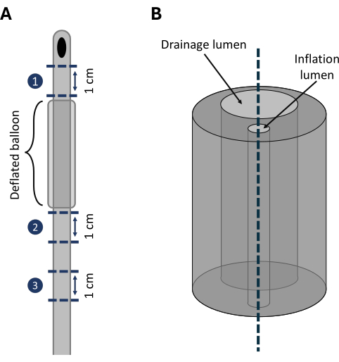
Figure 2: Sectioning of urinary catheters to process biofilm-associated cells. Example points on a urinary catheter for dissection for downstream biofilm quantification. (A) Catheters are first sectioned latitudinally in the 8 cm region proximal to the tip/eyeholes (where most biofilm formation is observed). 1 cm sections are removed with a scalpel, generally avoiding the region containing the deflated catheter balloon due to difficulties in processing reproducibility (1-3). (B) Once catheters are initially sectioned into 1 cm pieces, another longitudinal cut is made down the central lumen to expose any biofilm present for further biofilm quantification, i.e., CFU/mL enumeration of crystal violet staining. Dashed lines represent dissection points. Please click here to view a larger version of this figure.
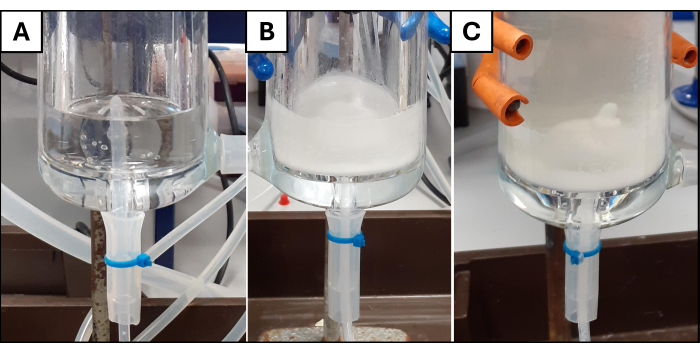
Figure 3: Stages of an in vitro bladder model experiment. Images of the in vitro bladder model central chamber, throughout different stages of experimentation. (A) AUM in the in vitro bladder model central chamber prior to inoculation. Media in the central chamber is completely transparent, indicating successful sterilization, and the catheter tip and eyeholes are visible above the line of media, indicating the catheter is successfully draining. (B) Media in the in vitro bladder model central chamber following inoculation with P. mirabilis (4 h post-inoculation). Successful inoculation is indicated by the increased turbidity of the media. The visibility of the catheter tip and eyeholes indicate that AUM is successfully draining through the catheter, and blockage has not occurred. (C) An example of a blocked in vitro bladder model. Inoculation with P. mirabilis and the subsequent biofilm formation and mineralization have resulted in the blockage of catheter eyeholes, preventing drainage and causing the level of media in the central chamber to rise above the catheter tip. Please click here to view a larger version of this figure.
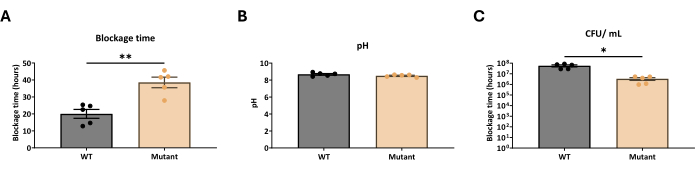
Figure 4: Endpoint measurements for an in vitro bladder model run until blockage. Example endpoint measurements at the time of blockage from an in vitro bladder model inoculated with a clinical strain of P. mirabilis, RS47 (WT), and a mutant strain with attenuated biofilm formation, RS47-2 (Mutant). (A) Blockage time (h) of WT and mutant strains of P. mirabilis. (B) pH of in vitro bladder models at the time of blockage. (C) CFU/mL of planktonic cells in in vitro bladder model chamber at the time of blockage. All data represent the mean of five biological replicates. Error bars show the standard error of the mean (SEM). *p ≤ 0.05; **p ≤ 0.01. This figure has been modified from Clarke et al.46. Please click here to view a larger version of this figure.
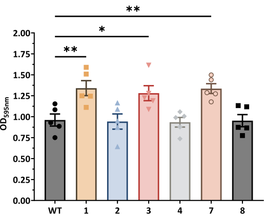
Figure 5: Biofilm-associated cell measurements from the in vitro bladder model. Quantification of P. mirabilis biofilm formation, comparing wild-type strain HI4320 to adapted populations passaged with or without chlorhexidine (CHD) via crystal violet staining and measurement of optical density (OD) at 595 nm. WT: P. mirabilis HI4320; 1, 3, 5: passage controls with no CHD exposure; 2, 4, 6: HI4320 passaged in 4 µg/ mL CHD. All data represent the mean of five biological replicates. Error bars show SEM. *p ≤ 0.05; **p ≤ 0.01. Please click here to view a larger version of this figure.
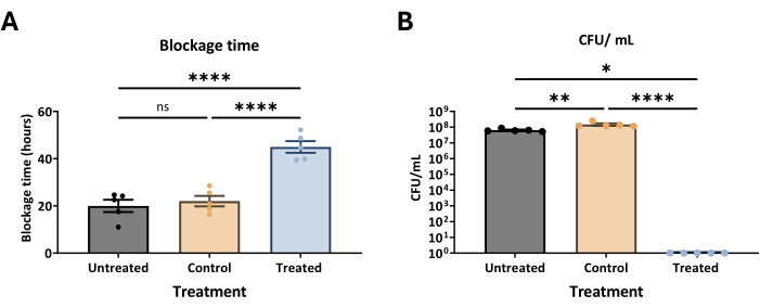
Figure 6: Measurements from in vitro bladder models following antimicrobial treatment. Planktonic samples were taken from an in vitro bladder model inoculated with P. mirabilis, following irrigation with a 0.02% chlorhexidine antimicrobial solution (100 mL) or a 0.9 % saline control. (A) Blockage time of in vitro bladder models following irrigation. (B) Planktonic CFU/mL immediately following irrigation treatment. All data represent the mean of five biological replicates. Error bars show SEM. *p ≤ 0.05; **p ≤ 0.01; ****p ≤ 0.0001. Please click here to view a larger version of this figure.
Discussion
The catheterized urinary tract represents a complex biological niche that can be challenging to replicate in a laboratory environment. The in vitro bladder model was designed for the purpose of simulating CAUTI, and utilizes standard urinary catheters to replicate the sterile, closed drainage system used in clinical practice. The catheterized urinary tract is highly susceptible to infection, as is evidenced by urinary catheters being the single biggest risk factor for the development of CAUTI6,55. Commensurately, careful aseptic technique is critical to the success of running in vitro bladder models. Turbidity of AUM is a clear indicator of sterility, as contaminated or expired media will become cloudy over time, requiring all replicates to be discarded. Careful aseptic technique should also be utilized when inoculating and sampling the in vitro bladder model, as opening the central chamber to add in the prepared inoculum breaks the sterile, closed drainage system, making the in vitro bladder model susceptible to contamination. To avoid contamination of the central chamber, triple-lumen catheters can be utilized to sample the planktonic cell population (by clamping off the waste lumen and draining via the additional lumen). Some washout is expected at the 4-h timepoint, but troubleshooting may be required if no viable planktonic cells are enumerated at this point. If no viable cells are enumerated after 4 h the inoculum density or AUM formulation utilized may need to be optimized to better support bacterial colonization. Additionally, inoculum washout may indicate an issue with the media circuit; kinks within the tubing could prevent adequate drainage, resulting in the buildup of toxic metabolites which may impede bacterial growth, whilst any residual disinfectant used to flush through media tubing could have a bactericidal effect.
This protocol details the preparation of AUM as previously described by Nzakizwanayo et al., as it has previously been validated for use in the in vitro bladder model with prominent CAUTI pathogens, including Proteus mirabilis and Escherichia coli43,45,46,48. However, there is scope to modify the AUM formulation utilized in the protocol to select for the growth of other pathogens. Numerous diverse AUM formulations have been published and could instead be used in the media circuit28,56,57,58,59,60. Additionally, AUM in the media reservoir can be supplemented with nutrients or antibiotics to select for the growth of specific pathogens. Historically, in vitro bladder models have also been supplied with pooled human urine, however, the large volumes of media required to run an in vitro bladder model replicate can make collection of human samples impractical53,54. Furthermore, the composition of human urine can vary greatly between patient samples, affecting experiment reproducibility, as differences in the concentration of hormones, salts, and proteins as well as hydration status, have been shown to affect bacterial growth and biofilm formation38,39,61,62.
The above protocol details the inoculation of the in vitro bladder model with ~1010 CFU/mL replicating late-stage CAUTI. However, the inoculation site of the in vitro bladder model could be modified in future assays to better replicate the earlier stages of CAUTI, i.e., initial colonization of the urinary catheter and ascension into the bladder. Manually contaminating Foley catheters with a known bacterial inoculum prior to the setup of in vitro models could provide insight into the early stages of CAUTI. Indeed, inoculum density has been modified in previous work to replicate earlier-stage infection; for example, Nzakizwanayo et al. inoculated in vitro bladder models with a lower starting CFU/ mL (ca. 103 CFU/mL) to replicate earlier-stage infection47. It is, however worth noting that altering inoculum density may result in extended time to blockage and requires additional validation prior to experimentation.
Additionally, whilst the protocol details inoculation of the in vitro model with bacterial monoculture, there is scope to inoculate models with fungal pathogens. Past work has suggested that up to 8% of CAUTIs result from infection with Candida spp., which are able to produce biofilms which are often recalcitrant to antifungal treatment27,63. Moreover, the in vitro bladder model can be modified to accommodate the study of polymicrobial CAUTI. Indeed, it has been suggested that up to 86% of CAUTIs are polymicrobial, and these infections are often associated with more severe symptoms and a higher rate of mortality than monospecies infection27,64,65,69. In vitro models can be inoculated with multiple species to study the formation of polymicrobial biofilms and the progression of CAUTI, but careful validation of polymicrobial communities and modification of inoculum density should be performed prior to inoculation. Previous studies have utilized the in vitro bladder model to monitor differences in biofilm formation in single species P. mirabilis CAUTI compared with dual species CAUTI with E. faecalis, highlighting the metabolic interplay between the strains, which resulted in enhanced biofilm formation compared with single species infection70.
A major advantage of using the in vitro bladder model to study CAUTI, is the ability to observe biofilm formation on the surface of a standard urinary catheter. The formation of biofilms in CAUTI is often initiated by the formation of conditioning films on the catheter lumen, comprised of organic components of urine to which uropathogens can adhere to, before proliferating and forming mature biofilms2,18,71,72,73. Standard assays used to study biofilm formation often utilize standard bacterial growth media rather than AUM and are often based on adherence to polystyrene microplates or pegs, i.e., the Calgary device; however, past work has suggested that the nutrient environment, pH, and oxygen levels of the system, as well as physical properties such as flow and difference in surface structure (silicone vs plastics), can affect biofilm formation36,37,74,75,76,77,78. Additionally, culturing bacteria with extensive urease activity, such as P. mirabilis in a closed system in AUM, can result in the buildup of toxic metabolites, which can result in cell death78,79. This is mitigated in the in vitro bladder model as the AUM in the central chamber is constantly replaced with fresh media via the media circuit, allowing for the study of CAUTI biofilms under more clinically relevant conditions45. The day-to-day running of the in vitro bladder model and the endpoint selected for models largely depend on whether models are inoculated with urease-positive or negative organisms. Theoretically, in vitro models could run indefinitely if supplied with an adequate volume of AUM and if the sterility of the model is maintained; however, it is recommended that an endpoint is selected prior to experimentation, whether that be a blockage or an arbitrary number of days. It is generally recommended that in vitro models inoculated with urease-negative organisms are run for ≤7 days to simulate the clinical environment, as this is typically sufficient to observe biofilm formation in most organisms, whilst keeping in line with clinical interventions, as standard best practice indicates that catheters should be replaced once visibly infected1,2,3,47,48. Although urease-positive organisms are likely to produce crystalline biofilms, there is a great deal of variation within urease-positive strains in time to in vitro bladder model blockage, with some strains of Proteus mirabilis causing catheter blockage in under 10 h, and others taking over 30 h to block39,46. A standard urease test could be used prior to in vitro bladder model experimentation to determine whether to run models to blockage or a pre-defined endpoint80.
Perhaps the main limitation of using the in vitro bladder model to study biofilm formation is that biofilm measurements are an endpoint reading, as they necessitate the removal and dissection of the urinary catheter. Conversely, planktonic samples can be taken from the in vitro bladder model at any point during experimentation. Additionally, once biofilm samples are taken from the in vitro bladder models, numerous extra downstream techniques can be used to better quantify biofilm production. Past studies have utilized flame photometry to quantify the amount of calcium and magnesium present in crystalline biofilms, and more complex stains can be used to accurately identify components of the EPS matrix of urease-negative biofilms45,73,81,82,83,84.
This work also describes the application of the in vitro bladder model to study the effects of therapeutics aimed at treating CAUTI. The in vitro bladder model simulates important aspects of the CAUTI niche, such as the use of AUM, as well as clinically relevant biofilm formation and increased organic load seen in CAUTI. Therapeutics such as antibiotics can be added directly to the AUM tank to simulate clinically relevant drug bioavailability, and past studies have utilized repurposed drugs such as thioridazine and fluoxetine to demonstrate an antibiofilm effect81. Additionally, minimum inhibitory concentration assays and bacterial suspension tests are often used to infer the antimicrobial activity of biocides, though there is little evidence to suggest that assays correlate to real-world biocide susceptibility41,85,86,87,88. The in vitro bladder model is advantageous for the testing of biocidal products aimed at treating or preventing CAUTI, as the presence of the urinary catheter and more representative simulation of the CAUTI niche mean that products such as antimicrobial lubricant gels and irrigation solutions can be tested as per manufacturer's instructions. Past studies have also used the in vitro bladder model to determine the effectiveness of phage therapy, and demonstrated that phage treatment could extend P. mirabilis time to block the in vitro bladder model by over 100 h47. The use of a standard-sized Foley catheter in the in vitro bladder model also means novel, coated catheters can be assayed for antimicrobial activity. Past work by Slate et al. demonstrated that catheters coated in a theranostic ciprofloxacin coating could extend P. mirabilis time to blockage by ~60 h in an in vitro bladder model48. This highlights the potential application of the in vitro bladder model in the study of novel therapeutics aimed at treating CAUTI, allowing for a more accurate assessment of antimicrobials in a biologically relevant simulated CAUTI niche.
Disclosures
The authors have nothing to disclose.
Acknowledgements
We thank Dr. Jonathan Nzakizwanayo and Dr. Anthony J. Slate for their shared expertise in developing this protocol. Work at the University of Bath was funded by The Medical Research Council GW4 Biomed DTP as a studentship to V.B. (MR/N0137941/1). B. V. J. is also supported by funding from the Dunhill Medical Trust (RPGF1906\171). Research by O.E.C. was funded by Kidney Research Northwest (RCN: 1144798). Figure 1 is created with Biorender.com.
Materials
| Name | Company | Catalog Number | Comments |
|---|---|---|---|
| 313 Rapid-Load Pumphead Extension | Watson-Marlow | 313 | Peristaltic pump - extra pumphead extension |
| 323S/D manual control variable speed pump | Watson-Marlow | 323S/D | Peristaltic pump |
| 3L Bed bags, sterile with lever taps | Great Bear Healthcare | GDW173 | |
| 3mm inner diameter platinum cured silicone tubing | Fisher | 10111801 | Small AUM circuit tubing (connect pump tubing to models) |
| 5mm inner diameter platinum cured silicone tubing | Fisher | 10539201 | Medium AUM circuit tubing (connect pump tubing to AUM reservoir) |
| ammonium chloride (NH4Cl) | Fisher | 11473713 | |
| anhydrous sodium sulfate (Na2SO4) | Fisher | 11498717 | |
| Azlon Tapered connector | Fisher | 10712807 | Used to connect different lengths of tubing in AUM circuit |
| Bed bag hangers | Great Bear Healthcare | 10705P | |
| Bochem pinch cock tube clamps, 25 mm diameter | Fisher | 11805101 | |
| calcium chloride (CaCl2) | Fisher | 10021681 | |
| Citric acid monohydrate | Merck | C1909-500G | Used to remove crystalline biofilm from models post-experimentation |
| Fisherbrand Glass Graduated Class B Type 1 Pipettes | Fisher | 11992168 | Used to supply in vitro blader models with AUM via bored bung |
| gelatine | VWR | 24360.233 | |
| Graphpad prism 10 | GraphPad | V10.0 | Analytical software used for graphing in vitro bladder model results and statistical analysis. |
| Hanna HI 2211-02 pH benchtop meter | Phillip Harris | PP00057414 | |
| In vitro bladder model | - | - | Bespoke equipment as previously described by Nzakizwanayo et al. (2019). |
| Instillagel 6 mL lubricant gel | mydoctorshop.co.uk | 40-006 | Lubricant gels used in section 7.1.; other suppliers are available. |
| Kartell Plastilab Uneven tapered connector | Fisher | 11914289 | Used to connect tubing ofdifferent diameters |
| MacConkey agar without salt (dehydrated) | Fisher | 10472643 | For the enumeration of single colonies of swarming bacteria i.e. Proteus mirabilis |
| magnesium chloride hexahydrate (MgCl2.6H2O) | Fisher | 10386743 | |
| monopotassium phosphate (KH2PO4) | Fisher | 10573181 | |
| Nalgene Y-shaped connector | Fisher | 10186070 | Used to run multiple AUM circuits from one AUM reservoir (1-4 models per tank) |
| potassium chloride (KCl) | Fisher | 10375810 | |
| Pyrex Aspirator Bottle with Ground Glass Side Socket, Capacity: 5000 mL | Fisher | 11347884 | AUM reservoir |
| Rubber Stoppers With a Bore, Diameter Outer: 37mm, Red, Diameter: 37mm | Fisher | 11517612 | Used in AUM reservoir and to supply in vitro models with AUM |
| Size 14 French 100 % silicone Foley catheter with sterile syringe of water for inflation | Great Bear Healthcare | 4833-0514 | |
| sodium chloride (NaCl) | Fisher | 10735921 | |
| sodium oxalate (Na2C2O4) | Fisher | 10442211 | |
| Teknon Biocleanse concentrate biocidal cleaner 5L | Fisher | 12447580 | Used to sterilize models post-experimentation |
| trisodium citrate (Na3C6H5O7) | Fisher | 11434993 | |
| tryptone soya broth | Fisher | 10198002 | |
| urea (CH4N2O) | Fisher | 10404185 | |
| Uro-Tainer M NaCl 0,9% | B.Braun | FB99833 | Saline bladder irrigation pouch used in section 7.2. |
| Watson-Marlow Pumpsil 1.6mm inner diameter platinum cured silicone tubing | Fisher | 12406280 | Thick AUM circuit tubing (for use in perstaltic pump) |
References
- Smith, D. R., et al. Epidemiology and health-economic burden of urinary-catheter-associated infection in English NHS hospitals: A probabilistic modelling study. J Hosp Infect. 103 (1), 44-54 (2019).
- Stickler, D. J. Bacterial biofilms in patients with indwelling urinary catheters. Nat Clin Pract Urol. 5 (11), 598-608 (2008).
- Feneley, R. C., Hopley, I. B., Wells, P. N. Urinary catheters: History, current status, adverse events and research agenda. J Med Eng Technol. 39 (8), 459-470 (2015).
- Lapides, J., Diokno, A. C., Silber, S. J., Lowe, B. S. Clean, intermittent self-catheterization in the treatment of urinary tract disease. J Urol. 107 (3), 458-461 (1972).
- Suetens, C., et al. Prevalence of healthcare-associated infections, estimated incidence and composite antimicrobial resistance index in acute care hospitals and long-term care facilities: Results from two European point prevalence surveys, 2016 to 2017. Euro Surveill. 23 (46), 1800516(2018).
- Nicolle, L. E. Catheter associated urinary tract infections. Antimicrob Resist Infect Control. 3, 23(2014).
- Jacobsen, S. M., Stickler, D. J., Mobley, H. L., Shirtliff, M. E. Complicated catheter-associated urinary tract infections due to Escherichia coli and Proteus mirabilis. Clinical Microbiol Rev. 21 (1), 26-59 (2008).
- Trautner, B. W., Darouiche, R. O. Role of biofilm in catheter-associated urinary tract infection. Am J Infect Control. 32 (3), 177-183 (2004).
- Garibaldi, R. A., Burke, J. P., Dickman, M. L., Smith, C. B. Factors predisposing to bacteriuria during indwelling urethral catheterization. N Engl J Med. 291 (5), 215-219 (1974).
- Warren, J. W., et al. Fever, bacteremia, and death as complications of bacteriuria in women with long-term urethral catheters. JInfect Dis. 155 (6), 1151-1158 (1987).
- Walker, J. N., et al. High-resolution imaging reveals microbial biofilms on patient urinary catheters despite antibiotic administration. World JUrol. 38 (9), 2237-2245 (2020).
- Chant, C., Smith, O. M., Marshall, J. C., Friedrich, J. O. Relationship of catheter-associated urinary tract infection to mortality and length of stay in critically ill patients: A systematic review and meta-analysis of observational studies. Crit Care Med. 39 (5), 1167-1173 (2011).
- Clarke, K., et al. Catheter-associated urinary tract infections in adults: Diagnosis, treatment, and prevention. J Hosp Med. 15 (9), 552-556 (2020).
- Tambyah, P. A., Maki, D. G. The relationship between pyuria and infection in patients with indwelling urinary catheters: A prospective study of 761 patients. Arch Intern Med. 160 (5), 673-677 (2000).
- Flemming, H. C., et al. Biofilms: An emergent form of bacterial life. Nat Rev Microbiol. 14 (9), 563-575 (2016).
- Stewart, P. S., Costerton, J. W. Antibiotic resistance of bacteria in biofilms. Lancet. 358 (9276), 135-138 (2001).
- Stewart, P. S. Mechanisms of antibiotic resistance in bacterial biofilms. Int J Med Microbiol. 292 (2), 107-113 (2002).
- Donlan, R. M., Costerton, J. W. Biofilms: Survival mechanisms of clinically relevant microorganisms. Clin Microbiol Rev. 15 (2), 167-193 (2002).
- Sabir, N., et al. Bacterial biofilm-based catheter-associated urinary tract infections: Causative pathogens and antibiotic resistance. Am JInfect Control. 45 (10), 1101-1105 (2017).
- Touzel, R., Sutton, J., Wand, M. Establishment of a multi-species biofilm model to evaluate chlorhexidine efficacy. JHosp Infect. 92 (2), 154-160 (2016).
- Luo, Y., Yang, Q., Zhang, D., Yan, W. Mechanisms and control strategies of antibiotic resistance in pathological biofilms. J Microbiol Biotechnol. 31 (1), 1-7 (2021).
- Dincer, S., Uslu, F. M., Delik, A. Antibiotic resistance in biofilm. Bacterial biofilms. , IntechOpen. (2020).
- Soto, S. M. Importance of biofilms in urinary tract infections: New therapeutic approaches. Adv Biol. 2014 (1), 543974(2014).
- Fux, C. A., Costerton, J. W., Stewart, P. S., Stoodley, P. Survival strategies of infectious biofilms. Trends Microbiol. 13 (1), 34-40 (2005).
- Otto, S. B., et al. Privatization of biofilm matrix in structurally heterogeneous biofilms. MSystems. 5 (4), e00425-e00520 (2020).
- Wood, T. K., Knabel, S. J., Kwan, B. W. Bacterial persister cell formation and dormancy. Appl Environ Microbiol. 79 (23), 7116-7121 (2013).
- Gaston, J. R., Johnson, A. O., Bair, K. L., White, A. N., Armbruster, C. E. Polymicrobial interactions in the urinary tract: Is the enemy of my enemy my friend. InfectImmun. 89 (4), (2021).
- Griffith, D. P., Musher, D. Á, Itin, C. Urease. The primary cause of infection-induced urinary stones. Invest Urol. 13 (5), 346-350 (1976).
- Armbruster, C. E., Mobley, H. L. T., Pearson, M. M. Pathogenesis of Proteus mirabilis infection. EcoSal Plus. 8 (1), 1128(2018).
- Jacobsen, S. M., Shirtliff, M. E. Proteus mirabilis biofilms and catheter-associated urinary tract infections. Virulence. 2 (5), 460-465 (2011).
- Mobley, H. L., Warren, J. W. Urease-positive bacteriuria and obstruction of long-term urinary catheters. J Clin Microbiol. 25 (11), 2216-2217 (1987).
- Wasfi, R., Hamed, S. M., Amer, M. A., Fahmy, L. I. Proteus mirabilis biofilm: Development and therapeutic strategies. Front Cell Infect Microbiol. 10 (414), (2020).
- Broomfield, R. J., Morgan, S. D., Khan, A., Stickler, D. J. Crystalline bacterial biofilm formation on urinary catheters by urease-producing urinary tract pathogens: A simple method of control. J Med Microbiol. 58 (10), 1367-1375 (2009).
- Stickler, D. J. Clinical complications of urinary catheters caused by crystalline biofilms: Something needs to be done. J Intern Med. 276 (2), 120-129 (2014).
- Coffey, B. M., Anderson, G. G. Biofilm formation in the 96-well microtiter plate. Pseudomonas methods and protocols. , 631-641 (2014).
- Ceri, H., et al. The Calgary biofilm device: New technology for rapid determination of antibiotic susceptibilities of bacterial biofilms. J Clin Microbiol. 37 (6), 1771-1776 (1999).
- Peeters, E., Nelis, H. J., Coenye, T. Comparison of multiple methods for quantification of microbial biofilms grown in microtiter plates. J Microbiol Methods. 72 (2), 157-165 (2008).
- Bouatra, S., et al. The human urine metabolome. PloS one. 8 (9), e73076(2013).
- Holling, N., et al. Elucidating the genetic basis of crystalline biofilm formation in Proteus mirabilis. Infect Immun. 82 (4), 1616-1626 (2014).
- Lawrence, E., Turner, I. Materials for urinary catheters: A review of their history and development in the UK. Med Eng Phys. 27 (6), 443-453 (2005).
- Stickler, D. J. Chlorhexidine resistance in Proteus mirabilis. J Clin Pathol. 27 (4), 284-287 (1974).
- Fraise, A. P. Biocide abuse and antimicrobial resistance-a cause for concern. J Antimicrob Chemother. 49 (1), 11-12 (2002).
- Pelling, H., et al. Derepression of the smvA efflux system arises in clinical isolates of proteus mirabilis and reduces susceptibility to chlorhexidine and other biocides. Antimicrob Agents Chemother. 63 (12), e01535(2019).
- Stickler, D. J., Morris, N. S., Winters, C. Simple physical model to study formation and physiology of biofilms on urethral catheters. MethodsEnzymol. 310, 494-501 (1999).
- Nzakizwanayo, J., Pelling, H., Milo, S., Jones, B. V. An in vitro bladder model for studying catheter-associated urinary tract infection and associated analysis of biofilms. Proteus mirabilis: Methods and Protocols. , 139-158 (2019).
- Clarke, O. E., et al. Lipopolysaccharide structure modulates cationic biocide susceptibility and crystalline biofilm formation in Proteus mirabilis. Front Microbiol. 14, 1150625(2023).
- Nzakizwanayo, J., et al. Bacteriophage can prevent encrustation and blockage of urinary catheters by proteus mirabilis. Antimicrob Agents Chemother. 60 (3), 1530-1536 (2015).
- Slate, A. J., et al. Infection-responsive coatings to reduce biofilm formation and encrustation of urinary catheters. J Appl Microbiol. 134 (6), lxad121(2023).
- Maierl, M., Jörger, M., Rosker, P., Reisner, A. In vitro dynamic model of a catheterized bladder and biofilm assay. Bio Protoc. 5 (2), e1381(2015).
- Stickler, D., Clayton, C., Chawla, J. The resistance of urinary tract pathogens to chlorhexidine bladder washouts. J Hosp Infect. 10 (1), 28-39 (1987).
- King, J., Stickler, D. An assessment of antiseptic bladder washout solutions using a physical model of the catheterized bladder. J Hosp Infect. 18 (3), 179-190 (1991).
- King, J., Stickler, D. The effect of repeated instillations of antiseptics on catheter-associated urinary tract infections: A study in a physical model of the catheterized bladder. Urol Res. 20, 403-407 (1992).
- Morris, N., Stickler, D. Encrustation of indwelling urethral catheters by Proteus mirabilis biofilms growing in human urine. J Hosp Infect. 39 (3), 227-234 (1998).
- Stickler, D., Morgan, S. Observations on the development of the crystalline bacterial biofilms that encrust and block foley catheters. J Hosp Infect. 69 (4), 350-360 (2008).
- Stamm, W. E. Catheter-associated urinary tract infections: Epidemiology, pathogenesis, and prevention. Am J Med. 91 (3, Supplement 2), S65-S71 (1991).
- Sarigul, N., Korkmaz, F., Kurultak, I. A new artificial urine protocol to better imitate human urine. Sci Rep. 9 (1), 20159(2019).
- Brooks, T., Keevil, C. A simple artificial urine for the growth of urinary pathogens. Lett Appl Microbiol. 24 (3), 203-206 (1997).
- Ipe, D. S., Horton, E., Ulett, G. C. The basics of bacteriuria: Strategies of microbes for persistence in urine. Front CellI Infect Microbiol. 6 (14), 3389(2016).
- Ipe, D. S., Ulett, G. C. Evaluation of the in vitro growth of urinary tract infection-causing gram-negative and gram-positive bacteria in a proposed synthetic human urine (SHU) medium. J Microbioll Methods. 127, 164-171 (2016).
- Chutipongtanate, S., Thongboonkerd, V. Systematic comparisons of artificial urine formulas for in vitro cellular study. Anal Biochem. 402 (1), 110-112 (2010).
- Saude, E. J., Adamko, D., Rowe, B. H., Marrie, T., Sykes, B. D. Variation of metabolites in normal human urine. Metabolomics. 3, 439-451 (2007).
- Andersen, S., et al. Proteomes of uropathogenic Escherichia coli growing in human urine and in j82 urinary bladder cells. Proteomes. 10 (2), 15(2022).
- Uppuluri, P., Dinakaran, H., Thomas, D. P., Chaturvedi, A. K., Lopez-Ribot, J. L. Characteristics of Candida albicans biofilms grown in a synthetic urine medium. J Clin Microbiol. 47 (12), 4078-4083 (2009).
- Gaston, J. R., et al. Enterococcus faecalis polymicrobial interactions facilitate biofilm formation, antibiotic recalcitrance, and persistent colonization of the catheterized urinary tract. Pathogens. 9 (10), 835(2020).
- Warren, J. W., Tenney, J. H., Hoopes, J. M., Muncie, H. L., Anthony, W. C. A prospective microbiologic study of bacteriuria in patients with chronic indwelling urethral catheters. The JI Infect Dis. 146 (6), 719-723 (1982).
- Armbruster, C. E., et al. Genome-wide transposon mutagenesis of Proteus mirabilis: Essential genes, fitness factors for catheter-associated urinary tract infection, and the impact of polymicrobial infection on fitness requirements. PLoS Pathog. 13 (6), e1006434(2017).
- Armbruster, C. E., et al. The pathogenic potential of Proteus mirabilis is enhanced by other uropathogens during polymicrobial urinary tract infection. Infect Immun. 85 (2), e00808(2017).
- Siegman-Igra, Y., Kulka, T., Schwartz, D., Konforti, N. Polymicrobial and monomicrobial bacteraemic urinary tract infection. J Hosp Infect. 28 (1), 49-56 (1994).
- Nye, T. M., et al. Microbial co-occurrences on catheters from long-term catheterized patients. Nat Commun. 15 (1), 61(2024).
- Hunt, B. C., et al. Metabolic interplay between Proteus mirabilis and Enterococcus faecalis facilitates polymicrobial biofilm formation and invasive disease. mBio. 15, e02164(2024).
- Stickler, D., Ganderton, L., King, J., Nettleton, J., Winters, C. Proteus mirabilis biofilms and the encrustation of urethral catheters. Urol Res. 21, 407-411 (1993).
- Donlan, R. M. Biofilms: Microbial life on surfaces. Emerg Infect Dis. 8 (9), 881-890 (2002).
- Habash, M., Reid, G. Microbial biofilms: Their development and significance for medical device-related infections. J Clin Pharmacol. 39 (9), 887-898 (1999).
- Ommen, P., Zobek, N., Meyer, R. L. Quantification of biofilm biomass by staining: Non-toxic safranin can replace the popular crystal violet. J Microbiol Methods. 141, 87-89 (2017).
- Wilson, C., et al. Quantitative and qualitative assessment methods for biofilm growth: A mini-review. Research and Reviews: J Eng Technol. 6 (4), 30214915(2017).
- Niebergall, A. K., et al. Influence of polymerized siloxane coating on growth and biofilm formation of aerobic grown nosocomial bacteria. J Cell Biotechnol. 3 (2), 107-115 (2018).
- Ionescu, A., Brambilla, E., Sighinolfi, M. C., Mattina, R. A new urinary catheter design reduces in vitro biofilm formation by influencing hydrodynamics. J Hosp Infect. 114, 153-162 (2021).
- Pelling, H., et al. Bacterial biofilm formation on indwelling urethral catheters. Lett Appl Microbiol. 68 (4), 277-293 (2019).
- Schaffer, J. N., Pearson, M. M. Proteus mirabilis and urinary tract infections. Microbiol Spectr. 3 (5), 1128(2015).
- Konieczna, I., et al. Bacterial urease and its role in long-lasting human diseases. Curr Protein Pept Sci. 13 (8), 789-806 (2012).
- Nzakizwanayo, J., et al. Fluoxetine and thioridazine inhibit efflux and attenuate crystalline biofilm formation by Proteus mirabilis. Sci Rep. 7 (1), 12222(2017).
- Strathmann, M., Wingender, J., Flemming, H. C. Application of fluorescently labeled lectins for the visualization and biochemical characterization of polysaccharides in biofilms of Pseudomonas aeruginosa. J Microbiol Methods. 50 (3), 237-248 (2002).
- Van Den Driessche, F., Rigole, P., Brackman, G., Coenye, T. Optimization of resazurin-based viability staining for quantification of microbial biofilms. J Microbiol Methods. 98, 31-34 (2014).
- Trafny, E. A., Lewandowski, R., Zawistowska-Marciniak, I., Stępińska, M. Use of MTT assay for determination of the biofilm formation capacity of microorganisms in metalworking fluids. World J Microbiol Biotechnol. 29, 1635-1643 (2013).
- Chapman, J. S. Biocide resistance mechanisms. Int Biodeterior Biodegrad. 51 (2), 133-138 (2003).
- Cloete, T. E. Resistance mechanisms of bacteria to antimicrobial compounds. Int Biodeterior Biodegrad. 51 (4), 277-282 (2003).
- Maillard, J. Y. Resistance of bacteria to biocides. Microbiol Spectr. 6 (2), 1128(2018).
- Maillard, J. Y. Bacterial resistance to biocides in the healthcare environment: Should it be of genuine concern. J Hosp Infect. 65, 60-72 (2007).
Reprints and Permissions
