Here, the protocol explains step-by-step the technique of harvesting an upper arm allograft at the trans humeral or gleno-humeral level during an arm allotransplantation.
A subscription to JoVE is required to view this content. Sign in or start your free trial.
Method Article
Vascularized Composite Upper Limb Allograft Harvesting for Proximal Arm Allotransplantation
In This Article
Summary
Abstract
In vascularized composite allograft (VCA) for upper limb reconstruction, upper arm-level or transhumeral transplantations are performed less frequently than forearm and hand transplantations. Transplantation is technically more feasible at this proximal level, largely owing to the use of macrovascular anastomoses. Despite these challenges, the outlook for arm allotransplantation remains encouraging, and this protocol provides a standardized technique for harvesting a vascularized upper arm composite allograft, ensuring both optimal outcomes and minimal tissue trauma. In the case of upper arm transplant, the technique varies according to the level of transplantation: supracondylar, transhumeral at a proximal arm level, or through the shoulder with the donor humeral head. A circumferential skin incision at mid-arm, or depending on the level at the origin of the upper limb, is made. A skin flap, such as a deltoid flap, can be harvested from the donor for skin closure at proximal levels. Dissection of the cephalic vein and deeper the brachial or axillary artery and veins is required. Then, the surgeon must identify the nerves depending on the level (terminal branches of brachial plexus or anteromedial and anterolateral cords for proximal transplantation) and transect the biceps, brachialis, and triceps muscles. The coracobrachialis and the deltoid muscles may also be dissected depending on the amputation level. Finally, the surgeon must make a transhumeral osteotomy or, if needed, harvest the donor humeral head for shoulder reconstruction. In this case, a peroneus longus graft for the suspension ligamentoplasty can also be harvested. The goal of this protocol is to standardize the procedure of harvesting and preparing an upper arm allotransplant.
Introduction
Since the first bilateral arm transplant in 2008 in Germany1, upper arm allotransplantation has been performed only 18 times2,3. The idea of transplantation at this level comes from a study showing good functional results for same-level replantation4,5,6. These 18 patients opened a new perspective to treating above-elbow amputees. The benefits of arm vascularized composite allograft (VCA) compared to prosthesis are essentially the recovery of sensation and complete body image7,8. The first studies of the outcomes were encouraging and increased interest in transplantation at proximal levels9,10.
Arm allotransplantation is less frequent than hand transplantation, and the explanation is, amongst others, the greater distance for nerve regeneration and the possibility of less effective function4. Hand transplantation is better studied with more data on results and detailed protocol for surgery11,12,13,14. Surgical rehearsal is necessary to prepare for the probable augmentation of arm VCA in the future. Hence, protocols for harvesting and preparing the graft are needed.
This protocol details all the processes from the installation to the closure of the donor limb during an upper arm VCA. It explains the steps to harvest the graft at a proximal humerus level or through the shoulder using a second method. The procurement level depends on the recipient stump’s condition and whether glenohumeral joint reconstruction is required.
The overall goal of this method is to standardize the harvest protocol for arm allotransplantation. Indeed, this procedure has been performed very few times in the world and can only be realized by an experienced, multidisciplinary team to successfully carry out the graft and the postoperative follow-up, which involves very lengthy rehabilitation. In addition, this procedure can only be performed in authorized centers or under a national hospital clinical research program.
Upper limb transplantation and prosthetic reconstruction should not be considered as competing options but rather as two treatment modalities with different risk-benefit profiles and indications8. Limb transplantation is a unique option for the restoration of sensation, body image, and function. The long-term evaluation of these patients (4 to 20 years) shows that functional outcomes and quality of life are very good overall, with a progressive improvement during the first three years before stabilizations3. In France, the approved indications for upper limb vascularized composite allotransplantation are restricted to bilateral transplantation, specifically in cases of bilateral traumatic amputation.
Access restricted. Please log in or start a trial to view this content.
Protocol
The Anatomy Laboratory of the Faculty of Medicine of Nice, France, generously provided the specimens and material used for the study. The French National Ethics Committee approved this study (approval number 83.2024), which was conducted following the Helsinki Declaration.
1. Preoperative care
- In a vegetative state patient who is an organ donor, install the patient half sitting at 30° inclination, with slightly flexed hips and the heel suspended, on a custom cushion. The arm is positioned on a Trimano to be easily mobilized.
NOTE: Ideally, the procurement should be performed under beating-heart conditions to minimize ischemic time and prior to the retrieval of other organs to preserve optimal tissue quality without adversely affecting the outcome of the remaining grafts. - Prepare and drape the upper limb, including the sternoclavicular joint, the mastoid, the bi-mammary line, and the scapula. Secure the sterile fields with staples. Also, drape both lower limbs to harvest, if needed, as well as femoral vessels, fibula, fibular tendons, and sural nerves, for potential grafts.
NOTE: The harvest needs to be as proximal as possible; hence, using a tourniquet is impossible. Precise timing between donor and recipient teams is essential; harvest teams usually begin 1 h before the recipient incision.
2. Donor upper limb harvesting for transhumeral transplantation (Figure 1)
NOTE: All structures were identified, isolated, and tagged during the recovery procedure.
- Make a circumferential fish mouth incision with a surgical scalpel blade n°15 proximal to the deltoid humeral insertion.
NOTE: Keep a deltoid and an axillary skin flap on the graft side to interdigitate with the recipient's volar and dorsal skin flap. - Raise the volar proximal skin flap using fine needle cautery and a pair of Adson tissue forceps, ligating proximally and dividing the cephalic veins.
NOTE: Ligate the structures with a 0 braided absorbable suture or hemostatic clips. An incision at the deltopectoral interval may be useful for dissecting the cephalic vein proximally. - Detach the pectoralis major from the humerus with monopolar diathermy to expose the coracobrachialis. Recline the deltoid with a scalpel from the clavicula and the acromion to access the scapular insertion of the long portion of the biceps brachii.
NOTE: The deltoid flap of the graft must be myocutaneous to keep the skin vascularized by the muscular perforator of the circumflex artery of the deltoid. - Isolate and detach, with a scalpel, the long portion of the biceps brachii and its short portion, along with the coracobrachialis. Also, detach the pectoralis minor from the coracoid to access the brachial plexus.
- Dissect and transect with scissors the median, ulnar, radial, and musculocutaneous nerves in their proximal third or at the cord level. Cut the axillary nerve to allow muscular involution of the deltoid.
NOTE: The medial brachial and antebrachial cutaneous nerves can also be transected at the same level for possible reparation. - Expose the brachial artery and veins up to the axillary vessels with scissors and tag them.
NOTE: If needed, a clavicular osteotomy can be performed to facilitate the dissection of the axillary artery and veins the more proximal possible. Ligate the brachial artery proximal to the origin of the profunda brachii artery. - Make a longitudinal mark along the bicipital groove to facilitate accurate rotational alignment during osteosynthesis. Make a transverse humerus osteotomy with an oscillating saw at the preoperative planned level. The level can be adjusted according to the remaining humerus on the recipient residual limb.
NOTE: Depending on the osteotomy level, it may be necessary to transect the deltoid and detach from their humeral insertion the tendons of the latissimus dorsi and teres major. Make sure to keep the tendons on the humerus as long as possible for future tenorrhaphy. Transection of the subscapularis can also be useful to gain a good exposition of the proximal extremity of the humerus. - Raise the dorsal proximal skin flap using fine needle cautery and a pair of Adson tissue forceps and clip proximally any superficial veins with hemostatic clips.
- Isolate and detach, with a scalpel, from their proximal origins the long and lateral heads of the triceps brachii.
- Ligate proximally the axillary vessels and divide them.
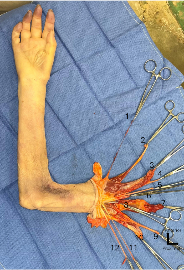
Figure 1: Transhumeral graft. (1) Cephalic vein, (2) Long portion of biceps brachii, (3) Short portion of biceps brachii with coracobrachialis, (4) Musculocutaneous nerve, (5) Median nerve, (6) Axillary artery, (7) Lateral head of triceps brachii, (8) Axillary vein, (9) Radial nerve, (10) Long head of triceps brachii, (11) Medial antebrachial cutaneous nerve, (12) Ulnar nerve. Please click here to view a larger version of this figure.
3. Packaging of the graft
- Catheterize the axillary artery and secure the cannula with suture ligation.
- Flush the limb via a universal irrigation set until clear venous outflow, with a preservation fluid such as University of Wisconsin (UW) solution at 4° C, Histidine-Tryptophan Ketoglutarate (HTK) infused with 5000 units of unfractionated heparin, or IGL-1 to preserve the graft.
- Package the graft as standard solid organ transplantation (SOT) protocol, wrap the limb with sterile towels, then place it in sterile bags, with the third one filled with ice water. Place the bags in a wheeled cooler.
4. Management of the donor stump
- Check for any bleeding and do hemostasis with bipolar diathermy.
- Close the incision with a suture or staples to obtain a water-tight seal.
- Put in place the prosthesis to restore the form.
5. Second technique of harvesting: Donor upper limb harvesting for transplantation at the glenohumeral level (with shoulder reconstruction) (Figure 2 and Figure 3)
NOTE: Same preoperative care as described in section 1.
- Make a double ogival-shaped incision with the proximal extremity at the coracoid level and the base 10 cm more distal, with a superior acromio-clavicular flap delimited by the deltopectoral interval and an inferior axillary flap. Extend proximally the skin incision to the sterno-clavicular joint, then 10 cm up to the posterior border of the sternocleidomastoid muscle.
NOTE: Preserve the cephalic vein in the deltopectoral interval at this level. - Detach the three chiefs of the deltoid from the clavicula, acromion, and scapula with a scalpel. Cut the posterior chief perpendicular to the scapula spine 10 cm from the acromion.
- Detach the pectoralis minor and the short portion of biceps brachii with a scalpel, along with the coracobrachialis from the coracoid.
- Detach the clavicular insertion of the pectoralis major and transect through the muscle belly of the pectoralis major using monopolar diathermy, keeping all the tendons on its humeral insertion plus 10 cm of muscle.
- Perform a clavicular osteotomy to access proximally to vessels and brachial plexus.
- Dissect and put vessel loops around the subclavian artery and vein at a proximal level.
- Identify the lateral, medial, and posterior cords of the brachial plexus and transect them, with scissors, proximally to the origins of their terminal branches.
NOTE: Axillary and radial nerves will be separately anastomosed on the recipient residual limb and need to be individualized. - Dissect, with scissors, from proximal to distal the subclavian vessels down to the axillary vessels and tag them.
NOTE: If, despite the proximal harvest of vessels, the anastomoses are not possible, harvest of femoral vessels is possible to realize a vascular bypass. - Ligate all the collateral arteries and veins from the axillary vessels destined to the thorax with hemostatic clips or braided absorbable sutures.
NOTE: This step allows the reclining of all the neurovascular bundles laterally and exposes the subscapularis. - Isolate and detach, with a scalpel, from its proximal origin the long portion of the biceps brachii for future humeral tenodesis.
- Incise with a scalpel, the tendon of the subscapularis, and the glenohumeral joint capsule at the neck of the scapula. Transect at the same level the tendons of the supraspinatus and infraspinatus and teres minor muscles posteriorly to the joint. Disarticulate the glenohumeral joint to harvest the entire humeral head with the graft.
NOTE: Be careful to leave 2-3 cm of rotator cuff tendons on the tuberosities of the humerus. - Mobilize laterally the humerus and detach, with a scalpel, from its proximal origin the long head of the triceps. Detach from their humeral insertions the latissimus dorsi and teres major.
- Ligate proximally the axillary/subclavian vessels and divide them.
NOTE: Ligate the axillary vein proximally to the ending of the cephalic vein. The same packaging of the graft and management of the donor stump are needed. Fibula and fibular tendons can be harvested to help in osteosynthesis and shoulder ligamentoplasty.

Figure 2: Infraclavicular dissection. (1) Tendon of the pectoralis major, (2) Deltoid, (3) Short portion of biceps brachii with coracobrachialis, (4) Long portion of biceps brachii, (5) Cephalic vein, (6) Lateral cord of brachial plexus, (7) Subclavian artery, (8) Subclavian vein, (9) Posterior cord of brachial plexus, (10) Medial cord of brachial plexus, (11) Long head of triceps brachii. Please click here to view a larger version of this figure.
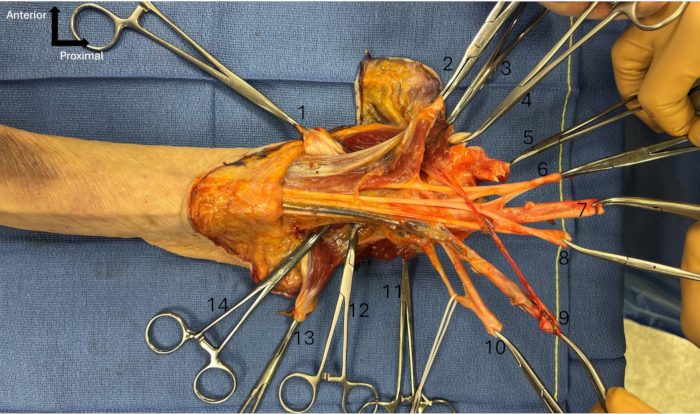
Figure 3: Total arm graft. (1) Tendon of the pectoralis major, (2) Short portion of biceps brachii with coracobrachialis, (3) Deltoid, (4) Long portion of biceps brachii, (5) Subscapularis, (6) Lateral cord of brachial plexus, (7) Subclavian artery, (8) Medial cord of brachial plexus, (9) Subclavian vein with cephalic vein, (10) Posterior cord of brachial plexus with axillary nerve in a white loop, (11) Tendons of supraspinatus, infraspinatus and teres minor, (12) Tendon of teres major, (13) Long head of triceps brachii, (14) Tendon of latissimus dorsi. Please click here to view a larger version of this figure.
Access restricted. Please log in or start a trial to view this content.
Results
This protocol allows the allotransplantation of an upper arm with a prepared graft harvested with sufficient tissue. The humerus is exposed for osteosynthesis with plate3,9 or intramedullary nail15. In the case of shoulder reconstruction, the glenohumeral joint capsule can be sutured to the joint capsule of the glenoid cavity15. Another option is a suspension ligamentoplasty using a peroneus longus graft
Access restricted. Please log in or start a trial to view this content.
Discussion
Some critical steps need particular attention. For transhumeral harvesting, depending on the level of humeral osteotomy, the detachment of the distal insertion of the pectoralis major, latissimus dorsi, and teres major can be necessary3. For nerve transection, the level depends on the recipient residual limb. Anastomose can be realized 7 cm proximal to the elbow joint15. Also, medial brachial and antebrachial cutaneous nerves can be dissected and harvested if needed for rep...
Access restricted. Please log in or start a trial to view this content.
Disclosures
The authors have no disclosures.
Acknowledgements
The authors wish to express their sincere gratitude to the individuals who generously donated their bodies to science, thereby enabling anatomical research. The insights gained from such studies have the potential to significantly advance scientific knowledge and improve patient care. As such, these donors and their families deserve our deepest appreciation.
Access restricted. Please log in or start a trial to view this content.
Materials
| Name | Company | Catalog Number | Comments |
|---|---|---|---|
| 11.5” Medium and Large Premium Surgiclip II Auto Suture vessel clip applier | Covidien | N/A | |
| 2-0 and 0 silk suture | Any brand is sufficient | ||
| Adson Forceps | MPM | 106-2112A | |
| Bipolar Coagulation Forceps | Olsen | 20-1320I | |
| Custodiol HTK Solution for limb perfusion | Essential Pharmaceuticals Inc. | https://www.custodiol.com/hansjb/wp-content/uploads/2023/03/Custodiol-HTK-IFU.pdf | off-label use |
| Cysto/ Bladder Irrigation Set | Baxter Healthcare Corp. | https://ecatalog.baxter.com/ecatalog/loadproduct.html?pid=1018837&lid=10039&cid=10032 | |
| Disposable Scalpel #15 | Sklar | https://www.sklarcorp.com/ | |
| DLP 3 mm vessel cannula blunt tip | Medtronic Inc | 30003 | |
| Fine needle cautery | Cormedica | https://www.cormedica.fr/en/cormedica-en/ | |
| Forceps Dilators | WPI | 15910 | |
| IV stopcock | N/A | N/A | Any brand is sufficient |
| Micro scissors | WPI | 504492 | |
| Monopolar Diathermy | Valleylab | N/A | |
| Oscillating saw | GPC Medical | N/A | |
| Saline solution 0.9% | GenDepot | S0600-101 | |
| Strabismus scissors | Surtex | 102-4109 | |
| Surgical marking pen | Cardinal health | 212PR | |
| Sutures Ethilon 4.0 and 3.0 | Ethicon | 1667G | |
| Syringue 10 mL | Agilent | 9301-6474 | |
| Three sterile procurement plastic bags, and three sterile zip ties | N/A | N/A | |
| Tissue Forceps | MPM | 106-0511 | |
| Vessel loop | Deroyal | 30-711 |
References
- Tuffs, A. Munich surgeons perform first transplantation of whole arms. BMJ. 337 (2), a1162-a1162 (2008).
- Iglesias, M., et al. Functional outcomes 18 months after total and midarm transplantation: a case report. Transplant Proc. 50 (3), 950-958 (2018).
- Lahlali, S., et al. Proximal bilateral arm transplantation with left shoulder reconstruction: outcomes at 24 months. Plast Reconstr Surg Glob Open. 12 (6), e5884-e5884 (2024).
- Jones, N. F., Schneeberger, S. Arm transplantation: prospects and visions. Transplant Proc. 41 (2), 476-480 (2009).
- Chew, W. Y., Tsai, T. M. Major upper limb replantation. Hand Clin. 17 (3), 395-410 (2001).
- Chen, Z. W., Yu, H. L. Current procedures in China on replantation of severed limbs and digits. Clin Orthop. 215, 15-23 (1987).
- Salminger, S., et al. Functional and psychosocial outcomes of hand transplantation compared with prosthetic fitting in below-elbow amputees: a multicenter cohort study. PLoS One. 11 (9), e0162507-e0162507 (2016).
- Kubiak, C. A., et al. Prosthetic rehabilitation and vascularized composite allotransplantation following upper limb loss. Plast Reconstr Surg. 143 (6), 1688-1701 (2019).
- Cavadas, P. C., Ibáñez, J., Thione, A., Alfaro, L. Bilateral trans-humeral arm transplantation: result at 2 years. Am J Transplant. 11 (5), 1085-1090 (2011).
- Kaczmarzyk, J. Result of arm-level upper-limb transplantation in two recipients at 19- and 30-month follow-up. Ann Transplant. 17 (3), 126-132 (2012).
- Hartzell, T. L., et al. Surgical and technical aspects of hand transplantation: is it just another replant. Hand Clin. 27 (4), 521-530 (2011).
- Mendenhall, S. D., et al. Technique for rapid hand transplant donor procurement through the elbow. Hand. 16 (3), 391-396 (2021).
- McClelland, B., et al. Proximal forearm transplantation for below elbow amputations: rationale and surgical technique. Vasc Compos Allotransplantation. 2 (1), 26-28 (2015).
- Cetrulo, C. L., Kovach, S. J. Procurement of hand and arm allografts. Tech Hand Up Extrem Surg. 17 (4), 232-238 (2013).
- Iglesias, M., et al. Anatomical and microsurgical implications in total and midarm transplantation. J Reconstr Microsurg Open. 2 (2), e94-e102 (2017).
- Salminger, S., Roche, A. D., Sturma, A., Mayer, J. A., Aszmann, O. C. Hand transplantation versus hand prosthetics: pros and cons. Curr Surg Rep. 4 (2), 8-8 (2016).
- Agnew, S., Ko, J., De La Garza, M., Kuiken, D., Dumanian, G. Limb transplantation and targeted reinnervation: a practical comparison. J Reconstr Microsurg. 28 (1), 63-68 (2012).
- Lupon, E., et al. Vascularized composite allografts in France: An update. Ann Plast Surg. 70 (2), 140-147 (2024).
- Shores, J. T., Brandacher, G., Lee, W. P. A. Hand and upper extremity transplantation: an update of outcomes in the worldwide experience. Plast Reconstr Surg. 135 (2), 351e-360e (2015).
- McFarland, L. V., Winkler, S. L. H., Heinemann, A. W., Jones, M., Esquenazi, A. Unilateral upper-limb loss: satisfaction and prosthetic-device use in veterans and servicemembers from Vietnam and OIF/OEF conflicts. J Rehabil Res Dev. 47 (4), 299(2010).
- Wells, M. W., Rampazzo, A., Papay, F., Gharb, B. B. Two decades of hand transplantation: a systematic review of outcomes. Ann Plast Surg. 88 (3), 335-344 (2022).
Access restricted. Please log in or start a trial to view this content.
Reprints and Permissions
